



Non-technical Summary
Herein we describe new specimens of a paracrinoid, which are stalked fossil echinoderms, from the Neuville Formation of Québec, Canada. Paracrinoids are restricted to the Late Ordovician (455–450 million years ago) and though globally rare, can be a locally abundant component in diverse echinoderm assemblages. Paracrinoids can vary significantly in their overall anatomy but share distinctive features including an asymmetry between the stalk and mouth along with distinctive feeding structures. An oddly preserved specimen of paracrinoid was described previously from the same locality the body outline was significantly distorted, and the preservation would likely make it difficult to compare to fossils that were preserved in a more stereotypical manner. This paracrinoid specimen was noted for having feeding structures that were unusual in their length and delicate nature, although many aspects of the animal remained unknown. Examination of this original specimen indicates many key features of the body and feeding appendages are also present in the newly discovered specimens, which allows us to more completely describe its anatomy and place it within a new genus while retaining the original species name, Quebecocystites gorgo.
The new specimens are exceptionally preserved showing features from the attachment structure, stalk, body, and feeding appendages, which are often unpreserved within paracrinoids. This allows us to more confidently place Q. gorgo in an evolutionary tree of paracrinoids. Groups of paracrinoids have been previously defined by their respiratory structures, but the new evolutionary tree including Q. gorgo supports these features evolving four separate times, thus indicating that they are less reliable features for defining these groups.
Finally, the exceptional preservation of these new specimens provides a wealth of ecological information about this animal. Examination of the well-preserved feeding structures provided a rare opportunity to quantify the hypothetical range of food particle size for this paracrinoid, thus enabling a comparison with co-occurring filter feeding echinoderms (e.g., crinoids). Interestingly, there are also imperfections in the fossil that provide further ecological details. One of the feeding appendages tapers dramatically, indicating that it was regenerated, which has been interpreted as related to predation attempts in other echinoderms. There are also several instances of swellings associated with pits on the body of the organism that may represent parasitism.
Introduction
Paracrinoidea are a small yet morphologically disparate group of Paleozoic echinoderms that are temporally and spatially limited (Parsley and Mintz, Reference Parsley and Mintz1975; Deline et al., Reference Deline, Thompson, Smith, Zamora, Rahman, Sheffield, Ausich, Kammer and Sumrall2020; Limbeck et al., Reference Limbeck, Bauer, Deline and Sumrall2024). They are primarily known from the Middle to Late Ordovician of Laurentia (Parsley and Mintz, Reference Parsley and Mintz1975), although there are several species that have been assigned to Paracrinoidea from Baltica and Gondwana based on features of their feeding appendages (Prokop and Petr, Reference Prokop and Petr1986; Rozhnov, Reference Rozhnov2017). Paracrinoids have been described to have the theca of cystoids and pinnuliferous arms of crinoids (Kesling, Reference Kesling and Moore1968). This group, which originally was classified by Jaekel (Reference Jaekel1900) and named by Regnéll (Reference Regnéll1945), is distinguished from other blastozoan echinoderms by asymmetry of the mouth and stem and uniserially plated brachioles that arise from the left side of the ambulacra. Alternatively, Rozhnov (Reference Rozhnov2017) placed several taxa (Achradocystites and Heckerites) into Paracrinoidea with biserially plated brachioles based on models of growth and homology. Their inclusion within paracrinoids has yet to be examined quantitatively in a broad phylogenetic analysis, but their potential inclusion within the group illustrates the difficulties of assigning taxa and exploring anatomical trends within this small but morphologically disparate and plastic clade. Even with the synapomorphies within Paracrinoidea noted by Jaekel (Reference Jaekel1900), Regnéll (Reference Regnéll1945), and later by Kesling (Reference Kesling and Moore1968) and Parsley and Mintz (Reference Parsley and Mintz1975), there are a variety of morphologies that can make understanding relationships among the species challenging. The clade varies in the number of ambulacra present, overall thecal shape, and in the type of respiratory structures, if they are even present (Bassler, Reference Bassler1950; Parsley and Mintz, Reference Parsley and Mintz1975; Frest et al., Reference Frest, Strimple and McGinnis1976, Reference Frest, Strimple and Witzke1980; Frest and Strimple, Reference Frest and Strimple1982; Parsley, Reference Parsley and Sprinkle1982; Sprinkle and Parsley, Reference Sprinkle, Parsley and Sprinkle1982; Guensburg, Reference Guensburg1991; Sumrall and Deline, Reference Sumrall and Deline2009; Rozhnov, Reference Rozhnov2017; Sheffield et al., Reference Sheffield, Limbeck, Bauer, Hill and Nohejlová2022; Paul et al., Reference Paul, Guensburg and Darrough2025). Given the temporal and spatial limitations of paracrinoids, their morphological and ecological disparity is consistent with other related blastozoan groups suggesting rapid evolutionary and morphological change (Deline et al., Reference Deline, Thompson, Smith, Zamora, Rahman, Sheffield, Ausich, Kammer and Sumrall2020; Novack-Gottshall et al., Reference Novack-Gottshall, Sultan, Smith, Purcell and Hanson2022, Reference NOVACK-GOTTSHALL, PURCELL, SULTAN, RANJHA, DELINE and SUMRALL2024).
Three new paracrinoid specimens were found in the Upper Ordovician Neuville Formation (Grondines Member) from two localities, east of Québec City, Québec, Canada. Prior to the discovery of these new specimens, a single paracrinoid, which is preserved as a glauconitic film (Fig. 1), had been reported from the Neuville Formation (Sinclair, Reference Sinclair1948). Sinclair (Reference Sinclair1948) briefly described this single poorly preserved specimen and tentatively placed it within Amygdalocystites? given that it was the only known paracrinoid in the broader region. Sinclair designated a species name (A.? gorgo) for reference but never designated a formal type and the only known specimen was still in his private collection at time of publication. Furthermore, Sinclair (Reference Sinclair1948) expressed doubt given the mode of preservation that more stereotypically preserved specimens could be confidently linked to this specimen. This specimen was donated to the Geological Survey of Canada after publication, where it is currently reposited. Examinations of this specimen along with the newly discovered specimens confirm that they share diagnostic characteristics and can be placed within the same species. In addition, this paracrinoid significantly differs from Amygdalocystites Billings, Reference Billings1854, based on the shape of the theca, absence of respiratory structures, reduction in the prominence of the floor plates, and the lack of thecal plate ornamentation, thus is placed into a new genus. Further discussion and diagnosis of this organism follows in the species description.
Lectotype (GSC13587) of Quebecocystites gorgo n. gen. n. comb. from the Neuville Formation of Québec, Canada, originally discussed and figured by Sinclair (Reference Sinclair1948). Specimen is preserved as a thin glauconitic film lacking any original calcite. (1) Full specimen showing the theca, brachioles, and proximal stem. Scale bar is 10 mm. (2) Details of the thecal plating. Note the taphonomically altered curvature of the ambulacra aligned with a crease through the middle of the theca. Scale bar is 10 mm.
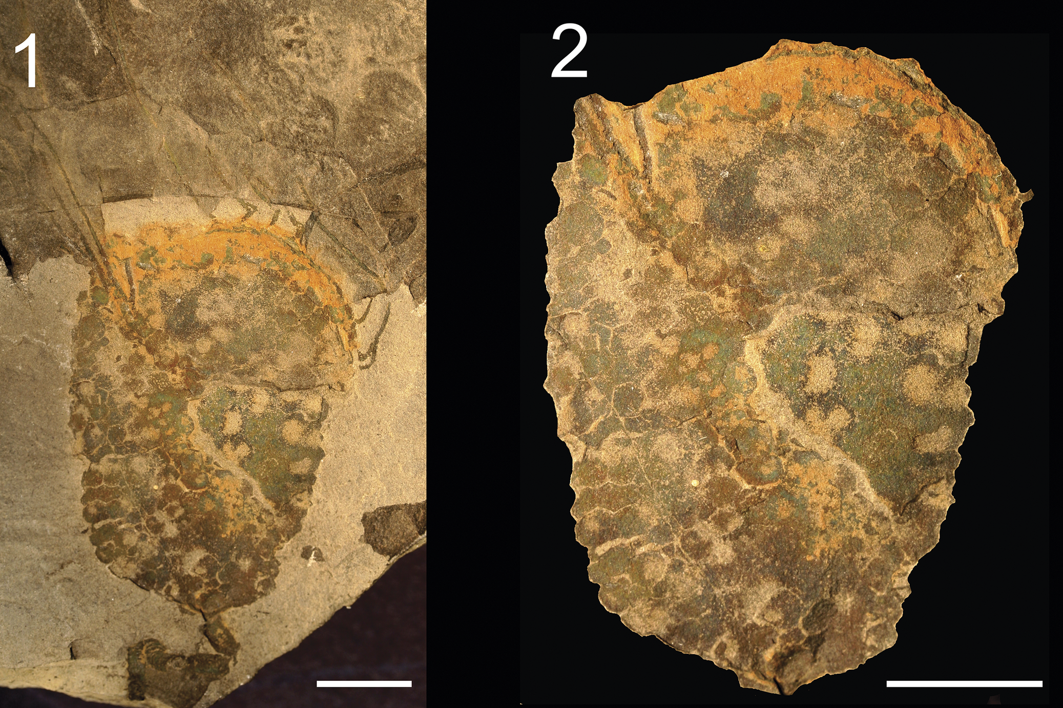
Figure 1. Long description
Panel 1 on the left displays the full fossil specimen embedded in rock, with theca at the top, brachioles extending from the upper region, and a short proximal stem at the bottom. The fossil is preserved as a thin, greenish-brown glauconitic film lacking original calcite. The scale bar at the bottom right represents 10 millimeters. Panel 2 on the right is a close-up of the theca, showing detailed polygonal plating and a central crease running vertically, with the ambulacra visibly curved and aligned along this crease. The taphonomic alteration is evident in the curvature of the ambulacra. The scale bar at the bottom right also represents 10 millimeters.
With few exceptions, paracrinoids are often preserved lacking features beyond the theca. Of the 11 currently known Laurentian paracrinoid genera, only three have well-preserved details of the feeding appendages and stem including Comarocystites (Billings, Reference Billings1854), Amygdalocystites (Raymond, Reference Raymond1921; Parsley and Mintz, Reference Parsley and Mintz1975; Guensburg, Reference Guensburg1991), and Implicaticystis (Paul et al., Reference Paul, Guensburg and Darrough2025). This lack of morphological knowledge dramatically limits the use of characters beyond the theca in phylogenetic analyses. This is particularly problematic given the lack of standardized or presumably genetically controlled thecal plating. Pairing this taphonomic bias with the limited temporal range and rapid evolutionary change produces clear obstacles in reconstructing evolutionary patterns within the clade. Furthermore, the stem and feeding apparatus contain a wealth of ecological and functional information needed to better understand the biology of this class of echinoderms (see Parsley and Mintz, Reference Parsley and Mintz1975, and Guensburg, Reference Guensburg1991, for discussions of the biology and feeding posture of paracrinoids).
The exceptional preservation of these newly discovered paracrinoid specimens allows a more reliable placement of this taxon within the clade. In addition, the unusual filtration fans of bilaterally symmetrical paracrinoids suggest a different feeding ecology compared with the parabolic or circular fans of most stalked echinoderm (Parsley and Mintz, Reference Parsley and Mintz1975; Guensburg, Reference Guensburg1991), thus well-preserved specimens can provide direct insight into their autecology and allow further testing of paracrinoid feeding postures and hydrodynamics. Finally, the detailed preservation provides information regarding this animal’s role within the ecosystem during the Great Ordovician Biodiversification Event of increased competition among filter feeding organisms and radiation of both predators and zooplankton (Servais et al., Reference Servais, Owen, Harper, Kröger and Munnecke2010). Herein, the new specimens are described, the evolutionary relationships of paracrinoids are analyzed with this new taxon incorporated, and the paleoecology of this organism is explored.
Materials and methods
The new specimens of paracrinoid are from two adjacent quarries northeast of Québec City (Fig. 2), both of which are privately owned and actively mined. The first two specimens, MPEP706.69a (Figs. 3 and 4) and MPEP706.69b (Fig. 3), found on the same limestone slab, were collected in the Saint-Joachim quarry (47.0741, −70.8553) while the third specimen, MPEP1523.2 (Fig. 5), was collected in the Château-Richer quarry (46.9768, −71.0171).
Locality map. (1) Map of Québec with indication of the sample localities as represented by the black box. (2) Enlargement of the study region indicating the quarries where the new paracrinoid specimens were discovered in relation to Québec City.
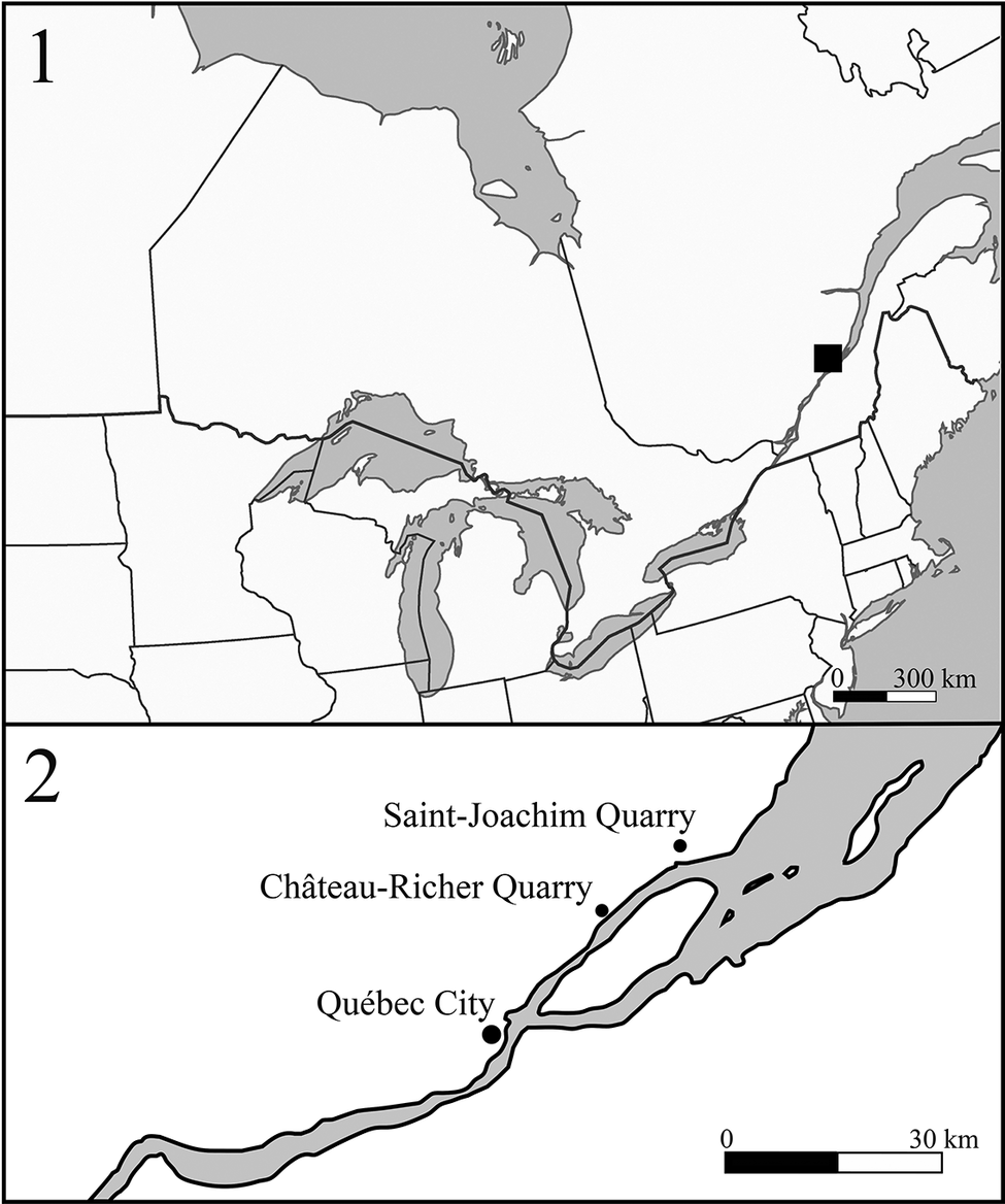
Figure 2. Long description
The top panel displays a regional map of eastern Canada and the northeastern United States, with a black square marking the study area in southern Québec. The bottom panel is an enlarged map of the marked region, oriented with north at the top. It shows the river running from southwest to northeast. Three locations are labeled with black dots: Québec City at the southwestern end, Château-Richer Quarry northeast of Québec City, and Saint-Joachim Quarry further northeast. A scale bar at the bottom right indicates 0 to 30 kilometers.
New specimen of Quebecocystites gorgo n. gen. n. comb. from the Neuville Formation of Québec, Canada. (1) View of complete theca (MPEP706.69a) and brachioles along with a partial specimen (MPEP706.69b) showing the thecal summit and proximal brachioles. (2) Details of the proximal brachioles on the left side of the specimen along with the steles of a co-occurring undescribed solutan echinoderm. (3) Details of the distal brachioles on the right of the specimen showing regeneration. (4) Details of the basal plates and the stem facet. (5) Details of slightly disrupted proximal brachioles showing the width of the food groove. (6) Close up of the thecal plating of Q. gorgo n. comb. (7, 8) Details of small irregularities on the thecal plate surfaces on the left side of the specimen. In both cases there are raised swellings with 1–2 distinctive pores. Scale bars: (1, 6) 10 mm; (2) 2 mm; (3, 5, 7, 8) 1 mm; (4) 5 mm. MPEP706.69a and MPEP706.69b.
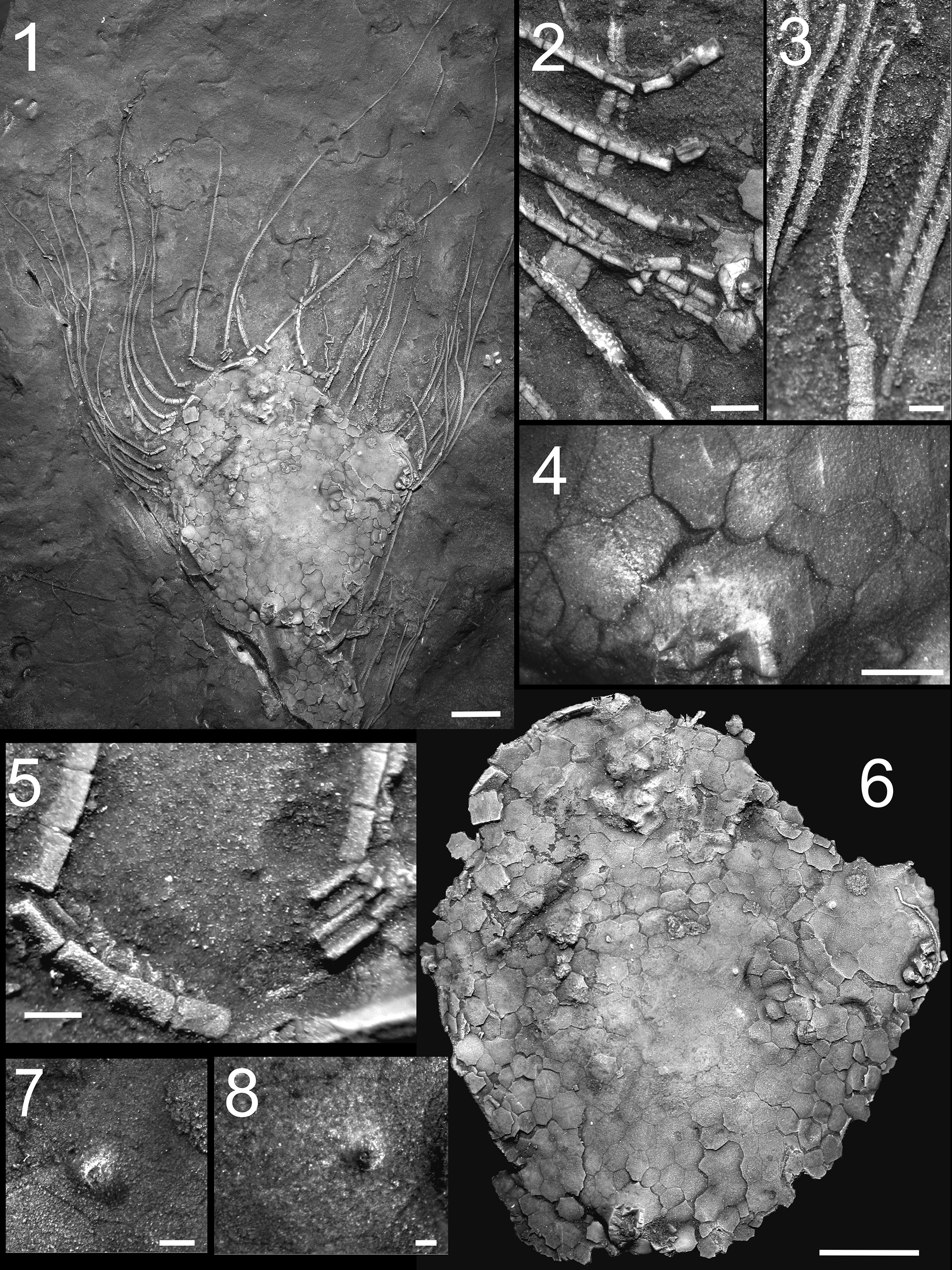
Figure 3. Long description
Panel 1 at the top left shows a complete theca with radiating brachioles and a partial specimen, scale bar 10 mm. Panel 2, top center, details proximal brachioles on the left side and includes steles of a co-occurring solutan echinoderm, scale bar 2 mm. Panel 3, top right, highlights distal brachioles on the right side with evidence of regeneration, scale bar 1 mm. Panel 4, center right, presents the basal plates and stem facet, scale bar 5 mm. Panel 5, bottom left, shows slightly disrupted proximal brachioles and the width of the food groove, scale bar 1 mm. Panel 6, bottom center, is a close-up of the thecal plating, scale bar 10 mm. Panels 7 and 8, bottom right, display small irregularities on the thecal plate surfaces on the left side, each with raised swellings and one to two distinctive pores, scale bars 1 mm. All panels are grayscale and highlight different structural and surface features of the fossil specimens.
Camera lucida drawings of the exceptionally preserved new specimen (MPEP706.69a) of Q. gorgo n. comb. showing details of its anatomy including the (1) stem facet, (2) brachiole facets, (3) brachiole cover plates, (4) brachiole food groove, (5) oral summit, and (6) a regenerating brachiole. Scale bar: 10 mm.
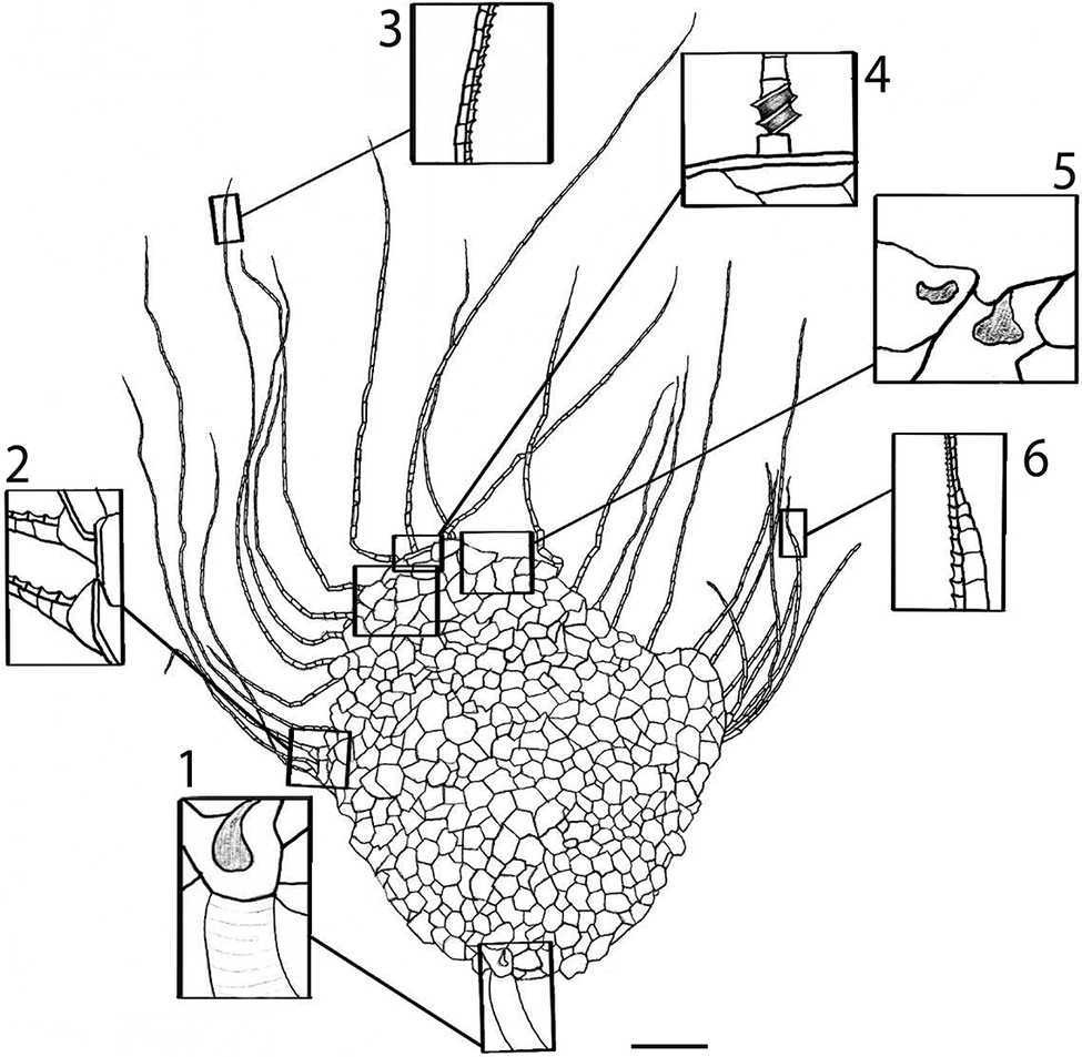
Figure 4. Long description
At the base is the stem facet, shown in callout 1, depicting the attachment point with internal structure. Moving leftward, callout 2 details the brachiole facets, illustrating their articulation with the main body. Callout 3, positioned above, shows the segmented structure of the brachiole cover plates. Callout 4, to the upper right, highlights the brachiole food groove with its internal ridges. Callout 5, further right, displays the oral summit with two distinct openings. Callout 6, at the far right, focuses on a regenerating brachiole, showing its segmented pattern. The main body is composed of polygonal plates, with elongated brachioles radiating outward. The scale bar at the bottom right represents 10 millimeters.
Additional specimen (MPEP1523.2) of Quebecocystites gorgo n. gen. n. comb. from the Neuville Formation of Québec, Canada. (1) Full specimen showing the theca, proximal brachioles, stem, and holdfast (shown above the crinoid, Ectenocrinus). (2) Details of the theca and proximal stem. (3) Details of the terminal floor plates and brachioles on the right side of the specimen. (4) Details of the stem showing the heteromorphic columnals along with the crenulated articulations. (5) Details of the distal stem and cemented holdfast. Scale bars: (1, 2) 10 mm; (3) 5 mm; (4, 5) 2 mm.
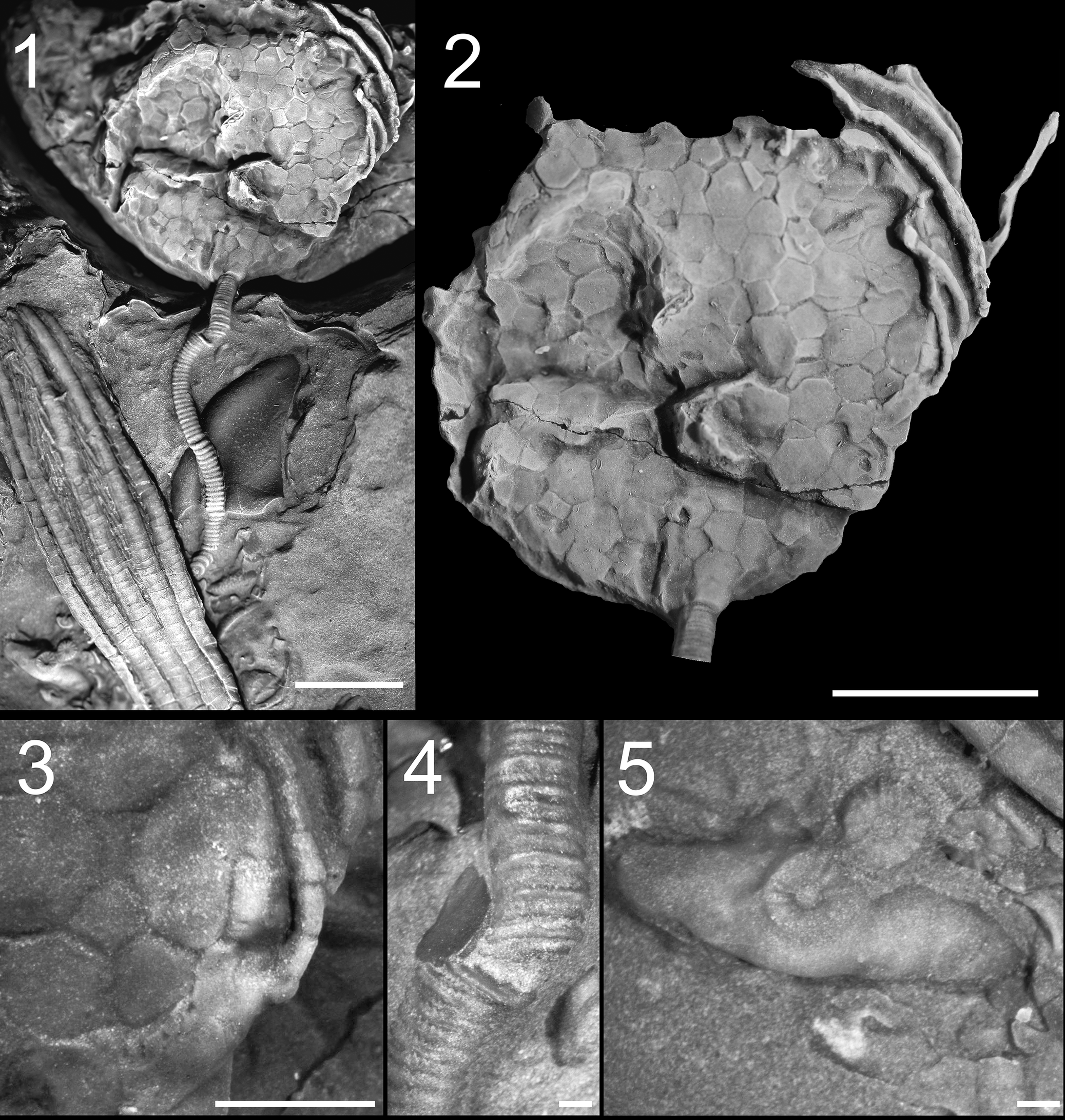
Figure 5. Long description
Panel 1 at top left shows the full fossil specimen embedded in matrix, with theca at the top center, a segmented stem extending downward, and a cemented holdfast at the bottom. To the left of the stem is a crinoid columnal for comparison. Panel 2 at top right presents a close-up dorsal view of the theca, displaying polygonal plate structure and the base of the stem at the lower edge. Panel 3 at bottom left magnifies the right side of the theca, highlighting terminal floor plates and attached brachioles. Panel 4 at bottom center focuses on the stem, revealing heteromorphic columnals with crenulated articulations. Panel 5 at bottom right details the distal stem and the cemented holdfast, showing attachment to the substrate. Scale bars indicate 10 millimeters for panels 1 and 2, 5 millimeters for panel 3, and 2 millimeters for panels 4 and 5.
The Neuville Formation (Trenton Group) is comprised of, in stratigraphic succession, the Saint-Casimir and Grondines members. Specimens described herein are from the upper Grondines Member. The Grondines Member of the Neuville Formation contains an extensive and well-studied conodont fauna (Globensky and Jauffred, Reference Globensky and Jauffred1971), which is dominated by the Phragmodus undatus assemblage and thus indicates a latest Sandbian to early Katian age (Globensky and Jauffred, Reference Globensky and Jauffred1971; Bergström and Ferretti, Reference Bergström and Ferretti2017). This unit is comprised of thinly (~10 cm) interbedded carbonate mudstones and wackestones showing a stratigraphically upward increase in bed thickness (Lavoie, Reference Lavoie1993). This unit has been interpreted as a transgressive sequence representing relatively deep outer-ramp deposits (Lavoie, Reference Lavoie1993). There are few sedimentary structures apart from rare large hummocky cross-stratification and associated small burrows, which suggest a depth close to storm wave base (Lavoie, Reference Lavoie1993).
Fossil preservation is extremely variable within the Grondines Member ranging from exceptionally preserved, articulated, and in-situ specimens (Ramirez-Guerrero et al., Reference Ramirez-Guerrero, Alghaled, Bateman, Cournoyer and Cameron2026) to extensively reworked and disarticulated skeletal remains consistent with infrequent storm transport (Lavoie, Reference Lavoie1993). Despite the variability in preservation, recent work has shown extraordinary preservation of a diverse community including bryozoans, brachiopods, trilobites, and echinoderms. The diverse echinoderm assemblage includes crinoids, rhombiferans, solutans, stylophorans, cyclocystoids, and asteroids in addition to the new paracrinoid described herein (Alghaled, Reference Alghaled2019).
A phylogenetic hypothesis for Paracrinoidea was estimated with the addition of this new taxon using the characters and data developed and published by Limbeck et al. (Reference Limbeck, Bauer, Deline and Sumrall2024) using the outgroup Cheirocystis fultonensis Sumrall and Schumacher, Reference Sumrall and Schumacher2002, a glyptocystid rhombiferan. Paul et al. (Reference Paul, Guensburg and Darrough2025) discussed potential outgroups for paracrinoids, including rhombiferans, eocrinoids, and Columbocystis Bassler, Reference Bassler1950, whose taxonomy has been controversial (Sprinkle, Reference Sprinkle1973; Parsley and Mintz, Reference Parsley and Mintz1975; Paul et al., Reference Paul, Guensburg and Darrough2025). The selection of C. fultonensis as outgroup in this current analysis enables a direct comparison of results with Limbeck et al. (Reference Limbeck, Bauer, Deline and Sumrall2024). Maximum likelihood trees were computed using PAUP*4.0b10 (Swofford, Reference Swofford2003); characters were treated as unordered and unweighted in this analysis. A heuristic search was completed for the analysis; bootstrap support was calculated using stepwise addition to assess the robustness of nodes within the tree. Tree scores can be found in supplemental materials.
Repositories and institutional abbreviations
The new specimens mentioned in this study, with the prefix MPEP, are deposited in the Musée de paléontologie et de l’évolution, Montréal, Québec, Canada. The specimen with the prefix GSC is deposited in the Organic Materials Collections at the Natural Resources Administration of Canada.
Systematic paleontology
Remarks
The systematic paleontology of paracrinoids needs to be re-evaluated based on recent phylogenetic analyses (Sheffield et al., Reference Sheffield, Limbeck, Bauer, Hill and Nohejlová2022; Limbeck et al., Reference Limbeck, Bauer, Deline and Sumrall2024). Current order and family diagnoses are utilized where appropriate while outlining the difficulties in assigning taxa within the current system. Terminology follows Kesling (Reference Kesling and Moore1968) and Parsley and Mintz (Reference Parsley and Mintz1975). Paul et al. (Reference Paul, Guensburg and Darrough2025) proposed referring to the feeding appendages of paracrinoids as pseudopinnules to differentiate these structures from brachioles observed in other blastozoans as well as from pinnules found within crinoids. The terminology of anatomical structures across classes of stalked echinoderms has caused significant issues in phylogenetics and nomenclature (see Sumrall and Waters, Reference Sumrall and Waters2012). Herein, the feeding structures are referred to as brachioles aligning the terminology with recent literature. However, this choice does not imply direct homology with other feeding appendages within blastozoans and the terminology and homology of feeding appendages across blastozoans should be reviewed in a similar manner as was recently done with respiratory structures (Sheffield et al., Reference Sheffield, Limbeck, Bauer, Hill and Nohejlová2022).
Subphylum Blastozoa Sprinkle, Reference Sprinkle1973
Class Paracrinoidea Regnéll, Reference Regnéll1945
Order Platycystitida Parsley and Mintz, Reference Parsley and Mintz1975
Remarks
The order is defined as paracrinoids without sutural pores, arms epithecal and typically branched, with thecal plates that are generally smooth with pustules. The new taxon generally fits within this description, but recent work by Sheffield et al. (Reference Sheffield, Limbeck, Bauer, Hill and Nohejlová2022) demonstrates that respiratory structures are highly convergent features within blastozoan echinoderms and are not reliably diagnostic. Furthermore, respiratory structures are also variable and often convergent within paracrinoids based on recent phylogenetic analysis (Limbeck et al., Reference Limbeck, Bauer, Deline and Sumrall2024), but specific and unique respiratory structures may be taxonomically informative (Paul et al., Reference Paul, Guensburg and Darrough2025).
Family incertae sedis
Remarks
Currently there are three established families within the Platycystitida: Platycystitidae (Parsley and Mintz, Reference Parsley and Mintz1975), Malocystitidae (Bather, Reference Bather1899), and Bistomiacystidae (Sprinkle and Parsley, Reference Parsley and Sprinkle1982). Even with key anatomical landmarks missing in this new species, we are confident that this new species does not meet the definitions of these established families. We are hesitant to erect a new family prior to a systematic re-evaluation of the class.
Genus Quebecocystites new genus
Type species
Quebecocystites gorgo (Sinclair, Reference Sinclair1948), by monotypy.
Diagnosis
Paracrinoid with numerous irregularly arranged, unornamented, polygonal thecal plates lacking respiratory structures, two ambulacra extending halfway down the theca lacking prominent thecal floor plates, and thin and widely separated uniserial brachioles.
Occurrence
Late Ordovician, Québec, Canada
Etymology
Named from the Canadian province of Québec, which is Algonquian in origin meaning ‘narrow passage’ thus referring to both the provenance of the genus as well as its distinctive and gracile feeding appendages.
Remarks
Sinclair (Reference Sinclair1948) noted the similarity of the original poorly preserved specimen (Fig. 1) to other paracrinoids such as Amygdalocystites and Platycystites based on the overall body outline and the feeding appendages. However, Sinclair (Reference Sinclair1948) decided to leave the genus indeterminate and referred it to Amygdalocystites? given that this was the only known paracrinoid at the time within the Trenton Group. Based on the new and better-preserved material, Quebecocystites n. gen. most closely resembles the other amygdaloidal paracrinoids (Amygdalocystites and Platycystites Miller, Reference Miller1889) but has some clear differences. The unornamented thecal plates lacking respiratory structures resemble Platycystites, while the small, unorganized thecal plating more so resembles Amygdalocystites. The reduced, narrow, epithecal ambulacral floor plates and gracile brachioles differentiate Quebecocystites from either genus. At present, Quebecocystites n. gen. is known only from the Neuville Formation in Québec, Canada.
Quebecocystites gorgo new combination
Amygdalocystites? gorgo Sinclair, Reference Sinclair1948, pl. 42, fig. 8, text fig. 6.
Type
Sinclair (Reference Sinclair1948) figured, but did not formally designate a type specimen, thus the singular specimen (GSC 13587) described by Sinclair is designated as the lectotype. This specimen is preserved as a glauconitic film and shows aspects of the theca, thecal plating, proximal stem, and feeding appendages. Specimen from the Late Ordovician Neuville Formation in Québec, Canada.
Material
Three additional specimens of Q. gorgo n. comb. preserved in calcite. MPEP706.69a and MPEP706.69b are on a single slab, the former is a largely complete specimen with the theca, feeding appendages, and stem facet, the latter is a partial specimen preserving the top of the theca and proximal feeding appendages. MPEP1523.2 is also a largely complete specimen with proximal feeding appendages, theca, stem, and holdfast.
Diagnosis
Characters as for genus.
Occurrence
Found only in the Neuville Formation, lectotype (GSC 13587) is from outside of Saint-Joachim, Québec and additional figured material is from Saint-Joachim and Château-Richer, Québec.
Description
Four specimens are known, three mostly complete and one partial, all specimens are likely mature based on the overall size. Specimens are taphonomically flattened, which likely exaggerates the amygdaloidal thecal shape of the individuals. The specimens all appear to be preserved in a roughly similar orientation given the apparent absence of the gonopore, hydropore, and anal pyramid, which are likely preserved on the opposite side of the specimens. Measurements are based on the most complete specimen (MPEP706.69a; Fig. 3).
Theca is large, amygdaloidal in shape, flattened perpendicular to the ambulacra plane (likely BC–DE) across the theca, slightly taller than wide. Stem attachment is slightly elevated, but oral summit is aligned with the top of theca. Complete specimen approximately 55 mm tall. Theca of mature specimen containing about 800 plates, thecal plates are small, unornamented, polygonal with 4–9 sides, irregularly arranged, lacking any circlets or columns. Small plates are often inserted around larger plates. Thecal plates are very thin. Average plate size consistent from base to summit. Thecal plates range in width from 1 mm to 4.6 mm. Plates lack specialized respiratory structures (pores or slits) and ornamentation. In rare instances, plates contain an anomalous pore that is slightly off center on the plate forming a small protrusion, although these pores are likely ecological rather than anatomical. Based on the differences in the orientation of flattening and creasing of the specimens (Figs. 1, 3, 5), it appears that the theca was taphonomically delicate compared to other paracrinoids, which have a more rigid and stable theca (e.g., Parsley and Mintz, Reference Parsley and Mintz1975; Parsley, Reference Parsley and Sprinkle1982). This may be the result of the increased number and thinness of the plates or a reduction in the articulation between thecal plates.
The stem facet is formed by three basals in largest specimen (Fig. 3.4), although there could be additional plates. The basal plates vary in size and shape with two larger plates and one small plate nested between the larger plates, indicating that these smaller plates might be inserted later in ontogeny. The largest basal plate articulates with approximately seven thecal plates above and is slightly wider than the largest thecal plates (Fig. 3.4). The smaller basal plates only articulate with one thecal plate above and are approximately 10% of the size of the larger plates. Two basal plates can be observed in the other specimen (Fig. 5), although the plates are more similar in size and shape further indicating that the structure and plating of the stem facet might change through ontogeny. Stem facet is round, approximately 3.3 mm in diameter. Column round, approximately twice the length of the thecal height in one complete specimen (Fig. 5), apparently relatively straight; borrowing from crinoid terminology, lumen irregular subcircular, columns very thin, approximately six times wider than high, with irregularly heteromorphic arrangement, with low convex epifacets, articular surfaces with crenulations (Fig. 5.4), holdfast oblong as preserved, apparently a single solid piece, attached to an unknown surface (Fig. 5.5).
Mouth at summit, aligned with stem facet offset from central axis of theca. Oral plating surrounding peristome unknown, but approximate location of the mouth at the thecal summit can be discerned from orientation of brachioles (i.e., pseudopinnules of Paul et al., Reference Paul, Guensburg and Darrough2025) and food groove. Hydropore and gonopore are unknown. Location and plating of periproct are also unknown.
Two non-bifurcating ambulacra, likely BC and DE (based on developmental models described by Sumrall and Wray, Reference Sumrall and Wray2007), roughly equal in length, extend approximately halfway down theca. Ambulacral floor plates not observed in the largest specimen and appear to be folded over such that they are on the downward side of the specimen. However, a ridge of thicker plates can be seen in cross section along the top edge of the specimen, which may represent the floor plates (Fig. 3.6). The orientation of the floor plates and the relative position of the facets along the ambulacra are unknown. The terminal floor plates can be observed in the smaller specimen (MPEP1523.2; Fig. 5.3). Ambulacral floor plates appear reduced compared to other paracrinoids, not raised significantly above the theca. Floor plates are epithecal overlying thecal plates consisting of a uniserial series of approximately 15 plates per ambulacrum (based on length of observed floor plates compared with the length of the ambulacrum), each bearing a single facet. Floor plates are approximately twice as wide (~3.5 mm) as high (~1.8 mm) and slightly thicker than other thecal plates, with a narrow food groove. Ambulacral floor plates are approximately equal in size from proximal to distal ends of ambulacra. Brachiole facets are approximately 0.7 mm wide, central to ambulacral floor plates, brachiole plates are polygonal, largely pentagonal but highly variable.
Approximately 12–15 brachiole appendages per ambulacrum, largest brachiole approximately 0.9 mm wide at base tapering distally to 0.2 mm. The most proximal brachiole ossicles are approximately 1.5 times taller than wide, each containing about 3–4 pairs of biserial cover plates covering a narrow food groove approximately 0.2–0.3 mm in width. Each cover plate bears a single thin spike (Fig. 3.5) approximately as long as the width of the cover plates with the size and orientation of the spikes consistent along the length of the brachiole. Longest preserved brachiole is 92 mm (1.67 × height of theca).
Remarks
Sinclair (Reference Sinclair1948) originally named the species based on a single specimen that was preserved as a thin glauconitic film (Fig. 1). Based on this single specimen, Sinclair (Reference Sinclair1948) gave a brief informal description, while noting the aspects of the fossil that are unknown and expressing his doubts in both designating a name and the inability to accurately compare this specimen to those preserved as recrystallized calcite (as is common within other described paracrinoid taxa). Within his brief description he focused on two aspects of morphology. First is the overall thecal outline, for which he noted the similarity with amygdaloidal paracrinoids while also suggesting that the outline was taphonomically altered and unlikely representative of the original animal. Secondly, he described the feeding structures, several of which are well preserved in the specimen. These feeding structures are as long as the height of the theca, thin, show little tapering, and indicate some degree of flexibility (Sinclair, Reference Sinclair1948). Further inspections of this original specimen (Fig. 1) indicate that the theca was likely distorted with a folded deformation down the middle corroborating Sinclair’s suggestion of the alteration of the original outline. This taphonomic fold contorts the ambulacra giving the appearance of a curve, which is common in other paracrinoids such as Canadocystis (Parsley and Mintz, Reference Parsley and Mintz1975). However, that superficial similarity is a byproduct of preservation, and details of the ambulacra are not preserved. In addition, the plating can be observed across the theca showing numerous, irregular, and smooth polygonal plates lacking any suggestion of ornamentation or respiratory structures. Overall, the anatomy of this specimen matches that of the newly discovered specimens, which allows us to more completely describe this species.
Traditionally, paracrinoid genera have been differentiated based on ambulacral symmetry varying from a 2-1-2 blastozoan pseudopentaradial oral system at the base of the clade or within the outgroup (depending on the phylogenetic position of Columbocystis) to 1-0-1 bilateral oral system, thecal shape ranging from globular to bilaterally flattened, the number, size, and regularity of theca plating, and the type and distribution of thecal respiratory structures (Parsley and Mintz, Reference Parsley and Mintz1975). Based on these characteristics Quebecocystites gorgo n. comb. most closely resembles Platycystites Miller and Amygdalocystites Billings sharing an amygdaloidal and flattened theca and similar ambulacral arrangement. Additionally, Q. gorgo is like Platycystites in the absence of thecal respiratory structures and having unadorned and gently convex thecal plates but is notably different in the plating and structure of the theca with numerous, small, variably shaped, and randomly articulated plates. Similarly, Quebecocystites gorgo n. comb. resembles Amygdalocystites florealis Billings, Reference Billings1854, in thecal construction with both taxa having numerous polygonal plates, however Q. gorgo lacks prominent respiratory structures and thecal ornamentation. In addition, the long, slender, and laterally separated brachioles are distinctive compared to other species of paracrinoid that have stouter, shorter, and more closely spaced brachioles.
Quebecocystites gorgo n. comb. is also distinctive from both taxa in terms of its overall body size. Body size can be a problematic metric to use in differentiating taxa given that body size can vary significantly between populations caused by genetic differences as well as differences in environmental factors (i.e., ecophenotypy) (e.g. Huntley et al., Reference Huntley, Schiffbauer, Avila and Broce2018). In addition, in taxa known from a limited number of specimens it is difficult to establish ontogenetic series along with estimates of average and maximum body size. However, body size is also a fundamental biologic property and important both ecologically and evolutionarily (Smith et al., Reference Smith, Payne, Heim, Balk and Finnegan2016). Therefore, body size should not normally be the sole factor to establish a new taxon but can be a useful metric to consider associated with other morphological differences. Many paracrinoids are locally abundant, thus their range and average body size can be established. For instance, within the Bromide Formation, Parsley (Reference Parsley and Sprinkle1982) reported 1228 specimens of Oklahomacystis bibrachiatus Parsley, Reference Parsley and Sprinkle1982, and 1781 specimens of Platycystites levatus Bassler, Reference Bassler1943, with most ranging from 1–3 cm in thecal height (based on examinations of the collections at the Sam Noble Museum, University of Oklahoma). The largest specimen of Q. gorgo is significantly larger at 5.5 cm in thecal height. This size places Q. gorgo n. comb. alongside the largest paracrinoids such as Comarocystites (Parsley and Mintz, Reference Parsley and Mintz1975) and Bistomiacystis (Sumrall and Deline, Reference Sumrall and Deline2009), which are comparable in size to Late Ordovician camerate crinoids (e.g., Brett et al., Reference Brett, Deline, MCLAUGHLIN, Ausich and Webster2008). However, there are a small number of specimens known within these larger paracrinoids (e.g., Quebecocystites n. gen., Comarocytites, and Bistomiacystis), thus it is difficult to discern the average and range of body sizes within the species and whether the taxa described are outliers within the population.
Results
The maximum likelihood analysis for paracrinoids, including the newly described Q. gorgo n. comb., retained four trees, with the best fit tree having a −lnL score of 283.436. Thirty-six characters were included in the analysis, two characters (30 and 34) were excluded because they could not be assessed for the included taxa. A maximum likelihood analysis for paracrinoids that was estimated prior to the description of Q. gorgo from Limbeck et al. (Reference Limbeck, Bauer, Deline and Sumrall2024) is also included for comparison. The resulting trees demonstrate largely resolved relationships with a few polytomies present (Fig. 6).
Phylogenetic hypotheses estimated for select paracrinoids (1) and paracrinoids including Q. gorgo n. comb. (2). Both were calculated using maximum likelihood strict consensus. Terminal nodes are color coded to indicate similar groups based on type of respiratory structure and ambulacral structure referred to in the text body. Taxa in olive and yellow, Comarycystites and Implicaticystis, respectively, both have four ambulacra, and three of the four species share the same respiratory structures. The taxon in green, Columbocystis, has five ambulacra and no respiratory structures. The taxa colored in tan, Bistomiacystis, Canadocystis, Malocystites, and Wellerocystis have s-shaped curvature to their ambulacral systems and no thecal respiratory structures. Taxa in pink, Oklahomacystis, Sinclairocystis, and Amygdalocystites have the same respiratory structures (with the exception of Sinclairocystis, which has Comarocystites-type respiratory structures) and two to three ambulacra that extend down the theca. Because there is discussion over the importance of respiratory structure type and ambulacral structure, we chose to include Sinclairocystis in this pink group based on similarity of ambulacral structure. Taxa in blue, Platycystites and Globulocystites, have no respiratory structures and two ambulacra that extend down the theca. The taxon in purple, Quebecocystites n. gen., although similar in feature to those taxa in blue, is separated out to indicate the first inclusion of this genus in a phylogenetic analysis. Major groups remain relatively stable between the two trees. Tree data for this study with Q. gorgo n. comb. can be found in the supplement to this paper, please see Limbeck et al. (Reference Limbeck, Bauer, Deline and Sumrall2024) for other tree data. Species-level taxa shown in Figure 6 not otherwise noted in the text are Amygdalocystites radiatus Billings, Reference Billings1854; Oklahomacystis spissus Frest, Strimple, and Witzke, Reference Frest, Strimple and Witzke1980; Oklahomacystis tribrachiatus Bassler, Reference Bassler1943; Oklahomacystis trigonis Guensburg, Reference Guensburg1984; Sinclairocystis praedicta Bassler, Reference Bassler1950; Platycystites faberi Miller, Reference Miller1889; Globulocystites rotundatus Frest, Strimple, and Coney, Reference Frest, Strimple and Coney1979; Globulocystites cristatus Bassler, Reference Bassler1943; Globulocystites infundus Frest, Strimple, and Coney, Reference Frest, Strimple and Coney1979; Malocystites murchisoni Billings, Reference Billings1858; Canadocystis tennesseensis Parsley and Mintz, Reference Parsley and Mintz1975; Canadocystis emmonsi (Hudson, Reference Hudson1905); Bistomiacystis schrantzi Sumrall and Deline, Reference Sumrall and Deline2009; Bistomiacystis globosa Sprinkle and Parsley, Reference Sprinkle, Parsley and Sprinkle1982; Implicatycystis shumardi (Meek and Worthen, Reference Meek and Worthen1868); Comarocystites tribrachius Parsley and Mintz, Reference Parsley and Mintz1975; Comarocystites punctatus Billings, Reference Billings1854; Columbocystis ovata Parsley, Reference Parsley1975; and Columbocystis typica Bassler, Reference Bassler1950.
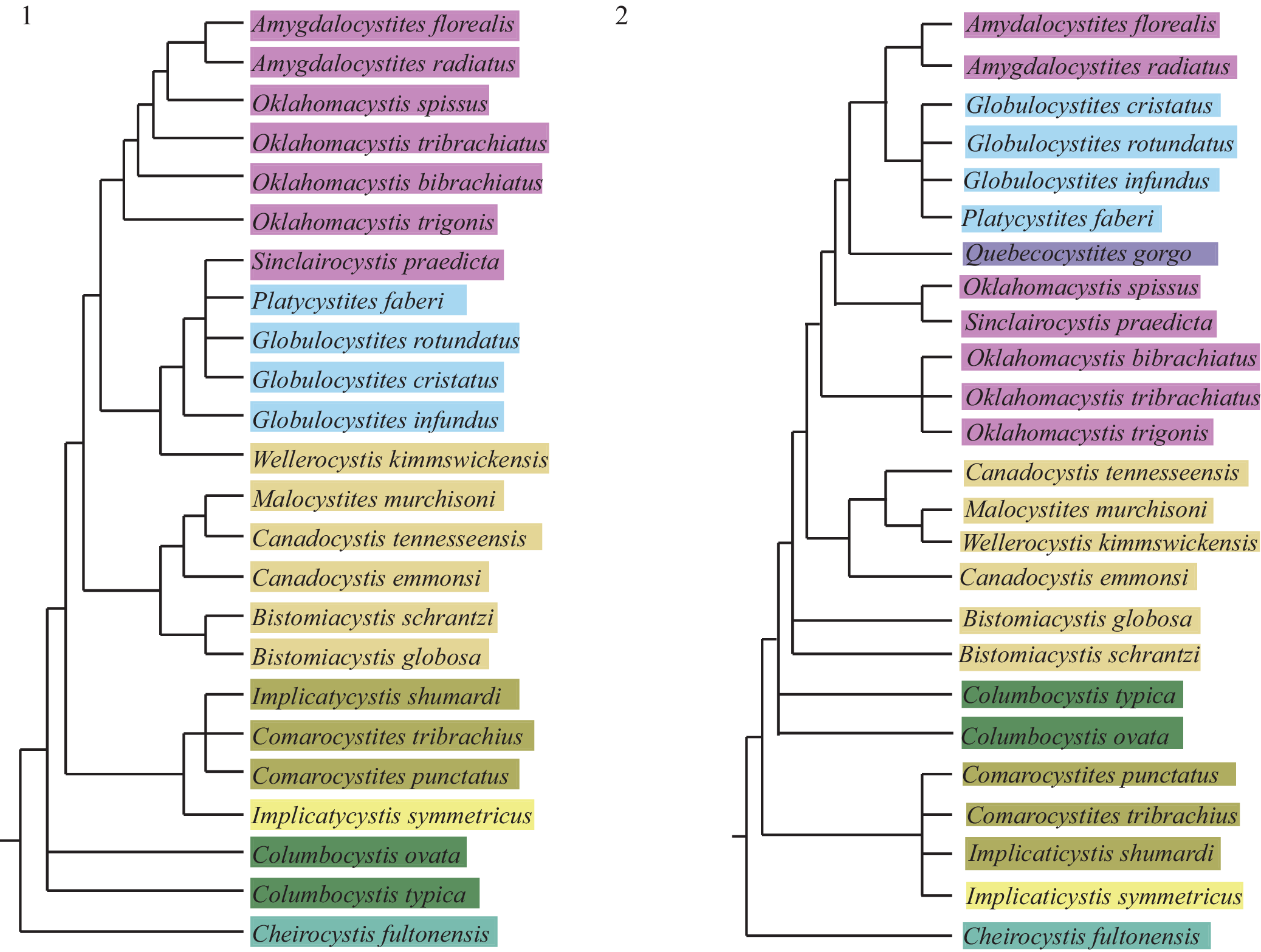
Figure 6. Long description
This is a two-panel phylogenetic tree diagram. Panel 1 on the left and Panel 2 on the right each show branching relationships among paracrinoid taxa, with terminal nodes labeled by species names. In both panels, taxa are color-coded: purple for Amygdalocystites and Oklahomacystis species, blue for Platycystites and Globulocystites, olive for Comarocystites, yellow for Implicaticystis, green for Columbocystis, and tan for Bistomiacystis, Canadocystis, Malocystites, and Wellerocystis. In Panel 2, Quebecocystites gorgo is added in blue, branching near Platycystites and Globulocystites. The branching order and groupings are nearly identical between panels, with only minor rearrangements and the addition of Quebecocystites in Panel 2. The color groups correspond to shared respiratory and ambulacral structures as described in the caption. All terminal taxa are labeled, and the branching structure is consistent with maximum likelihood strict consensus analysis.
The broad scale relationships in the tree that includes Q. gorgo n. comb. are congruent with other paracrinoid phylogenies estimated using maximum parsimony, maximum likelihood, and Bayesian methods (Limbeck et al., Reference Limbeck, Bauer, Deline and Sumrall2024). Included at the base of both trees are two well-resolved groups (Fig. 6.1 and 6.2), one including Comarocystites and Implicaticystis, and one including Canadocystis, Malocystites, and Wellerocystis Foerste, Reference Foerste1920. Comarocystites and Implicaticystis both possess four ambulacra, and three of the four species share the same type of respiratory structure. In the phylogenetic hypothesis including Q. gorgo n. comb. (Fig. 6.2), these two genera are represented as a polytomy whereas in the hypothesis from Limbeck et al. (Reference Limbeck, Bauer, Deline and Sumrall2024), I. symmetricus Frest and Strimple, Reference Frest and Strimple1982, is sister taxon to a polytomy made of the other three species included. Canadocystis and Malocystites have a similar relationship in both trees, however, in the tree estimated for this study, W. kimmswickensis Foerste, Reference Foerste1920, is sister taxon to M. murchisoni Billings, Reference Billings1858. Beyond these two resolved groups, Bistomiacystis and Columbocystis have different positions and relationships in each tree. In the hypothesis presented from Limbeck et al. (Reference Limbeck, Bauer, Deline and Sumrall2024), both Bistomiacystis species are sister taxa with strong nodal support (Fig. 6.1), however, in the hypothesis from this study (Fig. 6.2), Bistomiacystis taxa form a polytomy in the reported majority rule tree. Lastly, the newly estimated tree topology (Fig. 6.2) shifts the position of the two species of Columbocystis slightly crownward in the tree.
The remaining included taxa are all morphologically similar to each other, sharing amygdaloidal-shaped theca and two to three ambulacra that extend down the length of the theca. The major differences lie in presence or absence of respiratory structures and characters related to the shape of the thecal plates. These differences result in two separate groups on the tree estimated in Limbeck et al. (Reference Limbeck, Bauer, Deline and Sumrall2024) (Fig. 6.1). In the tree estimated for this study, these two groups are combined into one larger group that is sister taxon to the group containing Canadocystis (Fig. 6.2). Quebecocystites gorgo n. comb. is part of one of the largest groups on the tree, nested as sister taxon to a larger group containing Amygdalocystites, Globulocystites Frest et al., Reference Frest, Strimple and Coney1979, and Platycystites. With the exception of Amygdalocystites, all these genera lack respiratory structures; a character that appears to be relevant in separating these amygdaloid-shaped paracrinoids.
Discussion
A comparison of the phylogenetic estimation herein (Fig. 6.2) to those proposed by Limbeck et al. (Reference Limbeck, Bauer, Deline and Sumrall2024) (Fig. 6.1) demonstrates that the broad scale relationships in the tree are congruent with each other and the major relational differences are seen in more-basal taxa. In the phylogenetic analysis without Q. gorgo n. comb. (Fig. 6.1), the group of amygdaloid-shaped paracrinoids (Amygdalocystites, Platycystites, Globulocystites, Sinclairocystis Bassler, Reference Bassler1950, and Wellerocystis) are split into two sister taxa groups based on the presence and absence of respiratory structures, except for Sinclairocystis. In contrast, the updated phylogenetic hypothesis that includes Q. gorgo n. comb. (Fig. 6.2) no longer has the same two, larger groups. Rather, several smaller groups were still split based on the presence or absence of respiratory structures and the placement of Wellerocystis to the group with Canadocystis and Malocystites. There are three groups that have respiratory structures present, with a single group, and Q. gorgo, that do not have respiratory structures between both stages of evolution. This would indicate that even with the addition of Q. gorgo n. comb. to Paracrinoidea, respiratory structures possibly evolved independently four times, as suggested by Limbeck et al. (Reference Limbeck, Bauer, Deline and Sumrall2024). In addition, the thin plates of Quebecocystites n. gen. could have supported potential respiration by diffusion through thin stereom.
The placement of Q. gorgo n. comb. as sister taxon to the group that contains Amygdalocystites and the Platycystitidae (Platycystites and Globulocystites) is not surprising given the thecal morphology and lack of respiratory structures. Like many blastozoan echinoderms, orders of paracrinoids have been defined based on the presence and absence of respiratory structures, Comarocystitida and Platycystitida (Parsley and Mintz, Reference Parsley and Mintz1975), respectively. However, the addition of Q. gorgo n. comb. to this phylogenetic hypothesis disrupts one of the previously distinct groups made by the presence and absence of these respiratory structures (Amygdalocystites and Oklahomacystis), seemingly reducing the taxonomic significance of respiratory structures in this estimation. This is not a new finding, but rather supports other phylogenetic hypotheses of blastozoan echinoderm groups that respiratory structures should not be used alone to define groups (Nardin and Bohatý, Reference Nardin and Bohatý2012; Sheffield and Sumrall, Reference Sheffield and Sumrall2019; Sheffield et al., Reference Sheffield, Limbeck, Bauer, Hill and Nohejlová2022). The relationships presented in this phylogenetic hypothesis (Fig. 6.2) also highlights another taxonomic hypothesis, defining orders of paracrinoids based on whether or not the ambulacra are recumbent or erect, Varicata and Brachiata (Kesling, Reference Kesling and Moore1968), respectively. These two orders appear as two distinct sister groups in both trees (Fig. 6). Both taxonomic schemes laid out by Kesling (Reference Kesling and Moore1968) and Parsley and Mintz (Reference Parsley and Mintz1975) highlight features of paracrinoid morphology that are influential in determining their generic relationships, but each incompletely captures these relationships.
The relationships demonstrated among Q. gorgo n. comb., the Platycystitidae, and Amygdalocystites are of particular interest. Upon initial assessment of the new specimens, the morphological similarities to Amygdalocystites were substantial—laterally compressed theca, numerous small plates, and thin brachioles. However, upon further investigation and discussion, the diagnostic features of Amygdalocystitidae and Platycystitidae (Parsley and Mintz, Reference Parsley and Mintz1975) did not fully capture the characteristics of Q. gorgo n. comb. Paracrinoidea, as a clade, has long been challenging to describe due to its unusual and disparate morphologies (Kesling, Reference Kesling and Moore1968; Parsley and Mintz, Reference Parsley and Mintz1975; Limbeck et al., Reference Limbeck, Bauer, Deline and Sumrall2024). This has resulted in significant taxonomic challenges. These taxonomic challenges materialize here in the form of some of the polytomies seen in both phylogenetic hypotheses and determining how to diagnose this new taxon.
In addition to the taxonomic challenges highlighted, the different arrangements of basal taxa presented in these two phylogenetic hypotheses (Fig. 6.1 and 6.2) underscore the need to address both long-discussed, and newly suggested, differing taxonomic ideas on several genera that have been included as paracrinoids in this study. For example, there are two sets of orders that have been proposed for paracrinoids, the Brachiata and Varicata (Kesling, Reference Kesling and Moore1968) and the Comarocystitida and Platycystitida (Parsley and Mintz, Reference Parsley and Mintz1975). The orders Brachiata and Varicata are defined by differences in ambulacral structure, members of Brachiata have erect ambulacra while Varicata members have recumbent ambulacra. In contrast, the orders suggested by Parsley and Mintz (Reference Parsley and Mintz1975) are designated based on the presence (Comarocystitida) or absence (Platycystitida) of respiratory structures. Thus far, these two taxonomic schemes and their respective implications for the organization and recognition of paracrinoids are discussed in the literature (Limbeck et al., Reference Limbeck, Bauer, Deline and Sumrall2024; Paul et al., Reference Paul, Guensburg and Darrough2025), although no quantitative analyses specifically addressing this taxonomy have been completed.
Additionally, the genus Columbocystis and its relationship to paracrinoids requires further study. Columbocystis has been suggested to be a member of Paracrinoidea (Sprinkle, Reference Sprinkle1973), not a member of Paracrinoidea (Parsley and Mintz, Reference Parsley and Mintz1975), and sister group to Paracrinoidea (Limbeck et al., Reference Limbeck, Bauer, Deline and Sumrall2024; Paul et al., Reference Paul, Guensburg and Darrough2025). In this most recent phylogenetic hypothesis, Columbocystis is nested farther within the paracrinoid tree, rather than continuing to be sister taxa with paracrinoids. A taxonomic revision of Paracrinoidea is critically needed and is forthcoming. Additional future work prompted by these two phylogenetic hypotheses includes expanding the character suite to better capture homologies among the included genera and to include several genera of eocrinoids and rhombiferans to better assess the relationship of Paracrinoidea to other blastozoan echinoderm groups.
As discussed earlier, the vast majority of paracrinoid specimens are preserved lacking features beyond the theca (Brett et al., Reference Brett, Moffat and Taylor1997; Limbeck et al., Reference Limbeck, Bauer, Deline and Sumrall2024). Therefore, the exceptional preservation of Q. gorgo n. comb. that include aspects of the holdfast, stalk, and feeding appendages can provide insights into the paleoecology of paracrinoids. Extensive research has focused on filter morphology, tiering, competition, and niche differentiation in Paleozoic crinoids (Ausich, Reference Ausich1980; Bottjer and Ausich, Reference Bottjer and Ausich1986; Kammer and Ausich, Reference Kammer and Ausich1987; Cole et al., Reference Cole, Wright and Ausich2019; Higdon and Cole, Reference Higdon and Cole2025). Blastozoans often are significant components in Paleozoic marine assemblages with paracrinoids being locally abundant within the Late Ordovician (e.g., Parsley, Reference Parsley and Sprinkle1982), but the scarcity of preserved feeding appendages makes their inclusion into studies of competition and paleoecology difficult (but see Novack-Gotshall et al., 2024). A recent study on the Bromide Formation (Late Ordovician, Oklahoma) found that paracrinoids occupied a constrained niche that was distinctive and discrete from both crinoids and rhombiferans based on ecomorphological characteristics suggesting that competition between paracrinoids and other stalked echinoderms was unlikely (Higdon and Cole, Reference Higdon and Cole2025).
The excellent preservation of Q. gorgo n. comb. allows direct comparison with co-occurring echinoderms such as the crinoid Ectenocrinus (Fig. 4), although a detailed study of Neuville crinoid fauna is yet to be conducted. The stem of Q. gorgo is shorter than common Katian crinoids from Laurentia (Brett et al., Reference Brett, Deline, MCLAUGHLIN, Ausich and Webster2008), but with a larger body size and long brachioles it would be in a similar-tiered position above the sea floor. The slight disarticulation in the brachioles (Figs. 3 and 4) allows for direct measurement of the width of the food groove in the proximal region of the brachioles. The width of the food groove in modern crinoids controls the maximum size of food particles able to be captured and consumed (Meyer and Lawrence, Reference MEYER, LAWRENCE and Lawrence1982) and can be used in fossil echinoderms to explore whether co-occurring taxa are competing for similar food resources (Meyer et al., Reference Meyer, Miller, Holland and Dattilo2002; Brower, Reference Brower2007). The food groove in Q. gorgo n. comb. (Figs. 3.5 and 5.3) is ~0.3 mm, which is similar to many Late Ordovician crinoids such as Iocrinus trentonensis Walcott, Reference Walcott1883 (0.45 mm, Trenton Formation, New York) and Ectenocrinus simplex Hall, Reference Hall1847 (0.26 mm, Galena Group, Iowa) (Brower, Reference Brower2007). Therefore, even with a unique and distinctive feeding apparatus, Q. gorgo was likely feeding on similar-sized food particles at similar heights above the sea floor as co-occurring crinoids, thus suggesting potential competition between filter-feeding echinoderms in terms of the targeted food particles. Overall, the detailed preservation of diverse and co-occurring filter-feeding echinoderms including paracrinoids, rhombiferans, crinoids (Fig. 5.1), and potentially juvenile solutans (Fig. 3.2; see Lefebvre and Lerosey-Aubril, Reference Lefebvre and Lerosey-Aubril2018, and Savage and Rahman, Reference Savage and Rahman2025, for discussions regarding solutan feeding ecology) within the Neuville Formation presents an ideal community to explore broad feeding ecology and potential competition across echinoderms, as was recently examined in the Bromide Formation of Oklahoma (Higdon and Cole, Reference Higdon and Cole2025).
Echinoderms, primarily crinoids, have been a model system illuminating Paleozoic patterns of predation and parasitism based on rates of regeneration in feeding appendages (Gahn and Baumiller, Reference Gahn and Baumiller2010). Although there are examples of regeneration within blastozoans (e.g., eocrinoids, Gahn and Baumiller, Reference Gahn and Baumiller2010), blastozoans show a much lower rate of regeneration compared to crinoids. This could be the result of several biasing factors including a lower rate of preservation of brachioles compared to crinoid arms or more rapid regeneration within the thinner and simplistic feeding appendages found within blastozoans. Alternatively, this difference could arise from primary biological factors such as potentially lower amounts of soft tissue contained within the feeding structures of blastozoans or a lower frequency of commensal organisms within blastozoans making them less advantageous predatory targets. Alternatively, this difference could relate to the ability of some crinoids to autotomize their arms as an antipredator defense mechanism (Baumiller, Reference Baumiller2008) or as a means of adding additional arm branches during ontogeny (Breimer, Reference Breimer, Moore and Teichert1978), although the syzygial articulations related to autotomy in crinoids likely originated much later in geologic time than the specimens reported herein (Salamon et al., Reference Salamon, Zatoń and Gorzelak2008).
Differentiating among these causal mechanisms is challenging given overall taphonomy within blastozoans, thus documenting the rare instances of regeneration helps to capture if the difference in observed predation intensity across stalked echinoderms is taphonomic or related to predator preference. A clear example of regeneration can be observed in a single brachiole of Q. gorgo n. comb. (Figs. 3.3 and 4). The damage to a single brachiole might be related to the splayed and separated structure of the feeding apparatus in this taxon or reflect the feeding nature of the unknown predator. The high rates of predation on Paleozoic crinoids paired with the morphology of crinoid arms has been used to argue that crinoids were often collateral damage from predators targeting commensal organisms occupying crinoid filtration fans (Syverson et al., Reference Syverson, Brett, Gahn and Baumiller2018). This hypothesis is supported by the frequency of commensal organisms in modern crinoids, which can include more ideal prey taxa such as fish, crabs, and shrimp (Clark, Reference Clark1931; Hempson and Griffiths, Reference Hempson and Griffiths2008). However, recognizing commensal organisms associated with fossil crinoids is limited to mineralized taxa such as ophiuroids and parasitic gastropods (Baumiller and Gahn, Reference Baumiller and Gahn2002). Again, the exceptional preservation of Q. gorgo n. comb. provides insight into potential commensal interactions, given the delicate and gracile nature of the erect feeding appendages would be unlikely to support large commensal epibionts that could significantly increase predation intensity.
Lastly, in several locations across the theca of MPEP706.69a (Fig. 3.7 and 3.8) there are clearly defined pits associated with small, raised swellings in the plates. These swellings are in the middle to upper third of the theca on plates that are indistinguishable in size and shape from those nearby. The swellings are slightly off center and have 1–2 small pits at the summit. These swellings and pits are not present on any of the other specimens. The swellings have a superficial similarity to hydropores or gonopores, but the location on the theca makes this interpretation unlikely. Hydropores and gonopores, which are associated with the oral plates across blastozoans (Sumrall and Waters, Reference Sumrall and Waters2012; Kammer et al., Reference Kammer, Sumrall, Zamora, Ausich and Deline2013), have been documented in several paracrinoid species (e.g., Parsley, Reference Parsley and Sprinkle1982; Sumrall and Deline, Reference Sumrall and Deline2009). In addition, they do not resemble known respiratory structures within paracrinoids (Sheffield et al., Reference Sheffield, Limbeck, Bauer, Hill and Nohejlová2022) and their paucity across the theca makes this interpretation unlikely. Even so, these pits and swellings may represent a developmental abnormality or previously undescribed feature within paracrinoids. More likely, these swellings and associated pits are consistent with the ichnogenus Tremichnus (Brett, Reference Brett1985) in that they are small pits (0.3–0.4 mm) perpendicular to the plate surface, which do not penetrate through the plate. Tremichnus has been reported in echinoderms since the Miaolingian series of the Cambrian (Goñi et al., Reference Goñi, Monnet, De Baets, Topper, Régnier, Schröer, Cnudde, Jell and Clausen2025) and has been described throughout the Phanerozoic from various regions within crinoids including the stem, calyx, and arms (Brett, Reference Brett1985) as well as in several blastozoan groups, such as diploporitans (Paul, Reference Paul1971; Brett, Reference Brett1985). These structures have been interpreted as a parasitic or communalistic epibiont and their central location on thecal plates, lack of penetration of the plates, and the associated galling suggests a domichnia interpretation of the structures (Seilacher, Reference Seilacher1964; Brett, Reference Brett1985). Overall, the exceptional preservation within the Neuville Formation provides a wealth of complex paleoecological interactions previously unknown within Paracrinoidea.
Acknowledgments
Support for this project was provided to J. Baldwin and B. Deline from the University of West Georgia. J. Bauer was supported in part by NSF-EAR-2312212. We want to thank J. Iellamo, who collected, prepared, and made available the Saint-Joachim paracrinoid specimens. We want to thank K. Brett, who collected, prepared, and made available the Château-Richer paracrinoid specimen. Also, we want to thank M. Thornley and B. Cooper for additional preparation on the Château-Richer paracrinoid specimen. We would like to thank J. Jin and C. Tsujita for assistance in locating the specimen from Sinclair (Reference Sinclair1948) along with M. Coyne for providing access to the specimen. This manuscript was greatly improved by comments from G. Dix, T. Guensburg, B. Lefebvre, and C. Paul. Finally, we want to thank K. Lagacé, of Les Entreprises LT (Québec) for access to the Saint-Joachim quarry and D. Lachance for access to the Château-Richer quarry.
Competing interests
The authors declare none.
Data availability statement
Data available from the Dryad Digital Repository: https://doi.org/10.5061/dryad.rv15dv4m9.



