Introduction
Epistaxis is an extremely common problem that will occur at some point in 60 per cent of all people in the population. The majority of these nosebleeds are self-limiting and resolve spontaneously, but 6 per cent of people will seek medical attention.Reference Pallin, Chng, McKay, Emond, Pelletier and Camargo1,Reference Tunkel, Anne, Payne, Ishman, Rosenfeld and Abramson2 The severity of epistaxis is variable and dependent on the aetiology, as well as patient factors such as the use of anticoagulants or co-morbid coagulopathies. Management of epistaxis ranges from topical use of haemostatic medications, intranasal packing, to nasal cautery. When conservative treatment fails, more aggressive treatments such as surgery and embolisation must be considered.
Advances in endoscopic techniques have led to the endonasal ligation or cauterisation of the sphenopalatine artery for refractory severe posterior epistaxis.Reference Thakar and Sharan3 This has been shown to be safe and effective in the majority of cases. However, in a small percentage of patients, the anterior ethmoidal artery (or its branches) is the cause of refractory bleeding. This usually occurs in patients who have sustained facial trauma.Reference Tunkel, Anne, Payne, Ishman, Rosenfeld and Abramson2,Reference Pletcher and Metson4
An endoscopic endonasal approach to anterior ethmoidal artery ligation has been described, but it requires a pre-operative computed tomography scan and extensive resection of uninvolved sinuses. Furthermore, there is the risk of a cerebrospinal fluid leak when manipulating tissues close to where the anterior ethmoidal artery enters the lateral lamella and cribriform plate.Reference Jones and Woolford5 In addition, it has been shown that only 20 per cent of anterior ethmoidal arteries in cadaver heads can be adequately ligated transnasally.Reference Floreani, Nair, Switajewski and Wormald6 For these reasons, transorbital approaches for anterior ethmoidal artery ligation are preferred.
The traditional transorbital approach involves a Lynch-type incision, which is a transcutaneous incision made between the medial canthus and nasal bridge. It requires transection of the medial canthal tendon prior to reaching the bony medial orbital wall. The disadvantages of this approach include a visible scar, and risks of telecanthus, ectropion and epiphora.Reference Moe7,Reference Cornelis and Lubbe8 The transcaruncular approach to the medial orbit has been gaining popularity because of the decreased scarring, direct access and ease of use. It has been shown to be a safe and effective approach to the anterior ethmoidal artery, and has many different applications for use.Reference Cornelis and Lubbe8–Reference Miller, Bly and Moe13
Traditional techniques involve identifying anatomical landmarks such as the anterior lacrimal crest (with use of the 24-12-6 rule, in which the anterior ethmoidal artery is located 24 mm posterior to the anterior crest, the posterior ethmoidal foramina is 12 mm posterior to the anterior ethmoidal artery, and the optic nerve is 6 mm posterior to the posterior foramina), the posterior lacrimal crest, Horner's muscle and the fronto-ethmoidal suture line.Reference Cornelis and Lubbe8,Reference Lander and Terry14 However, in a previous cadaveric study, it was shown that the nasion is an excellent landmark for the level of the anterior ethmoidal artery, and can be used to safely avoid injury to the lacrimal system.
This paper presents a quick, easy, replicable technique for the transcaruncular approach to anterior ethmoidal artery ligation, in which the nasion is the only anatomical landmark. The technique avoids injury to the medial canthal tendon and lacrimal sac, and prevents breaching of the lamina papyracea or entering of the intracranial cavity. Written consent was obtained for the use of all patients’ clinical photographs.
Technical description
The transcaruncular approach is indicated for multiple situations, including: ligation of anterior and posterior ethmoidal arteries, access to the medial orbital wall for reconstruction, multiportal surgery for optic nerve decompression, removal of medial orbital lesions, access to the ipsilateral anterior cranial fossa, and access to medial cavernous sinus.
For ligation of the anterior ethmoidal artery, pre-operative imaging is not required. In addition, if no orbital pathology or visual problems are present, pre-operative ophthalmological examination is not necessary for anterior ethmoidal artery ligation.
The patient should be counselled on all possible complications, including: epiphora resulting from injury to the lacrimal system, orbital haematoma associated with injury to ethmoidal vessels, diplopia occurring because of injury to extraocular muscles, and blindness resulting from injury to the optic nerve. This technique is minimally invasive and requires little dissection, with no suturing required, and so early discharge from the hospital is possible. Because the dissection is outside the peri-orbital fascia, no ophthalmological assistance is required.
Anaesthesia preparation and considerations
Routine anaesthetic monitoring and airway preparations are indicated. The principal author prefers total intravenous anaesthesia, but haemostasis is essential in order for the patient to be normotensive. Peri-operative antibiotics (1 g intravenous cefazolin) are given on the induction of anaesthesia.
Surgical preparation
Patient positioning is similar to that required for routine endoscopic sinus surgery, with the patient supine and their head flat or flexed 15 degrees. The eye is cleansed prior to draping, using either normal saline or diluted Betadine® topical antiseptic. The surgical site is prepped with 0.2 ml local anaesthetic (1:80 000 adrenaline with 2 per cent lidocaine) injected in the caruncle. The cornea should be kept moist and protected with a corneal protector throughout the duration of the procedure, to prevent corneal ulceration or keratitis.
The following instruments should be available: camera system; 0 degree, 4 mm diameter, standard 18 cm length endoscope; sharp curved pointed iris scissors; fine forceps; ribbon retractors and dura retractors – ensure a variety of sizes are available (8 mm, 10 mm, 15 mm); small cat's paw retractor; Freer suction elevator; Freer elevator; bipolar forceps; and (with or without) a Ligaclip® (ligating clip) applicator.
Procedure
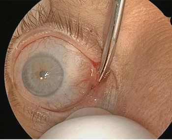
First, identify the caruncle of the eye and grasp it with toothed forceps to retract it laterally. Incise about one-third of the caruncle medially and aim the tip of sharp scissors towards the medial orbital wall (Fig. 1). Minor variations on the incision include the pre-caruncular or retro-caruncular incision. The pre-caruncular incision is made medial to the caruncle and lateral to the mucocutaneous junction, and provides shorter access to the bony wall, but has a greater risk of injury to the lacrimal system. The retro-caruncular incision is made lateral to the caruncle and medial to the plica semilunaris, but has an increased risk of injury to the medial rectus. Spread the scissors while palpating the bony wall to identify bone. Once bone is exposed, use a suction elevator or Freer elevator to dissect in a subperiosteal plane between bone and peri-orbital fascia. Use ribbon retractors to retract the orbital contents laterally.
Right eye showing initial transcaruncular incision.

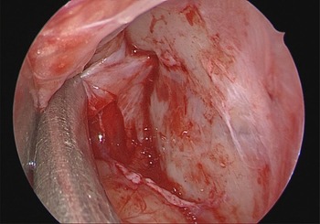
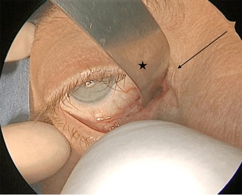
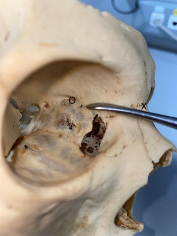
Once a subperiosteal plane is visualised, insert an endoscope into the surgical portal to confirm the correct plane (Fig. 2). The endoscope can initially rest on the ribbon retractor for stability. Continue the dissection superiorly towards the level of the nasion. The nasion is defined as the point in the midline of both the nasal root and nasofrontal suture. It represents the deepest bony point of the midline forehead. It is very important to stay at the same level as the nasion while dissecting posteriorly, as this will avoid injury to surrounding structures and lead directly to the anterior ethmoidal artery. Check the pupil for any changes in size or shape every 5 minutes while retracting, as this may indicate increased intra-ocular pressure. While dissecting, you may encounter Horner's muscle or the frontoethmoidal suture line, but this is not required. Once at the level of the nasion, proceed with the dissection posteriorly until the anterior ethmoidal artery is identified as a ‘tenting structure’ from the periosteum towards the bony orbital wall (Fig. 2). This should be about 25–45 mm posterior to the level of the nasion (Figs 3 and 4).Reference Cornelis and Lubbe8
Right orbital cavity with Freer elevator dissecting in the subperiosteal plane, showing tenting of the right anterior ethmoidal artery.

Right orbital retractor (asterisk), with nasion to the level of the anterior ethmoidal artery (arrow).

Right eye showing the nasion (‘X’) in relation to the anterior ethmoidal artery foramen (‘O’).

Use standard bipolar forces to cauterise the anterior ethmoidal artery or apply a Ligaclip. If necessary, the dissection can continue posteriorly until the posterior ethmoidal artery is identified about 12 mm posterior to the anterior ethmoidal artery. It is important to remember that there may be accessory ethmoidal arteries or the posterior ethmoidal artery may be absent. It is crucial that the optic nerve is not confused for the posterior ethmoidal artery. It is therefore important to ensure that only the tip of the bipolar forceps is in contact with the vessel, to avoid inadvertent thermal injury to the optic nerve.
Once the anterior ethmoidal artery is ligated, the surgical portal is irrigated with saline and nasal endoscopy is performed to check for haemostasis. The retractors are removed and a dry dressing may be applied. There is no need for sutures.
Discussion
Transorbital approaches have become an area of increased study and development within the field of otolaryngology. This novel area of surgery requires the examination and description of safe and effective surgical landmarks. This paper describes one such landmark to the medial orbital wall for the purposes of anterior ethmoidal artery ligation. Other approaches to the ethmoidal arteries are not always effective in safely identifying the artery, and often require extensive dissection of uninvolved sinuses or tissues.
Conclusion
The transcaruncular approach to the medial orbit, with anterior ethmoidal artery ligation, is a safe and effective technique for the treatment of severe refractory epistaxis. The nasion is the only anatomical landmark required to identify the anterior ethmoidal artery, safely avoiding injury to surrounding structures, with minimal scarring and a good post-operative recovery. A previous cadaver study found that the anterior ethmoidal artery can be found consistently about 12 mm from Horner's muscle and at the same level as the nasion.Reference Cornelis and Lubbe8 The frontoethmoidal suture is normally used as a guide to the anterior ethmoidal artery in the Lynch–Howarth approach, but this is not always visible through the transcaruncular approach and should not be depended upon.Reference Moe7,Reference Cornelis and Lubbe8
Competing interests
None declared.