Case presentation
A 24-year-old Hispanic woman received a heart transplant at 18 months of age for idiopathic dilated cardiomyopathy. She never received genetic testing for pathogenic cardiomyopathy variation. An unknown prior physician informed her that she was infertile owing to polycystic ovarian syndrome, fibroids, and the use of multiple medications, such as lisinopril, allopurinol, lansoprazol, everolimus, and tacrolimus. She has always had a normal QT interval. She discovered that she was pregnant around 3–4 weeks of gestation and stopped all medications except for everolimus and tacrolimus. The pregnancy was uneventful and her graft function was maintained. Fetal ultrasound at 22 weeks showed normal fetal anatomy with a normal cardiac view and normal cardiac function.
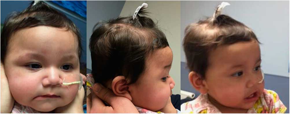
The infant was born at 37 weeks of gestation via normal spontaneous vaginal delivery. The birth weight of the infant was 2565 g. She did not receive a neonatal echocardiogram. She later presented with poor feeding and difficulty breathing at 5 months of age. On examination, she was alert, interactive, and appeared pink in room air. Facial features are shown in Figure 1. She had prominent fetal pads with otherwise normal fingers, thumbs, nails, and palmar creases. Her second toes were overlapping the first toes. Neurological examination revealed mild hypotonia and 1+ reflexes throughout.
Facial features of the infant. She was brachycephalic and plagiocephalic with one hair whorl to the right. The eyes were wide set. Epicanthal folds were present bilaterally. Her nasal bridge was depressed. Her ears were large with normal morphology without pits or tags. The lips were full and palate was intact. Pictures are published with the mother’s permission.

An electrocardiogram showed a normal QT interval for age. Echocardiography revealed a severely dilated left ventricle, moderate left ventricular non-compaction, severe left ventricular systolic dysfunction with an ejection fraction of 18%, and normal right ventricular function. She underwent pulmonary artery banding, followed by de-banding 7 weeks later with left ventricular assist device (Thoratec PediMag™, Pleasanton, California, United States of America) placement after failure of improvement with banding. She remained on this device, without complication, for 7 days, before transplantation.
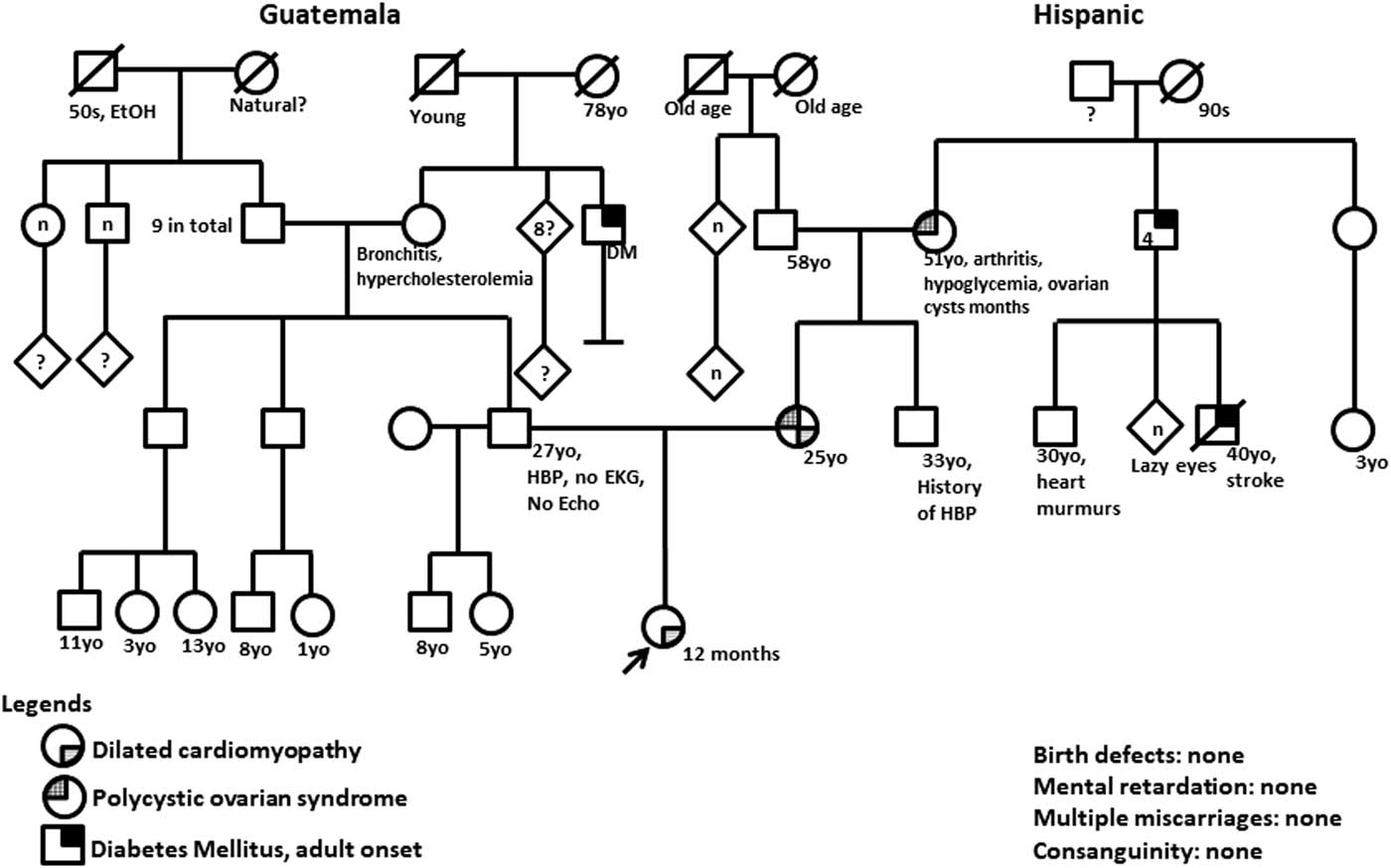
A four-generation pedigree is shown in Figure 2. The patient had a normal single-nucleotide polymorphism microarray. A cardiomyopathy genetic sequencing panel revealed heterozygous variants of uncertain significance in RYR2 (F1763S), TPM1 (R160H), and SCN5A (R1958Q). Targeted testing of the mother indicated that she harboured the TPM1 (R160H) and SCN5A (R1958Q) variants, but not the RYR2 (F1763S) variant. The R160H variant of TPM1was not observed in ~6500 individuals of European and African American ancestry in the NHLBI Exome Sequencing Project, indicating that it is not a common benign variant in these populations. This substitution occurs at a position that is highly conserved across species, and in silico analysis predicts that this variant is probably damaging to the protein structure and function.
Four-generation pedigree. DM=diabetes mellitus; EKG=electrocardiogram; EtOH=Alcohol; HBP=high blood pressure.
She eventually received a heart transplant at 7 months of age. She has been doing well since transplant on tacrolimus and sirolimus immunosuppressant medications. Her growth and development appear to be age appropriate.
Discussion
The effect of immunosuppressant medications on fertility and teratogenicity is controversial. Previously, sirolimus-associated infertility was observed in a young heart–lung transplant recipient with oligospermia.Reference Deutsch, Kaczmarek and Huber 1 No structural defects have been reported as a pregnancy outcome in transplant recipients while on immunosuppressant medications.Reference Sifontis, Coscia, Constantinescu, Lavelanet, Moritz and Armenti 2 A case of an unplanned pregnancy in a heart transplant patient on everolimus and cyclosporine was reported and resulted in a healthy infant.Reference Fiocchi, D’Elia and Vittori 3 Our patient was receiving both medications during the pregnancy. The infant had dysmorphic features, which can occur owing to a direct effect of a genetic mutation or indirectly via a genetic disturbance, such as gestational exposure to a teratogen.Reference Solomon and Muenke 4 Dysmorphic features suggesting teratogenecity include, but are not limited to, a tall and broad forehead, medial deficiency of eyebrows, infraorbital grooves, a broad nasal bridge, an anteverted nose, a thin upper lip, dysplastic ears, arachnodactyly, and clinodactyly.Reference Kini, Adab and Vinten 5 Dysmorphic features suggestive of teratogenicity have been studied in anti-epileptic medications; however, most reports of dysmorphic features owing to the use of immunosuppressant medications have been either nonspecific or unclassified.Reference Leroy, Rigot and Leroy 6 Therefore, the significance of dysmorphic features in this infant was uncertain.
The maternal and fetal risks during pregnancy and the postpartum period are great in heart transplant recipients. Decisions regarding pregnancy timing are often difficult and require a multidisciplinary team of healthcare providers.Reference Abdalla and Mancini 7 Management of maternal and fetal complications during pregnancy has been relatively well described.Reference Abdalla and Mancini 7 , Reference Wasywich, Ruygrok, Wilkinson, Gibbs and Coverdale 8 However, there is currently very limited information on genetic testing and monitoring for cardiac defects in fetal and neonatal offspring in order to determine recurrence risk. In our case, the mother was surprised that the baby had the same cardiac defect and also needed a heart transplant.
Genetic forms of dilated cardiomyopathy must be distinguished from the many acquired non-genetic causes of dilated cardiomyopathy. After exclusion of all acquired identifiable causes, dilated cardiomyopathy is traditionally referred to as idiopathic dilated cardiomyopathy, which includes genetic forms of dilated cardiomyopathy. When two or more closely related family members meet a formal diagnostic standard for idiopathic dilated cardiomyopathy, the diagnosis of familial dilated cardiomyopathy is made.
The genetic forms of dilated cardiomyopathy are diagnosed by family history and molecular genetic testing. The R160H variant of the TPM1 gene has been reported as a de novo variant in a patient with left ventricular non-compaction, whose father was diagnosed with ischaemic dilated cardiomyopathy.Reference Hoedemaekers, Caliskan and Michels 9 This variant has been classified as a likely pathogenic variant for primary dilated cardiomyopathy by ClinVar.Reference Landrum, Lee and Benson 10 As mother and baby both carry this variant, it is likely to be pathogenic for both the mother and child, rather than a variant of uncertain significance. Our patient’s father did not have sequencing performed.
The RYR2 gene encodes a protein for the ryanodine receptor found in cardiac sarcoplasmic reticulum. Mutations in this gene are associated with stress-induced polymorphic ventricular tachycardia and arrhythmogenic right ventricular dysplasia.Reference George, Higgs and Lai 11 The R1958Q variant in the SCN5A protein has been reported previously in one patient with long QT syndrome.Reference Tester, Will, Haglund and Ackerman 12 The mother was reported to have arrhythmia before transplant, although neither had a prolonged QT interval. At this point, we do not have sufficient evidence to classify either variant as pathogenic.
This case represented a missed opportunity to detect and promptly treat familial cardiomyopathy. It is necessary to provide prenatal and postnatal monitoring for both the heart transplant recipient parents and their offspring, especially screening for the familial dilated cardiomyopathy. Early genetic consultation and counselling may help determine the risk of recurrence and overall management. This should include a minimum of a three-generation pedigree and appropriate genetic testing for a suspected molecular cause. We also recommend routine prenatal evaluation for the fetus with a fetal echocardiogram and postnatal evaluation including a neonatal echocardiogram, followed by echocardiography every 3 months until 1 year of age, and then annually. Increased surveillance in the first year of life is prudent, given the increased incidence of dilated cardiomyopathy in this age group.Reference Towbin, Lowe and Colan 13 Counselling and postnatal testing should be carried out in the case of both male and female transplant recipients and their offspring.
Acknowledgements
The authors thank the patient and her family for their participation and contribution.
Financial Support
This research received no specific grant from any funding agency, commercial or not-for-profit sectors.
Conflicts of Interest
None.