Introduction
The Acanthocephala is a small, monophyletic group of obligate endoparasites comprising over 1200 species (Smales, Reference Smales and Schmidt-Rhaesa2015). Morphological and molecular evidence strongly suggest a close relationship exists between Acanthocephala and Rotifera (Garey et al., Reference Garey, Schmidt-Rhaesa, Near and Nadler1998; Near et al., Reference Near, Garey and Nadler1998; García-Varela & Nadler, Reference García-Varela and Nadler2006; Gazi et al., Reference Gazi, Sultana, Min, Park, García-Varela, Nadler and Park2012; Sielaff et al., Reference Sielaff, Schmidt, Struck, Rosenkranz, Mark Welch, Hankeln and Herlyn2016). A common morphological feature of the two phyla is the cement gland. Cement glands are associated with the male reproductive organ in acanthocephalan and in the toes of rotifers (Near et al., Reference Near, Garey and Nadler1998). Despite the morphological similarity, totally different functions have been ascribed to the cement glands in each phylum.
The morphology and phylogenetic significance of the cement glands were first reported in a detailed study by Van Cleave (Reference Van Cleave1949), where it was shown that cement glands vary in shape and structure as well as in number. According to Van Cleave (Reference Van Cleave1949), in Eoacanthocephala there is a single syncytial gland, in Archiacanthocephala and in Palaeacanthocephala there are as many as eight tubular or flask-shaped glands and there are eight tubular cement glands in Polyacanthocephala (Amin & Dezfuli, Reference Amin and Dezfuli1995). Centrorhynchus globocaudatus (Zeder, 1800) belongs to the class Palaeacanthocephala and has four elongate tubular glands and was described from birds of prey in different regions of the world (Lisitsyna & Greben, Reference Lisitsyna and Greben2015). Taxonomic studies were unable to provide the exact number of glands present in this acanthocephalan (e.g. Lisitsyna & Greben, Reference Lisitsyna and Greben2015). Cement glands were considered as the most conspicuous and distinctive elements in the genital apparatus of male acanthocephalans (Van Cleave, Reference Van Cleave1949). In Acanthocephala, the glands and their products have a fundamental role in the reproductive process (Abele & Gilchrist, Reference Abele and Gilchrist1977; Parshad & Crompton, Reference Parshad and Crompton1981). Very few studies are available on the ultrastructure and function of the cement apparatus of acanthocephalans (Asaolu, Reference Asaolu1981; Dezfuli et al., Reference Dezfuli, Onestini, Carcupino and Mischiati1998, Reference Dezfuli, Simoni and Mischiati2001; Dezfuli, Reference Dezfuli2000), although it has been reported that the main component the cement secretion is protein (Dezfuli et al., Reference Dezfuli, Onestini, Carcupino and Mischiati1998).
Copulatory structures of the male acanthocephalan consist of an eversible campanulate bursa, the penis and muscular Saefftigen's pouch (Miller & Dunagan, Reference Miller, Dunagan, Crompton and Nickol1985). Light microscopical and ultrastructural analyses of Saefftigen's pouch and the seminal vesicle of male acanthocephalan are lacking. The current study provides the first transmission electron micrographs of Saefftigen's pouch and seminal vesicle in Acanthocephala. Furthermore, data are presented describing the ultrastructure of cement glands and formation of granules within the gland of mature C. globocaudatus.
Materials and methods
Birds and sampling
On several occasions, moribund or dead birds of prey Falco tinnunculus and Buteo buteo from the province of Ferrara (northern Italy) were provided to the Experimental Zooprophylactic Institute of Ferrara for routine monitoring for public health in search of West Nile Virus or New Castle Disease, both of which use birds of prey as a vector. After dissection of the birds, the whole digestive tract was removed from each animal and examined for helminths. Numerous specimens of C. globocaudatus were obtained from the intestine of these birds of prey.
Transmission electron microscopy
Twenty-five mature male C. globocaudatus were selected and dissected, and the whole genital apparatus removed from the acanthocephalan body. The reproductive organs were fixed in 2.5% glutaraldehyde in 0.1 M cacodylate buffer, pH 7.2, post-fixed in 1% osmium tetroxide in the same buffer for 1.5 h, dehydrated in a graded acetone series and embedded in epoxy resin. Semi-thin sections of the embedded samples were cut on a Reichert OM2 ultramicrotome, Wien, Austria with glass knives, and stained with toluidine blue. Ultra-thin sections were obtained with a Reichert Ultracut ultramicrotome stained with uranyl acetate and lead citrate and observed with a Hitachi H-800 electron microscope (Hitachi, Tokyo, Japan). Light micrographs were obtained using an ECLIPSE 80i microscope (Nikon, Tokyo, Japan). Five female C. globocaudatus with attached copulatory cap were routinely processed for scanning electron microscopy (SEM) observation using a Cambridge Stereoscan 360, London, UK at an acceleration voltage of 20 kV.
Results
Species was identified by comparing our specimens with the description provided for C. globocaudatus by Lisitsyna & Greben (Reference Lisitsyna and Greben2015). Our measurements generally fell within the range of variation reported by these authors. Overall (eight females, 18 males), proboscis rounded apical, conical toward middle-basal part, short neck, proboscis with 30–36 longitudinal rows of 19–21 hooks per row, receptacle doubled-walled, lemnisci sacciform and longer than receptacle and both sexes with subterminal gonopore.
Females: trunk length ranging from 15.00 to 38.00 mm, width from 800 to 1300 mm, proboscis 900–1250 mm long, receptacle 900–1200 mm long, lemnisci 1600–2200 mm long, elongate-oval eggs.
Males: trunk length ranging from 13.00 to 25.00 mm, width from 800 to 1100 mm, proboscis 900–1200 mm long, proboscis with 30–34 longitudinal rows of 19–21 hooks per row, receptacle 900–1100 mm long, lemnisci 1500–2000 mm long, testes oval in shape, in tandem almost overlapping, cement glands 8.00–12.00 mm long, Saefftigen's pouch 1.8–2.5 mm long.
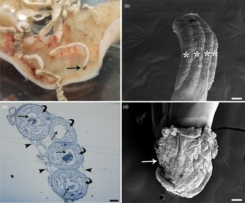
The current survey was carried out on moribund or recently deceased (less than 3 h before dissection) F. tinnunculus and B. buteo. The reproductive apparatus of 25 mature male specimens of C. globocaudatus and five females of the same species with copulatory cap still attached to the posterior end of the worm were evaluated. During dissection of the intestine of heavily infected birds, several females with a brownish-coloured cap were documented (fig. 1a). In some females, the cap was observed in another part of the worm body rather than on the genital pore. This cap was also found on the body surface of a few males. Males with extruded bursa were also commonly encountered. All acanthocephalans were alive during the initial inspection and most of them remained alive in saline solution at room temperature until four days post detachment from the host intestine.
(a) Photo of heavily infected intestine of Falco tinnunculus, several Centrorhynchus globocaudatus are attached to the intestinal wall, note brownish copulatory cap (arrow) at terminal part of body of alive female; (b) SEM micrograph shows four elongated cement glands (asterisks) of C. globocaudatus; (c) semithin section of four cement glands of an adult male surrounded by ligament sac (arrow heads), outer cytoplasmic layer (curved arrows) and gland's lumen (arrows) are visible; (d) SEM micrograph of copulatory cap (arrow) at terminal part of the female body. Scale bars: (b) 100 μm; (c) 50 μm; (d) 200 μm.

Four elongate tubular cement glands of male C. globocaudatus are situated posterior to the testes, cement glands are packed very closely one to other (fig. 1b) and are surrounded by a ligament sac. Borders of individual glands can only be seen in toluidine-blue-stained semithin sections (fig. 1c). The cement substance within the gland, duct lumen and cement reservoir also stains with toluidine blue. A semithin section through the cement gland shows two different parts within the gland: an outer ‘cytoplasmic layer’ and a luminal area (fig. 1c). The cytoplasmic layer contains a number of nuclei (see below) and surrounds the luminal area (fig. 1c). In living or fixed (glutaraldehyde or ethanol) males, any distinction between single glands was impossible using a light microscope. SEM observation of females with an attached cap showed the cap to be compact, with a rough surface and covering the posterior end of the female (fig. 1d).
Ultrastructure of the cement gland, cement reservoir and Saefftigen's pouch in mature C. globocaudatus
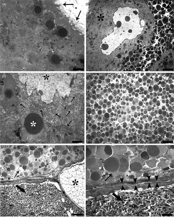
The glands of C. globocaudatus are surrounded by material of a fibrous nature, which appears as an extension of the posterior region of the ligament sac. Each gland has its own envelope, which is about 0.6 μm thick (fig. 2a). Beneath this envelope there is an outer cytoplasmic layer, with an approximate thickness variable from 22 to 26 μm, within which are found nuclei with diameter ranging from 20 to 22 μm (fig. 2b). Each nucleus has a lobed shape with an irregular outline (fig. 2b). In this layer, there is no indication of a plasmalemma between the nuclei of the cement gland. The cytoplasmic layer contains numerous mitochondria with lamellar cristae. Clusters of mitochondria were frequently found very close to the nucleus and cement granule (fig. 2c). Also, the cytoplasmic layer was filled with prominent free ribosomes (fig. 2c). In contrast, the Golgi apparatus and smooth and rough endoplasmic reticulum were rarely observed in the cytoplasmic layer. The granules progress from the cytoplasmic layer toward the luminal area, where residue of degenerated nuclei and undefined organelles was observed between electron-dense secretory granules (fig. 2b).
Transmission electron micrograph of cement apparatus, seminal vesicle and Saefftigen's pouch of male Centrorhynchus globocaudatus. (a) Micrograph shows the periphery of a cement gland, each gland is surrounded with an envelope (arrows), within the outer cytoplasmic layer some electron-dense cement granules are visible; (b) portion of cytoplasmic layer (asterisk) and luminal area of a cement gland, a big nucleus with irregular outline within the cytoplasmic layer, numerous electron-dense cement granules inside the lumen are evident; (c) cluster of mitochondria (arrows) close to nucleus (asterisk) within the cytoplasmic layer, note abundance of free ribosomes in cytoplasm, a cement granule (white asterisk) is in formation; (d) middle portion of gland's duct is filled with cement granules; (e) micrograph shows interface region between cement reservoir (arrow), seminal vesicle (thick arrow) and Saefftigen's pouch (asterisk), each organ is surrounded with its own envelope, close contact and narrow empty spaces between envelopes are visible; (f) high magnification of interface region between cement reservoir (arrow) and seminal vesicle (thick arrow), envelopes of both organs are in close vicinity (arrow heads). Scale bars: (a) 1.3 μm; (b, d) 4 μm; (c) 0.5 μm; (e) 2.5 μm; (f) 1.1 μm.

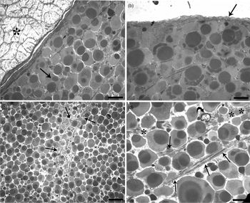
Electron microscopy of the mature C. globocaudatus cement glands gave us some insight into the probable mode of formation of secretory granules. The electron-opaque inclusions initially form from the outermost part of the cytoplasmic layer (fig. 2a), often in close proximity to the nucleus, surrounded by a single membrane (fig. 2c). A reduced electron-dense space exists between the granule membrane and its electron-dense content (fig. 2c), and this space is gradually filled with a fine, granular substance. Finally, the ‘mature’ secretory granules develop a closely associated membrane. Thus, within the ‘mature’ granules, two regions are immediately recognized: a narrow outer granular area encloses a wider, electron-dense, amorphous component. In many instances, the granules form in close proximity to the fully ‘mature’ cement granules (ranging from 1 to 1.3 μm in diameter). A cement duct arises from each gland and appears to lead into the genital sheath toward the posterior end of the male body. The thin wall of the duct averages 0.6 μm in thickness and numerous secretory granules fill the duct lumen (fig. 2d). The terminal part of the male's body was next evaluated with respect to the fine structure of the cement reservoir, seminal vesicle and Saefftigen's pouch, and the interface region between them (fig. 2e). All three structures have thin envelopes, which are in close apposition one to another (fig. 2e). Figure 2f shows the interface between cement reservoir and the seminal vesicle full of mature spermatozoa, where a very narrow space is appreciable (figs 2e, f). A similar interface is seen between the cement reservoir and Saefftigen's pouch, the latter of which has a spongy appearance (fig. 3a). The cement reservoir (thickness ranging from 0.4 to 0.8 μm) has a similar appearance to the cement duct structure and is filled with numerous larger electron-dense cement granules variable from 2 to 2.8 μm in diameter (fig. 3b). The periphery of the cement reservoir does not have empty spaces (fig. 3b), whereas, in deeper areas of the tissue, granules are loosely packed with some space between them. Interestingly, several long microtubules and residues of cytoplasm and undefined organelles were frequently observed in the middle area of the cement reservoir (respectively, figs 3c, d).
Transmission electron micrograph of cement reservoir and Saefftigen's pouch of male Centrorhynchus globocaudatus. (a) Interface region between cement reservoir (arrow) and Saefftigen's pouch (asterisk), note narrow empty space between envelopes of both organs; (b) high magnification of periphery of cement reservoir, envelope (arrow) and lack of empty spaces due to close packing of cement granules are visible; (c) low magnification of middle part of cement reservoir, empty spaces among granules are abundant, in the centre of the picture numerous microtubules (arrows) scattered among the granules can be seen; (d) high magnification of middle part of cement reservoir, microtubules (arrows) among granules, residue of undefined organelle (curved arrow) and cytoplasm (asterisks) are evident. Scale bars: (a) 1.7 μm; (b, d) 1.4 μm; (c) 5 μm.

Discussion
Our SEM evaluation of male C. globocaudatus allowed us to determine the exact number of cement glands in this species. The acanthocephalan has four long tubular cement glands, which are packed tightly against one another. The glands are surrounded with a tightly associated ligament sac, which is visible under light microscopy and obscures individual glands in both live and fixed males. Only the use of SEM and/or sections of embedded cement glands enabled an accurate determination of the number of glands. The occurrence of debris from nuclei, cytoplasm and undefined organelles in the gland lumen and cement reservoir confirmed that, as in Pomphorhynchus laevis (Dezfuli et al., Reference Dezfuli, Capuano, Pironi and Mischiati1999a) and Acanthocephalus anguillae (Dezfuli et al., Reference Dezfuli, Simoni and Mischiati2001), the glands of C. globocaudatus are also of holocrine type. Golgi apparatus were rarely observed in the cement glands of C. globocaudatus, a finding in agreement with an earlier study of this gland type (Patt & Patt, Reference Patt, Patt, Patt and Patt1969). Centrorhynchus globocaudatus, P. laevis and A. anguillae belong to the class Palaeacanthocephala, and all three show similarities with respect to gland structure, duct and cement reservoir. Nevertheless, some differences were noticed among these three species – for example, microtubules are present in the cytoplasmic layer of the gland of A. anguillae and prominent rough endoplasmic reticulum is found mainly in the gland of P. laevis.
Inside the clade, acanthocephalans are probably sisters of Seisonidea (Rotifera), as suggested by mitochondrial gene order (García-Varela & Nadler, Reference García-Varela and Nadler2006; Gazi et al., Reference Gazi, Sultana, Min, Park, García-Varela, Nadler and Park2012; Sielaff et al., Reference Sielaff, Schmidt, Struck, Rosenkranz, Mark Welch, Hankeln and Herlyn2016). Rotifers have a foot at the end of which are toes, pointed projections from which open cement glands secrete a sticky material used to anchor the animal during feeding (Buchsbaum et al., Reference Buchsbaum, Buchsbaum, Pearse and Pearse2013). The occurrence of toes with adhesive glands in rotifers has been questioned because the cement glands of acanthocephalans may be homologues to the adhesive glands of rotifers (Near et al., Reference Near, Garey and Nadler1998). Nonetheless, in Acanthocephala, gland secretion is fundamental for reproduction (Van Cleave, Reference Van Cleave1949; Abele & Gilchrist, Reference Abele and Gilchrist1977; Dezfuli & De Biaggi, Reference Dezfuli and De Biaggi2000). Cement glands appear in two general forms, as a single gland or as a group of glands, and both forms occur in Acanthocephala and Rotifera. Thus, cement glands can be considered a plesiomorphic character (Brooks & McLennan, Reference Brooks, McLennan, Ricklefs and Schluter1993).
Males of the phylum Acanthocephala might display very peculiar behavioural strategies to avoid eventual second insemination of the same female by another male (Parker, Reference Parker1970). One of these strategies is the formation of a cap from secretions of the male accessory gland (Parker, Reference Parker1970; Gillott, Reference Gillott2003). The male glands of acanthocephalan produce a proteinaceous substance named cement (Van Cleave, Reference Van Cleave1949). After insemination, the cement internally occludes the vagina and uterus (Dezfuli et al., Reference Dezfuli, Capuano, Pironi and Mischiati1999a; Dezfuli & De Biaggi, Reference Dezfuli and De Biaggi2000) and externally covers the surface of the female genital pore with a cap (Richardson et al., Reference Richardson, Martens and Nickol1997; Dezfuli et al., Reference Dezfuli, Capuano, Pironi and Mischiati1999a, Reference Dezfuli, Simoni and Mischiati2001; Dezfuli, Reference Dezfuli2000). The presence of a cap over the posterior end of female acanthocephalans as well as a misplaced cap on female and male body surface were mentioned in some earlier accounts (Parshad & Crompton, Reference Parshad and Crompton1981; Richardson et al., Reference Richardson, Martens and Nickol1997; Dezfuli et al., Reference Dezfuli, Capuano, Pironi and Mischiati1999a, Reference Dezfuli, Simoni and Mischiati2001). In several live female C. globocaudatus, a brownish-coloured cap was frequently encountered on the posterior end and a misplaced cap on either the male or female body was observed in some individuals.
With reference to cap behaviour in Acanthocephala, two functions were proposed by Abele & Gilchrist (Reference Abele and Gilchrist1977): (1) the cap avoids escape of spermatozoa from the female genital pore; and (2) the cap formation evolved in response to sexual selection and acts in preventing successive insemination. With respect to the second function, Crompton (Reference Crompton, Crompton and Nickol1985) expressed that the cap can have several advantages for the genes of the inseminating male. Our previous direct evidence on the occurrence of spermatozoa on or in the cement cap which was adhered to the female posterior in A. anguillae (Dezfuli et al., Reference Dezfuli, Simoni and Mischiati2001) agree with the two proposed functions.
Previous work on P. laevis (Palaeacanthocephala) discovered that the major component of cement is a 23 kDa protein (Dezfuli et al., Reference Dezfuli, Onestini, Carcupino and Mischiati1998). We purified this protein and produced a polyclonal antibody against it called anti-23PL (Dezfuli et al., Reference Dezfuli, Onestini, Carcupino and Mischiati1998). This antibody was applied to the paraffin sections of males belonging to other species within the class Palaeacanthocephala and to males of both Moniliformis moniliformis (Archiacanthocephala) and Neoechinorhynchus emydis (Eoacathocephala). The anti-23PL antigen was detected in paraffin sections of cement glands from all the species examined, suggesting that the p23 protein has been retained among the three classes of the taxon Acanthocephala (Dezfuli et al., Reference Dezfuli, Capuano, Giari, Pironi and Mischiati1999b). Therefore, it is reasonable to believe that the 23 kDa protein is also the main component of C. globocaudatus (Palaeacanthocephala) cement glands.
The essential role of the cytoskeletal system in cytoplasmic transport is well-established (Pathak et al., Reference Pathak, Thakur and Mallik2018). Microtubules serve as tracks for the transport of vesicles, organelles, messenger RNA and signalling molecules throughout the cytoplasm (Barlan & Gelfand, Reference Barlan and Gelfand2017; Balabanian et al., Reference Balabanian, Chaudhary and Hendricks2018). Herein, microtubules were identified in the glands and in greater numbers in the reservoir where they were scattered among the cement granules. An active role of microtubules in transporting vesicles and granules from storage sites to their final destination, the vaginal tract, could explain the greater number of tubules in the cement reservoir.
Saefftigen's pouch could be considered as a morphological character that arose with the acanthocephalans. It is enclosed in a sheath of circular muscle and the spongy medulla of the pouch contains fluid, which moves back and forth through the stalk of pouch into the apical portion of the bursa (Miller & Dunagan, Reference Miller, Dunagan, Crompton and Nickol1985). There are very few accounts that describe the structural organization of Saefftigen's pouch and seminal vesicles (e.g. Miller & Dunagan, Reference Miller, Dunagan, Crompton and Nickol1985). The study presented here is the first to provide a description of the fine structure of seminal vesicles and Saefftigen's pouch and their interface region with the cement reservoir in an acanthocephalan.
Acknowledgements
Thanks are due to D. Tartari and L. Borghi from LIPU (League for Birds Protection), Ferrara, Italy, for their help in collecting the birds of prey. We are indebted to E. Simoni, L. Giari and the Centre of Electron Microscopy of the University of Ferrara for their technical assistance.
Financial support
This work was supported by a local grant from the University of Ferrara (B.S.D., grant number FAR 2019).
Conflicts of interest
None.
Ethical standards
The authors assert that all procedures contributing to this study comply with the ethical standards of the relevant national and institutional guides.