Williams syndrome, also known as Williams–Beuren syndrome, is an autosomal dominant disorder of chromosome region 7q11.23.43. Most cases of Williams syndrome are caused by de novo deletion of this chromosome region, which codes for elastin. Williams syndrome occurs in approximately 1 in 10,000 live births, affecting an estimated 20,000–30,000 people in the United States. Williams syndrome can lead to multi-system abnormalities but most commonly is associated with behavioural, developmental, and cardiovascular problems.Reference Pober 1
Abnormalities in elastin production in patients with Williams syndrome often cause smooth muscle overgrowth, leading to vasculopathy within their medium and large arteries. This vasculopathy can manifest as stenoses of vital structures such as the supravalvular aorta, aortic arch, descending aorta, supravalvular pulmonary artery, peripheral pulmonary arteries, and coronary, renal, mesenteric, and intracranial arteries.Reference Pober, Johnson and Urban 2 Additionally, Williams syndrome has been associated with other cardiovascular abnormalities such as ventricular septal defects, valvar disease (e.g. aortic insufficiency, pulmonary insufficiency, and mitral valve prolapse), and QT interval prolongation.Reference McCarty, Tang, Swearingen and Collins 3 For some children with Williams syndrome and congenital heart disease, cardiac catheterisation may be necessary.Reference Pham, Moller, Hills, Larson and Pyles 4 We present the case of a 3-month-old infant with Williams syndrome who developed sudden cardiac arrest during a cardiac catheterisation procedure and ultimately died. We also present images of pathological specimens that provide valuable insight into the aetiology of this tragic event.
Case presentation
A female infant born at term was found to be hypertensive soon after birth. An echocardiogram performed at 1 week of age revealed mild supravalvular aortic stenosis, mild branch pulmonary arteries stenosis, mild left ventricular hypertrophy, mild right ventricular hypertrophy, and coarctation of aorta. Williams syndrome was highly suspected based on this constellation of findings and later confirmed by fluorescence in situ hybridisation testing.
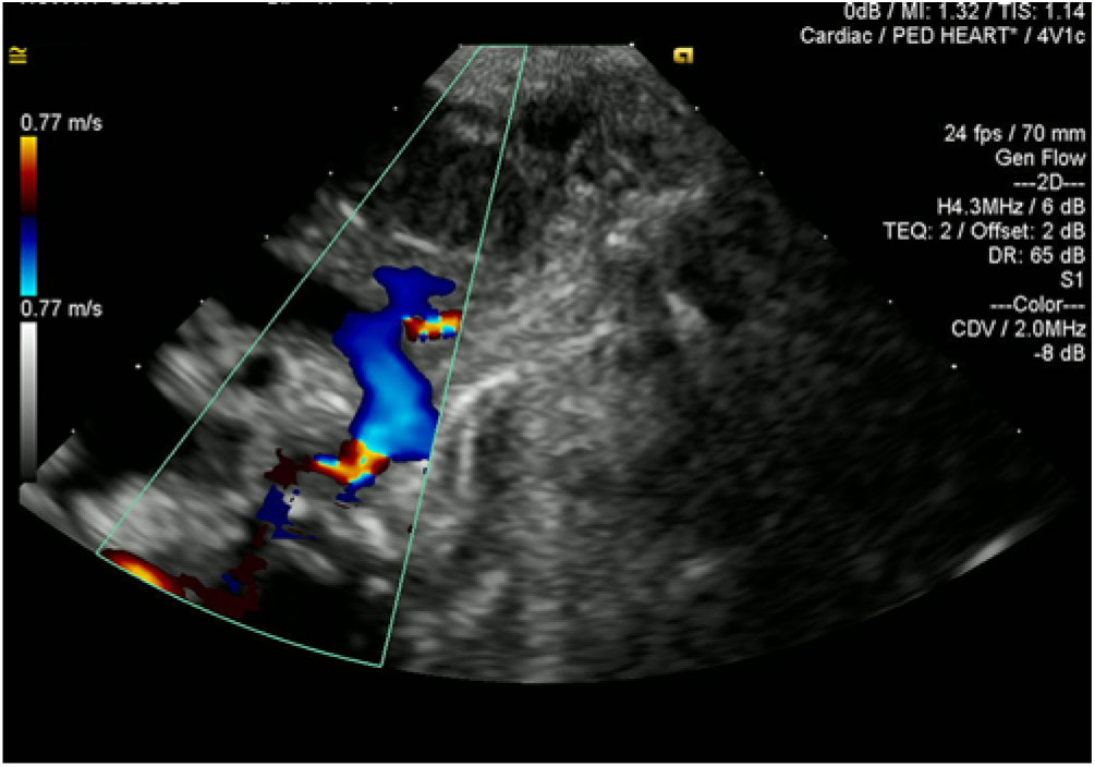
At 2 months of age, the patient underwent surgical repair for her aortic coarctation via posterior end-to-end anastomosis and left subclavian arterial patch angioplasty. A follow-up echocardiogram 1 month after surgery showed residual aortic coarctation, with a doppler peak systolic gradient across the coarctation of 110–120 mmHg (Fig 1). Also present were a diffusely small aortic arch, moderate branch pulmonary arteries stenosis (gradient 36 mmHg), and moderate right ventricular hypertrophy. Coronary arteries appeared normal, with normal origins and flow patterns. Electrocardiogram showed findings consistent with right ventricular hypertrophy, normal corrected QT interval, and no ST-segment changes.
Coarctation of aorta in a patient with William syndrome prior to cardiac catheterisation as seen by echocardiogram. The residual coarctation was considered clinically significant, measuring 2.5 mm with a gradient across the coarctation site of 69 mmHg.

At 3 months of age, the patient underwent balloon angioplasty of the coarctation. During the initial diagnostic portion of the cardiac catheterisation, she developed ST-segment depression followed by bradycardic cardiac arrest, which was suspected to have resulted from decreased coronary perfusion pressure related to anaesthesia. Prior to the arrest, no haemodynamic data had yet been obtained by catheterisation. She required 30 minutes of cardiopulmonary resuscitation per the Pediatric Advanced Life Support guidelines. After return of spontaneous circulation, nitroglycerin was initiated to mitigate the possibility of coronary vasospasm as a cause of the arrest and an epinephrine infusion was initiated for further post-arrest haemodynamic support. Due to critical condition of the patient after cardiac arrest, extensive cardiac catheterisation imaging and measurements were not obtained. Instead, the team decided to proceed directly with the balloon angioplasty. A peak-to-peak pull-back gradient was 69 mmHg from the ascending to descending aorta. No other baseline haemodynamics were obtained. To decrease renal contrast exposure, a baseline aortic angiogram was not performed. Instead, echocardiography was used to measure the aorta, which was narrowed to 2.5 mm at the aortic coarctation site prior to intervention. Distally, the descending aorta measured 6 mm. She underwent three consecutive balloon dilatations using a 7-mm × 2-cm Sterling balloon, after which the diameter of the coarctation site measured 5 mm with reduction in the gradient to 17 mmHg. Left ventricular function immediately post-cardiac arrest was moderate-to-severely diminished.
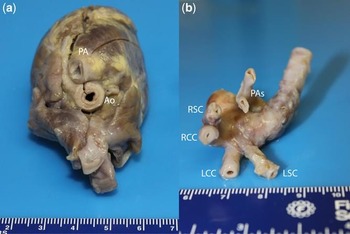
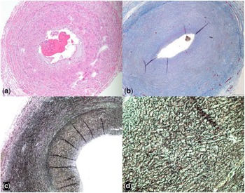
Following the procedure, she was admitted to the cardiovascular ICU endotracheally intubated on mechanical ventilation and appeared to be recovering with adequate haemodynamics on vasoactive medications. Admission of 12-lead electrocardiogram showed prolonged corrected QT interval (502 mseconds) and no ST depression or elevation. She was able to be weaned off epinephrine within 2 hours after admission to the cardiovascular ICU. However, ST depression was observed in lead II of her continuous telemetry monitoring for the first 12 hours post-procedure, and she continued to have prolonged corrected QT interval thereafter. Additionally, echocardiogram performed 4.5 hours after the cardiac arrest showed severe left ventricular hypertrophy with hyperdynamic left ventricular systolic function and normal right ventricular systolic function. After 48 hours post-cardiac catheterisation, she had another sudden onset bradycardic cardiac arrest accompanied by ST-segment elevation. Diastolic blood pressure during the 4 hours prior to the arrest ranged from 31 to 56. Cardiopulmonary resuscitation per Pediatric Advanced Life Support guidelines was initiated. Though return of spontaneous circulation was achieved intermittently during the resuscitation, it could not be sustained. After consultation with family, resuscitation efforts were stopped, and the patient expired. Post-mortem gross sections of the abnormal vascular anatomy are provided in Figure 2 and microscopic images are provided in Figure 3.
Gross anatomy of the heart and great vessels: (a) aorta and pulmonary trunk origin at the heart base (Ao: aorta; PA: pulmonary arterial trunk) and (b) Posterior views of the pulmonary trunk and aortic arch with their first branches. Diffuse arteriopathy can be seen, characterised by concentric and contiguous thickening of the arterial wall at the expense of the lumen (LCC = left common carotid artery; LSC = left subclavian artery; PAs = right and left pulmonary arteries; RCC = right common carotid artery; RSC = right subclavian artery).

Microscopic cross sections of the coronary arteries using (a) hematoxylin and Eosin stain, demonstrating thickened muscular wall with disorganised elastic fibres and narrow lumen; (b) trichrome stain showing thickened vessel walls comprised mostly of fibrous tissue, with markedly diminished amount of smooth muscle; (c) and (d) trichrome stain at 200x and 400x, respectively, demonstrating thickened, disorganised elastic fibres that also appear shortened and fractured.

Discussion
Pathophysiology of cardiac disease in Williams syndrome
The pathophysiology of Williams syndrome results from defects in the gene encoding elastin, which is responsible for providing the aorta with its high degree of reversible distensibility.Reference Collins 5 Smooth muscle cells in patients with Williams syndrome have reduced arterial elastin content, pathological alignment of the elastin fibres, and increased arterial stiffness, primarily in large vessels. The reduced elastin content in arterial walls also leads to increased proliferation and hypertrophy of smooth muscle cells, multi-layer thickening of the media of large arteries, and subsequent development of obstructive hyperplastic intimal lesions, most notable of which is supravalvular aortic stenosis.
Coronary artery abnormalities are also common in children with Williams syndrome.Reference Collins 5 Coronary ostial stenosis is the most common coronary artery abnormality reported, resulting from adhesion of the aortic leaflet edge to the narrowed sinotubular junction; obstruction to flow by the overhanging, stenosing supravalvular aortic stenosis ring; or direct occlusion by a thickened arterial wall. Alternatively, in patients with the discrete hourglass form of supravalvular aortic stenosis, high aortic pressure proximal to the supravalvular aortic stenosis is transmitted to the coronary arteries and may result in severe coronary artery dysplasia, narrowing, or dilatation.
Coronary artery abnormalities, along with alterations in ventricular mass, end-diastolic pressures, and duration of diastolic coronary blood flow that can also occur in children with Williams syndrome, can create a precarious balance between myocardial oxygen demand and delivery, increasing the risk for sudden cardiac death. The incidence of sudden cardiac death in patients with Williams syndrome has been reported to be 1 in 1000 patient-years, which is 25- to 100-fold higher than the incidence in the age-matched normal population.Reference Wessel, Gravenhorst, Buchhorn, Gosch, Partsch and Pankau 6 There have been many cases of sudden arrest and death previously reported in the literature, nearly all of which were associated with administration of sedation or general anaesthesia. The decrease in endogenous catecholamine production, which often leads to hypotension or myocardial depression, coupled with the risk of transient episodes of hypopnea and hypoxemia associated with sedation and anesthesia, is, in large part, likely responsible for these occurrences.
Sudden cardiac death and cardiac catheterisation
Patients with Williams syndrome often require sedation or anaesthesia for cardiac and non-cardiac procedures. Due to developmental delay and anxiety, some Williams syndrome children may require sedation for even simple imaging procedures (e.g. echocardiogram). In most children without Williams syndrome, sedation for imaging studies and many non-cardiac procedures is deemed to be low risk. However, episodes of sudden cardiac arrests have been reported in children with Williams syndrome undergoing tonsillectomy, placement of myringotomy tubes, orchiopexy, hernia repair, or dental procedures.Reference Wessel, Gravenhorst, Buchhorn, Gosch, Partsch and Pankau 6 – Reference Hornik, Collins and Jaquiss 14
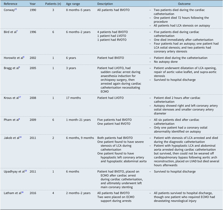
The most frequently reported site of sudden cardiac arrest for children with Williams syndrome, however, is during cardiac catheterisation. A summary of the current literature which includes 25 children with Williams syndrome who suffered cardiac arrests in association with cardiac catheterisation is provided in Table 1.Reference Pham, Moller, Hills, Larson and Pyles 4 , Reference Bird, Billman and Lacro 7 – Reference Bragg, Fedel and DiProsperis 9 , Reference Conway, Noonan, Marion and Steeg 15 – Reference Upadhyay, Seiden and Epstein 19 Notably, 20 of the 25 patients described in this summary had biventricular outflow tract obstruction, which has identified as a risk factor for sudden cardiac arrest in children with Williams syndrome.Reference Pham, Moller, Hills, Larson and Pyles 4 , Reference Bird, Billman and Lacro 7 , Reference Gupta, Tobias and Goyal 11 , Reference Hornik, Collins and Jaquiss 14 – Reference Latham, Ross, Eisses, Richards, Geiduschek and Joffe 16 In addition to the risk of anaesthesia and sedation, these procedures carry higher risk as unintended provocation of arrhythmia which, even transiently, could disrupt the tenuous balance of myocardial oxygen demand and delivery and could precipitate arrest.Reference Burch, McGowan, Kussman, Powell and DiNardo 20 Moreover, acute changes in aortic diastolic blood pressure that can occur when left ventricular obstructive lesions are relieved can compromise coronary perfusion, especially in the setting of stenotic or dysplastic coronary arteries, further decreasing the threshold for cardiac arrest.Reference Fricke, d’Udekem and Brizard 21 It is therefore important for cardiologists, interventional cardiologists, intensivists, and anaesthesiologists to understand the risk of sudden cardiac arrest and death in this patient population. Any procedure requiring sedatives or anaesthesia should be performed at tertiary care medical centres where subspecialists with expertise in paediatric cardiac care are available and where extracorporeal membrane oxygenation can be rapidly deployed. The risk of sudden cardiac arrest and the possibility of extracorporeal support should be fully explained to families prior to these procedures, and necessity of elective procedures should be carefully considered.
Reported cases of sudden cardiac arrest in children with William syndrome during or after cardiac catheterisation.

AS = aortic stenosis; BVOTO = biventricular outflow tract obstruction; ECMO = extracorporeal membrane oxygenation; LCA = left coronary artery; LVAD = left ventricular assist device; LVOTO = left ventricular outflow tract obstruction; RVOTO = right ventricular outflow tract obstruction
Cardiovascular lesions of Williams syndrome can also be progressive. Therefore, full cardiac evaluation should be performed shortly before the scheduled procedure. Evaluation should include a careful history to determine if symptoms of myocardial ischemia have occurred, measurement of systemic blood pressure in upper and lower extremities, and electrocardiogram and echocardiogram. Electrocardiogram should be used to evaluate the corrected QT interval and document findings that could represent ischemia such as ST-segment changes. Echocardiogram can be used to evaluate the degree of left ventricular hypertrophy, supravalvular aortic or pulmonary stenosis, anatomy and patency of the coronary arteries, and underlying cardiac function. Unfortunately, the extent of coronary artery and aortic involvement may not be evident until the cardiac catheterisation is conducted.Reference van Pelt, Wilson and Lear 22
During cardiac catheterisation procedures, appropriate haemodynamics should be maintained with special attention to preventing tachycardia and hypotension to ensure adequate coronary perfusion pressure and avoid increasing myocardial oxygen demand.Reference Burch, McGowan, Kussman, Powell and DiNardo 20 It is crucial that blood pressure, especially diastolic pressure, is closely monitored and continuous telemetry is carefully assessed for ST-segment changes. If hypotension and ST changes do occur, peri-procedure vasoactive infusions should be promptly started to ensure adequate coronary perfusion pressure. Phenylephrine and arginine vasopressin can improve blood pressure without increasing the heart rate and thus may be better suited for this clinical scenario. Beta-adrenergic agonists such as epinephrine should be used cautiously, as sudden tachycardia will be detrimental. Post-procedure, high-risk patients should be monitored postoperatively in an ICU setting, where meticulous attention to maintaining appropriate blood pressure and minimising tachycardia should be continued.
Conclusion
This case illustrates the risk of catastrophic cardiovascular event during cardiac catheterisation procedures in children with Williams syndrome. The gross specimens and microscopic images available also provide important histopathological evidence of why these events occur in this fragile patient population. Cardiac interventionalists as well as all other physicians caring for these children must be aware of these risks and appreciate the need for a comprehensive and cautious multi-disciplinary peri-procedural approach to minimise the risk of sudden cardiac death.
Author ORCIDs
Samer Abu-Sultaneh 0000-0002-2659-2070
Financial Support
This project received no specific grant from any funding agency, commercial or not-for-profit sectors.
Conflict of Interest
None.
Author Contributions
All authors were involved in the conception of the review, reviewed related literature, performed critical revisions of the manuscript, and granted final approval of the manuscript.
Ethical Standards
Not applicable.