Introduction
Paediatric heart failure is a clinical and pathophysiological syndrome that causes significant morbidity and mortality worldwide due to ventricular systolic or diastolic dysfunction 1. Treatment of heart failure in children is based on approaches used in adults 2. The clinical course and outcomes of paediatric heart failure are unique, and these differences cannot be predicted from studies conducted on adult heart failure patients. Recently, the use of newly introduced heart failure medications that are effective in adults has been limited in children. To date, levosimendan, dapagliflozin, and sacubitril/valsartan have demonstrated efficacy as monotherapies in a small number of paediatric heart failure trials. Reference Shaddy, Burch and Kantor1,Reference Newland, Law and Albers3–Reference Séguéla, Mauriat and Mouton9
Approximately 40–50% of patients with end-stage heart failure due to dilated cardiomyopathy either undergo heart transplantation or die within 2 years of diagnosis. Reference Séguéla, Mauriat and Mouton9–Reference McMurray, Packer and Desai10 This highlights the importance of the effectiveness of medications used in paediatric heart failure in preventing mortality, along with the use of left ventricular assist devices, long waiting lists for transplants, and the progression to heart transplantation.
Levosimendan, dapagliflozin, and sacubitril/valsartan are potent agents used in the clinical treatment of heart failure characterised by low ejection fraction Reference Shaddy, Burch and Kantor1–Reference Séguéla, Mauriat and Mouton9 . However, the limited efficacy of monotherapy and the lack of extensive clinical experience with triple combination therapy warrant further investigation.
This study aimed to evaluate the effects of triple therapy on left ventricular function in children with advanced heart failure whose left ventricular function had not improved despite standard heart failure treatment (furosemide, spironolactone, lisinopril, digoxin, bisoprolol, and ivabradine) and who remained dependent on inotropes (milrinone).
Materials and methods
Patients’ selection
Patients were selected from 78 inpatients and outpatients with dilated cardiomyopathy. The diagram for creating a patient community is shown in Figure 1. The study included children who were admitted to the hospital with advanced heart failure and remained inotrope-dependent (left ventricular ejection fraction < 35%) (Pedimacs 2: “Progressive decline”, Pedimacs 3: “Stable but inotrope dependent” groups) at mean 42 days after the initiation of standard heart failure treatment, and who subsequently began triple therapy while being treated at our hospital between January 1, 2024, and December 31, 2024. Of the 21 patients who received triple combination therapy, three were excluded from the study for structural reasons (two with significant aortic regurgitation and one with advanced left ventricular non-compaction). Patient data were collected retrospectively. Informed consent was obtained from the patients’ parents. The study was approved by the hospital ethics committee (No: 2025.02.54). This was conducted in accordance with the Helsinki Declaration of 1975.
The diagram for creating a patient community.

Patient exclusion criteria
Patients diagnosed with acute myocarditis within 1 month of the initial diagnosis, who responded to standard medical treatment, were not dependent on inotropes and could be discharged with this therapy, were excluded from the study. Additionally, patients with structural heart disease and left ventricular non-compaction were also excluded study.
Protocol for classical drug therapy
The doses of the drugs used were arranged as follows: milrinone 0.5 mcg/kg/minute (min); furosemide 1–2 mg/kg/day, intravenous or orally, 1–2 doses; spironolactone 1–3 mg/kg/day, orally, two doses, digoxin 3–5 mcg/kg/dose, two doses; bisoprolol 0.1 mg/kg/day, orally, single dose; lisinopril 0.1 mg/kg/day, single dose; aspirin 3–5 mg/kg/day, single dose. Ivabradine was started at 0.05 mg/kg/dose twice daily and then titrated to 0.1, 0.2, and 0.3 mg/kg/dose twice daily. In patients weighing over 40 kg, the dose was initiated at 2.5 mg twice daily and then titrated to 5, 7.5, 10, and 15 mg twice daily. Doses were titrated every 2 weeks. The dose of ivabradine treatment was increased to achieve a 20% reduction in heart rate without inducing bradycardia or its symptoms. Heart rates for bradycardia limits: <80 beats/min for patients aged 6 to 12 months; <70 beats/min for patients >1 year and <3 years; <50 beats/min for patients 3 to 18 years. If the patient develops bradycardia, the ivabradine dose is reduced to the previous dose. Reference Lipshultz, Sleeper and Towbin11
Protocol for triple combination therapy
The loading dose of levosimendan is 6–12 micrograms/kg over 30 minutes. The continuous infusion dose is 0.1–0.2 micrograms/kg/min. It is given once a week for at least 5 weeks. The starting dose of the sodium-glucose cotransporter-2 inhibitor (dapagliflozin) was 0.1 mg/kg once daily, and it was increased to 0.2 mg/kg/dose after 1 week. After 1 week, the dose was increased to 5 mg if the patient weighed less than 30 kg and to 10 mg if the patient weighed more than 30 kg. The angiotensin-converting enzyme inhibitor is stopped 36 hours before starting angiotensin receptor neprilysin inhibitor. The starting dose of angiotensin receptor neprilysin inhibitor was 0.8 mg/kg/dose twice daily. The dose was titrated to 1.6, 2.3, and 3.1 mg/kg/dose, administered twice daily every 2 weeks. When the sodium-glucose cotransporter-2 inhibitor and angiotensin receptor neprilysin inhibitor are at maximum dose, an additional dose of levosimendan is added once a week for at least 3 weeks, if the desired improvement in left ventricular ejection fraction is not achieved. Milrinone is discontinued when left ventricular ejection fraction is >40%. Patients with adequate left ventricular ejection fraction may be followed on an outpatient basis. If ejection fraction normalises after initiation of the sodium-glucose cotransporter-2 inhibitor, there is no need to add angiotensin receptor neprilysin inhibitor or to increase the dose of angiotensin receptor neprilysin inhibitor. Reference Shaddy, Burch and Kantor1–Reference Séguéla, Mauriat and Mouton9
Conventional echocardiography
Echocardiographic assessments were performed using the Philips EPIQ 7 CVx cardiovascular ultrasound with an X5-1 matrix array transducer (Philips, Andover, MA). All echocardiographic images obtained were digitally recorded. Two-dimensional greyscale harmonic images were captured at a frame rate of 60–80 frames per second in the left lateral decubitus position. M-mode echocardiography was performed from the parasternal region. Left ventricular end-systolic and end-diastolic dimensions, as well as left ventricular shortening fraction and ejection fraction, were measured from the parasternal long axis. Left ventricular ejection fraction (%) was calculated using the biplane Simpson method. Transmitral pulse wave Doppler early and late diastolic inflow velocities were assessed with a four-chamber view. Mitral annular plane systolic excursion (septal and free wall) was measured by placing the M-mode lever at the junction of the interventricular septum and left ventricular free wall annuli in the four-chamber view. The American Society of Echocardiography criteria were followed when obtaining conventional echocardiographic values. Reference Bonnet, Berger, Jokinen, Kantor and Daubeney12–Reference Zhu, Ye and Song13
Tissue Doppler imaging
According to the recommendations of the American Society of Echocardiography, tissue Doppler echocardiography was performed to determine the peak systolic, peak early diastolic velocities from a sample volume located at the basal left ventricular free walls and the atrioventricular annular junction. Reference Bonnet, Berger, Jokinen, Kantor and Daubeney12–Reference Dhillon14 Each variable was measured as the average of at least three consecutive beats.
Speckle tracking echocardiography
As previously reported, ventricular strain imaging was performed using two-dimensional speckle tracking echocardiography. Reference Castaldi, Cuppini and Fumanelli2,Reference Lipshultz, Sleeper and Towbin11,Reference Zhu, Ye and Song13 At least three beats were captured during the echocardiography, which was conducted at frame rates exceeding 40 Hz. The left ventricular global longitudinal strain was calculated by averaging the peak longitudinal strain from the apical two-, three-, and four-chamber views. An 18-segment model was used to calculate the regional left ventricular longitudinal strain from the apical two-, three-, and four-chamber views, with the results averaged across artificially created basal, mid, and apical segments. Manual adjustments were made to the regional data. Left ventricular global longitudinal strain for the four-, three-, and two-chamber views, as well as their averages, were automatically derived from these data.
Measurement of blood test
Complete blood count, liver function tests, kidney function tests, electrolytes, thyroid function tests, iron, iron-binding capacity, ferritin, folate, troponin, and pro-brain natriuretic peptide, as well as a complete urinalysis, were assessed during the follow-up visits.
Statistical analyses
Median and range are used for continuous data, while absolute values and percentages are used for categorical data. The Mann–Whitney U test was applied to compare some initial and final values of the groups. The Wilcoxon signed-rank test was employed to compare paired groups with dependent sequential measurements, and the Friedman test was utilised to compare groups with all dependent sequential measurements. Statistical analysis of the data was conducted using SPSS for Windows, version 22.0 (SPSS Inc., Chicago, IL, USA). A P-value of less than 0.05 was deemed statistically significant.
Results
The demographic characteristics of the patients are shown in Table 1. The study included 18 patients, 8 (44%) of whom were male. The median age was 47 months (range, 21.7–98.5), and the median body mass index was 0.64 (range, 0.43–0.89). The results of the cardiac MRI performed before classical treatment are shown in Table 1. The median left ventricular ejection fraction value of the patients on CMRI was 25% (21–28). There were three patients on the heart transplant list. As left ventricular ejection fraction values improved significantly in these patients after initiation of triple combination therapy, this treatment approach was initiated in other inotrope-dependent patients who could not be discharged. These patients demonstrated improvement in left ventricular ejection fraction during follow-up. This prevented the implantation of a left ventricular assist device or inclusion on the transplant list. These patients have started to be followed up on an outpatient basis. When the patients’ mitral regurgitation was graded (significant:4, moderate:3, mild:2, trace:1), the final mitral regurgitation values decreased statistically significantly compared to the initial mitral regurgitation values (median values, initial: 3(2.75−4); final: 2(1–2), p = 0.001 ).
The demographic characteristics and outcomes of CMR of the patients

CMR = cardiac magnetic resonance; LVEDV = left ventricular end-diastolic volume; LVEF = left ventricular ejection fraction; LVESV = left ventricular end-systolic volume.
Comparison of results of conventional echocardiography and tissue Doppler imaging, speckle tracking echocardiography
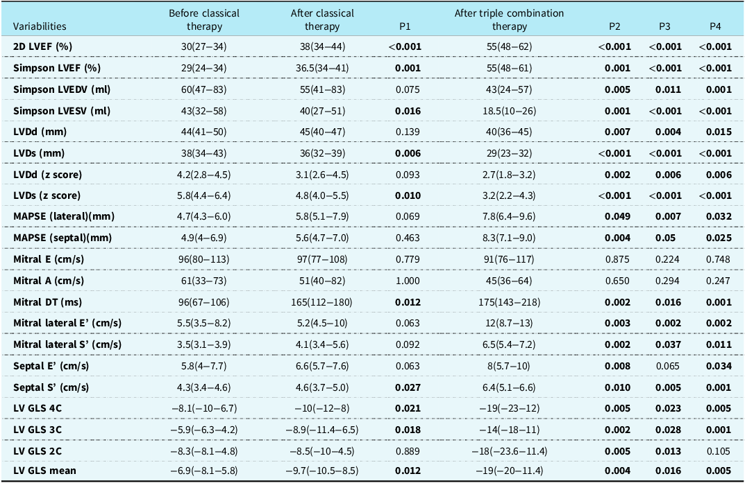
A comparison of all results before and after classical treatment, as well as after triple combination treatment, is shown in Table 2. Before and after classical treatment and after triple treatment, statistically significant improvement in two-dimensional left ventricular ejection fraction (%) (median values 30; 38; 55, respectively), left ventricular end-diastolic diameter (median values 44; 45; 40 mm), left ventricular end-systolic diameter (median values 38; 36; 29 mm), left ventricular end-diastolic diameter (z score) (median values 4.2; 3.2; 2.7), left ventricular end-systolic diameter (z score)(median values 5.8; 4.8; 3.2), Simpson left ventricular ejection fraction (%) (median values 29; 36.5; 55), Simpson left ventricular end-diastolic volume (median values 60; 55; 43 ml), left ventricular end-systolic volume (median values 43; 40; 18. 5 ml), mitral deceleration time (median values 96; 165; 175 ms), mitral lateral peak early diastolic velocity (median values 5.5; 5.2; 12 cm/s), mitral lateral peak systolic velocity (median values 3.5; 4.1; 6.5 cm/s), septal peak early diastolic velocity (median values 5.8; 6. 6; 8 cm/s), septal peak systolic velocity (median values 4.3; 4.6; 6.4 cm/s), left ventricular global longitudinal strain four-chamber (median values −8.1; −10; −19), left ventricular global longitudinal strain three-chamber (median values −5.9; −8.9; −14), and left ventricular global longitudinal strain mean (median values −6.9; −9.7; −19) values was observed ( all values p < 0.05 ). A statistically significant improvement in left ventricular global longitudinal strain two-chamber (median −8.3; −8.5; −18, respectively) was observed between pre-treatment and post-triple therapy and between pre-treatment and post-triple therapy ( all values p < 0.05 ) (Figure 2 a, b, c).
( a ) Echocardiographic findings before classical therapy, ( b ) after classical therapy, and ( c ) after triple combination therapy in one patient.

Comparison of all results before and after classic and triple combination therapy

P1 = comparison before and after classical treatment; P2 = comparison before classic and after triple therapy; P3 = comparison after conventional treatment and after triple therapy; P4 = comparison of all result. A = transmitral pulse wave Doppler late diastolic inflow velocity; C = chamber; DT = deceleration time; E = transmitral pulse wave Doppler early diastolic inflow velocity; E’ = peak early diastolic velocity by tissue doppler imaging; GLS = global longitudinal strain; LVDd = left ventricular end-diastolic diameter; LVDs = left ventricular end-systolic diameter; LVEDV = left ventricular end-diastolic volume; LVEF = left ventricular ejection fraction; LVESV = left ventricular end-systolic volume; MAPSE = mitral annular plane systolic excursion; S’ = peak systolic velocity by tissue doppler imaging.
Patients received a median of 9 doses of levosimendan (range, 5–20), 100 days of dapagliflozin (range, 65–150), and 71 days of sacubitril/valsartan (range, 37–102). The median time from first presentation to last follow-up was 107 days (57–170 days), the median duration of classical treatment was 20 days (15–105 days), and the median time from the start of triple therapy to recovery was 80 days (31–105 days). In the outpatient follow-up of 18 discharged patients, they continued on their respective treatments, including classic, dapagliflozin, and sacubitril/valsartan.
Etiology of dilated cardiomyopathy
One patient had a filamin C gene mutation, three patients developed dilated cardiomyopathy after chemotherapy, one patient had mitochondrial myopathy, one patient had a spectrin repeat containing nuclear envelope protein 2 gene mutation and co-infection with mycoplasma pneumoniae, and twelve patients had viral myocarditis-related dilated cardiomyopathy.
Drug side effects and approach
One girl and two boys were treated with antibiotics for urinary tract infections. Dapagliflozin was continued at half the dose in one patient after treatment for a urinary tract infection. In a male patient with normal left ventricular ejection fraction, treatment with dapagliflozin and sacubitril+valsartan was discontinued due to mildly elevated urea and creatinine levels. After the urinary tract infection was treated, these two drugs were resumed at half the dose. In the other patient, both drugs were continued without interruption. In another patient with non-specific dyspnoea, the dose of dapagliflozin was reduced by half, and sacubitril/valsartan was added. Subsequently, the same complaints were not observed, even though the dose of dapagliflozin was increased to the full dose. In another patient, renal function tests improved after treatment with full-dose dapagliflozin and medium-dose sacubitril/valsartan, prompting the discontinuation of both drugs. When renal function tests returned to normal, dapagliflozin and sacubitril/valsartan were resumed, along with the classical treatment. Hypoglycaemia was not observed in any patients, and anorexia was noted in a few patients. A patient sustained a fracture to the head of the humerus after falling while playing on the playground. Treatment with dapagliflozin has been stopped.
Comparison of complete blood count and biochemical values
A comparison of complete blood count and biochemical values was performed before and after classic and triple combination therapy. Statistically significant improvements were observed in troponin T (median, respectively, 91; 38; 27 ng/L), pro-brain natriuretic peptide (median, respectively, 24,095, 3077, 1129 pg/ml) before and after classical treatment and after triple therapy ( all values p < 0.05 ).
Discussion
The most significant and valuable findings of this study are presented below.
Triple combination therapy may enhance left ventricular systolic and diastolic function in children with advanced heart failure. It can eliminate patients’ dependency on inotropes and facilitate their discharge. This treatment may help to decrease the need for left ventricular assist devices or prevent progression to heart transplantation in children. Utilising multiple doses of levosimendan in children with low left ventricular ejection fraction who are inotrope-dependent and facing advanced heart failure may be beneficial, especially if they cannot be discharged.
Levosimendan is a calcium sensitiser for troponin and an adenosine triphosphate-sensitive potassium agonist. It improves ventricular function by sensitising myocardial cells to troponin calcium during the systolic phase and regulating intracellular calcium influx, which prevents cell damage and provides coronary vasodilation while preserving diastolic function. The maximum haemodynamic effect of levosimendan has been shown to occur 1–3 days after the start of infusion and to persist for at least 1 week. Levosimendan, which increases myocardial contractility without raising myocardial oxygen demand, is primarily used in decompensated heart failure and acute fulminant myocarditis that is refractory to standard therapy. Reference Dündar, Yılmaz and Argun7–Reference Séguéla, Mauriat and Mouton9 Séguéla et al. reported that levosimendan improved haemodynamics in children with dilated cardiomyopathy and end-stage heart failure, reduced the waiting time for heart transplantation, and, with repeated infusions, enabled several months to pass without the need for mechanical circulatory support. Reference Séguéla, Mauriat and Mouton9 These results align with our findings.
Sacubitril/valsartan is a first-in-class angiotensin receptor neprilysin inhibitor that simultaneously inhibits neprilysin through sacubitrilat (the active metabolite of sacubitril) and blocks the angiotensin II type 1 receptor via valsartan. Neprilysin, or neutral endopeptidase, is an enzyme responsible for the degradation of multiple peptides, including type atrial and brain natriuretic peptides and angiotensin II. The active metabolite of sacubitril inhibits neutral endopeptidase, resulting in increased brain natriuretic peptide longevity through diuresis and blood pressure reduction, but it also increases angiotensin II and its effects. This latter result can be avoided by adding valsartan, an angiotensin receptor blocker, to the formulation. In the PARADIGM-Heart Failure trial, sacubitril/valsartan proved superior to the angiotensin-converting enzyme inhibitor enalapril in reducing mortality and the risk of hospitalisation for heart failure in adults with chronic heart failure. Reference McMurray, Packer and Desai10 In the PANAROMA study, sacubitril/valsartan did not demonstrate superiority over enalapril in treating children with heart failure due to systemic left ventricular systolic dysfunction, using a pre-specified global endpoint. However, both treatment arms showed clinically significant improvements over 52 weeks. Reference Shaddy, Burch and Kantor1 Hale et al. reported that in children with heart failure, the 1-year incidence of heart transplantation or death was similar between those receiving sacubitril-valsartan and those receiving angiotensin-converting enzyme inhibitors or angiotensin receptor blockers. Reference Hale, Prichett, Jandu and Ravekes5
Dapagliflozin, a selective sodium-glucose co-transporter two inhibitor, is widely used in the treatment of type 2 diabetes mellitus and heart failure. Reference Dhillon14,Reference Kaze, Zhuo, Kim, Patorno and Paik15 The 2021 European Society of Cardiology guidelines for the diagnosis and treatment of acute and chronic heart failure recommend the use of sodium-glucose cotransporter-2 inhibitors as a standard treatment strategy for heart failure. Reference McDonagh, Metra and Adamo16 Sodium-glucose cotransporter-2 inhibitors decrease preload and overload by reducing plasma volume and vascular resistance. Reference Heerspink, Kosiborod, Inzucchi and Cherney17,Reference Tamargo18 They reduce preload, left ventricular wall tension, and myocardial oxygen demand by normalising elevated intracellular sodium through natriuresis and osmotic diuresis. Reference Verma and McMurray19,Reference Bertero, Prates Roma, Ameri and Maack20 It improves myocardial energetics by reducing fatty acid dependence and cardiac sodium-hydrogen exchanger 1 and increasing ketone body formation and utilisation. Reference Tamargo18,Reference Verma, Rawat and Ho21 Accordingly, cardiac efficiency and contractile function are improved. It provides cardiac remodelling by reducing inflammation/fibrosis, cardiac wall tension, and cardiomyocyte stiffness. Reference Heerspink, Kosiborod, Inzucchi and Cherney17,Reference Tamargo18,Reference van Heerebeek, Hamdani and Handoko22 Thus, it reduces blood pressure, Reference Heerspink, Kosiborod, Inzucchi and Cherney17 improves cardiac contractility, reduces left ventricular mass Reference Verma, Garg and Yan23,Reference Verma, Mazer and Yan24 and improves diastolic function. Reference Heerspink, Kosiborod, Inzucchi and Cherney17 In preclinical models, treatment with sodium-glucose cotransporter-2 inhibitors increased total cardiac adenosine triphosphate production by approximately 30%. Reference Verma, Rawat and Ho21 It improves cardiac function in dilated cardiomyopathy by reducing oxidative stress Reference Uthman, Li and Baartscheer25,Reference Zuurbier, Abbate and Cabrera-Fuentes26 and inflammation. Reference Park, Belcastro and Hasan27 As a result of all these effects, it provides protection against myocardial damage and heart failure.
Since cardiac Na transporters are activated in pathological conditions such as systemic inflammation, ischaemia, myocarditis, dilated cardiomyopathy, and heart failure, they can be modulated by sodium-glucose cotransporter-2 inhibitors. Reference Dyck, Sossalla and Hamdani28 The active metabolite of sacubitril inhibits neutral endopeptidase, ensures the longevity of brain natriuretic peptide and ultimately, like sodium-glucose cotransporter-2 inhibitors, potentiates the diuretic and antihypertensive effects. Sodium-glucose cotransporter-2 inhibitors potentiate angiotensin two receptor inhibition by reinforcing the same effect as Valsartan. Sodium-glucose cotransporter-2 inhibitors increase nitrite oxide, which enhances the coronary vasodilator effect of levosimendan and reduces ischaemia. Sodium-glucose cotransporter-2 inhibitors potentiate the effect of levosimendan by increasing its sensitivity to adenosine triphosphate, thereby enhancing adenosine triphosphate production. Sodium-glucose cotransporter-2 inhibitors contribute to cell regeneration by significantly improving the pathophysiology of damaged cells, thus enhancing the efficacy of levosimendan and sacubitril/valsartan in a synergistic manner. The targeted reduction in interstitial fluid observed with sodium-glucose cotransporter-2 inhibitors may limit the reflexive sympathetic activation associated with conventional diuretics. Reference Cherney, Perkins and Soleymanlou29 In our study, the initial sign of recovery was the normalisation of the left ventricular wall thickness, which had been very thin and visible at first. Overall, a more significant increase in left ventricular ejection fraction was noted when the dapagliflozin dose exceeded 0.2 mg/kg (single dose) and the sacubitril/valsartan dose exceeded 2.3 mg/kg (two doses). The earlier the treatment begins, the better the response in terms of increased ejection fraction. Consequently, our patients’ left ventricular ejection fractions improved with the triple combination therapy, and they no longer required heart transplants or ventricular assist devices.
This study is the first to demonstrate the superiority of combination therapy over monotherapy because of its synergistic effects in hospitalised, inotrope-dependent children with advanced heart failure whose left ventricular function is not adequately improved by classical drug therapy.
Study limitations
The main disadvantages and limitations of the study include the small sample size, the single-centre design, and the absence of a randomised control group. Triple combination therapy was not sufficiently effective in two inotrope-dependent patients with severe aortic regurgitation, severe left ventricular non-compaction, and left ventricular dysfunction who were not on the patient list. This was believed to be due to a structural disorder or delayed diagnosis and treatment. Such situations highlight the constraints of the new triple combination therapy. Additionally, a patient with advanced left ventricular dysfunction and severe aortic insufficiency, who was not on the list and dependent on inotropes, had her heart failure normalised with triple combination therapy following aortic valve repair. This case demonstrated that left ventricular function can be improved with triple therapy when structural heart issues are addressed.
Conclusions
It seems that in children admitted to the hospital with advanced heart failure, and whose left ventricular function does not improve sufficiently with classical drug treatment, triple combination therapy can be utilised to enhance left ventricular systolic and diastolic function, reduce inotropic dependence, and allow patients to be discharged. Triple treatment appears to decrease the need for left ventricular assist device or heart transplantation in children. It can be considered superior to monotherapy due to the synergistic interaction of the three drugs. Furthermore, it seems prudent to use the newly available drugs in combination with greater caution and confidence in children with advanced heart failure. More comprehensive studies on this subject are needed.
Acknowledgements
I would like to thank the paediatric heart transplant coordinators, Seda Safak and Ozlem Sarı, and the secretaries, Erdi Yildiz and Sadullah Coskun, for their contributions.
Author contributions
All authors contributed to the study conception and design. Material preparation and data analysis were performed by [Isa Ozyilmaz]. Data collection was performed by [Halise Zeynep Genc], [Senay Coban], [Nurullah Yilmaz] and [Ahmet Saki Oguz]. The first draft of the manuscript was written by [Isa Ozyilmaz], and review and editing were performed by [Ibrahim Cansaran Tanidir], [Erkut Oztürk], and [Ali Can Hatemi]. All authors commented on previous versions of the manuscript. All authors read and approved the final manuscript.
Financial support
The authors declare that no funds, grants, or other support were received during the preparation of this manuscript.
Competing interests
The authors have no relevant financial or non-financial interests to disclose.