




No CrossRef data available.
Article contents
Characterization of Trypanosoma cruzi TcRjl locus and analysis of its transcript
Published online by Cambridge University Press: 23 August 2004
Abstract
RJLs represent a recently described family of the Ras-related GTP-binding proteins. The Trypanosoma cruzi orthologue, TcRjl, was isolated and its locus was characterized in a region of almost 5 kb. Its 660 bp orf, predicting a protein of 24·13 kDa, is present as a single copy gene in T. cruzi I lineage, and from 1–2 copies in T. cruzi II lineage. TcRjl shares 73% aa sequence similarity with its closest identified orthologue, T. brucei TbRjl. RT–PCR experiments revealed that TcRjl is transcribed in mRNA in the 3 main life forms of the parasite, while Northern hybridization demonstrated that TcRjl is transcribed in T. cruzi epimastigotes as at least 2 transcripts, one of around 950 nt and the other of 1500 nt. Splice-leader addition was mapped to a single site at −69 bp upstream of TcRjl orf indicating that the two mRNA types may derive in differences at the 3′ of TcRjl mRNA. TcRjl locus presents considerable synteny with Rjl loci from Trypanosoma brucei and Leishmania major as available from their respective genome projects.
Information
- Type
- Research Article
- Information
- Copyright
- © 2004 Cambridge University Press
INTRODUCTION
The Ras superfamily consists of many small monomeric GTP-binding proteins that play key roles in the dynamic regulation of several cellular events, ranging from mitogenic signalling to vesicle budding and trafficking (reviewed by Takai, Sasaki & Matozaki, 2001). Genes belonging to this superfamily are further classified into at least 7 families: Ras, Rho, Rab, Ran, Arf/Sar, RGK and RJL. RJL genes constitute a recently described family of Ras-related GTP-binding proteins present in many protist lineages and also as chimeras fused to C-terminal DNAJ domains in deuterostome metazoans (Nepomuceno-Silva et al. 2004). Such genes seem to be absent in plants, fungi and protostome metazoans, and it has been suggested that a horizontal gene transfer event may have occurred between protists and deuterostome metazoans (Nepomuceno-Silva et al. 2004). Functional studies of Ras-related GTPases are usually carried out in yeast and higher eukaryotic models. However, a consistent body of knowledge has been growing regarding the function of Ras-related GTP-binding proteins in unicellular parasites. Ras-related proteins are recognized as powerful tools for studying parasite physiology, being also potential targets for therapeutic exploitation (reviewed by Field, Ali & Field, 1999).
The Trypanosomatidae family is composed of obligate flagellated protozoan parasites, some of pivotal medical and economical importance, including Trypanosoma cruzi, T. brucei and Leishmania species. T. cruzi is the aetiological agent of Chagas disease, for which estimates of infection prevalence point to over 11 million infected individuals (Dias, Silveira & Schofield, 2002).
Here we report the characterization of T. cruzi TcRjl transcript and the respective gene locus, as well as its comparison with counterpart loci in the genomes of two other model trypanosomatids: T. brucei and L. major.
MATERIALS AND METHODS
Parasites
Epimastigotes of Dm28c and of the Y strain of T. cruzi were maintained at 28 °C in liver infusion triptone medium supplemented with 10% heat-inactivated foetal calf serum (HI-FCS, Life Technologies, Inc.) and 0·025 mg/ml haemin (Sigma). Metacyclogenesis of Dm28c metacyclic trypomastigotes was carried out in TAU-3AAG medium as described (Contreras et al. 1988). Metacyclic trypomastigotes were added to 3T3/NIH fibroblasts growing at 37 °C with 5% CO2 in a humidified incubator, in DMEM supplemented with 10% HI-FCS. Infected fibroblasts were cultured at 37 °C with 2% HI-FCS with 5% CO2 in a humidified incubator. Intracellular amastigotes were obtained on the second and third day of culture and culture-derived trypomastigotes were obtained from the supernatant on the fourth and fifth days after infection.
Generation of the TcRjl probe
In order to detect Ras superfamily sequences from the T. cruzi genome, we employed an RT–PCR based approach using a sense primer (5′GCCGCCGAATTCTTATTGATACAGTTTCTG3′) corresponding to the miniexon region (added by trans-splicing to the 5′end of all trypanosome mRNAs), and an degenerated antisense primer (5′GACTGAATTCTTCYTGGCCDGCBGTRTCCCA3′) (note that Y corresponds to either C or T nucleotides, R to A or G, D to A, G or T, and B to C, G or T), specifically designed to anneal to the PM3 (WDTAGQE) corresponding conserved region of Ras-related GTPases (Bourne, Sanders & McCormick, 1991). cDNA was synthesized from RNA isolated from epimastigote cells using 100 pmol of random hexamer primers in the reaction. Ten ng of cDNA were used as template for PCR reactions. Each reaction mixture contained 20 mM dCTP, dGTP, dTTP, 10 mM dATP, 2·5 mCi of [α-32P]dATP, 1 mM MgCl2, 1× dilution of Amplitaq buffer (Perkin Elmer), 1 U of Amplitaq polymerase and 100 pmol of each primer in a final volume of 100 μl. These reactions underwent 5 cycles of 1 min at 94 °C, 1 min 48 °C and 1 min 72 °C, followed by 35 cycles of 1 min 94 °C, 1 min 55 °C and 1 min 72 °C. The PCR labelled products were dissolved in a 6% polyacrylamide DNA sequencing gel.
Amplified fragments were cloned in pKS-II+, introduced in XL1-Blue Escherichia coli strain and sequenced by the chain termination method, using the T7 Sequencing Kit (Amersham Pharmacia Biotech) following the procedures recommended by the manufacturer. After confirming the identity of the fragments as originating from Ras superfamily mRNAs, cloned fragments were used as probes for subsequent experiments.
Southern and Northern blot analysis
For Southern blot analysis, total DNA of the epimastigote stage of Dm28c Trypanosoma cruzi was extracted and digested (5 μg) with BamHI, BglII, EcoRI, NdeI, PstI and XbaI at 37 °C for 2 h. Total DNA of the Y strain was digested with EcoRI, BamHI, SalI, HindIII, PstI in the same conditions. The digestion products were separated on a 0·8% agarose gel. The DNA was blotted onto nitrocellulose membranes (Life Technologies, Inc.) using standard conditions (Sambrook, Fritsch & Maniatis, 1989). The hybridization was carried out at 42 °C in 2× SSC, 0·5% (w/v) SDS, 5× Denhardt's solution (5×: 1% Ficoll, 1% (w/v) polyvinylpyrrolidone, 1% (w/v) BSA), 0·1 mg/ml salmon sperm DNA, 50% formamide using the above-mentioned RT–PCR generated fragment, radiolabelled with [α-32P]dATP by random priming, as a probe. Membranes were washed in 0·1×SSC, 0·5% SDS for 5 min at room temperature, followed by two 15 min washes in 0·1×SSC, 0·5% SDS (w/v) at 55 °C and exposed to X-ray film for autoradiography.
Total RNA from 5×10⁷ Dm28c epimastigotes was extracted with TRIZOL reagent (Life Technologies, Inc.) following the procedure recommended by the manufacturer, fractionated on a 1·5% (w/v) denaturing agarose–formaldehyde gel and blotted onto nitrocellulose membranes (Life Technologies, Inc.), as described by Sambrook et al. (1989). The hybridization was carried out using the same procedure described above for Southern hybridization. RNA was also extracted from Dm28c amastigotes and culture-derived trypomastigotes in order to perform RT–PCR assays (as described below).
Library screening
An λ-EMBL3 Dm28c genomic library was kindly provided by Dr W. Degrave (DBBM, Fiocruz, Brazil) and propagated in E. coli strain KW252. Approximately 60000 independent recombinant phages were screened with the [α-32P]dATP-labelled TcRjl probe. Plugs containing positive plaques were selected and submitted to secondary and tertiary screens. After 3 rounds of selection, phage clones displaying positive hybridization signals were selected. One of them, named λTcRjl, was selected for the characterization of TcRjl.
Subcloning and sequencing of the TcRjl coding region
The λTcRjl clone was amplified in E. coli strain LE392. DNA from this clone was extracted and purified as described (Sambrook et al. 1989). A 5·7-kb EcoRI fragment was selected for cloning in pKS-II+, introduced in XL1-Blue E. coli strain and sequenced by the automated chain termination method in a MegaBACE 1000 sequencer (Amersham/Molecular Dynamics). DNA sequencing was carried out using commercially available T3 and T7 sequencing primers and the following primers designed according to the probe sequence: Tcrjl-sense (5′GTCGCCTGCGCCGCCC3′), Tcrjl-antisense (5′GAAATTCACCCGAAC3′), Tcrjl-start (5′AGATGAGTGCGAAGACC3′) and Tcrjl-end (5′TTACATGAATGCTTGCTGCCACC3′).
Mapping the TcRjl gene locus
A 5·7 kb cloned EcoRI fragment was digested with restriction enzymes with target sites occurring in the polylinker of the plasmid pKS-II+ or in the cloned insert. Digested plasmids were submitted to agarose gel electrophoresis followed by Southern blot hybridization against the TcRjl probe. Hybridizing fragments were compared to an ethidium bromide-stained digestion pattern. In order to obtain the maximum information about the TcRjl locus, physical mapping data were combined with automated sequencing data, with two genomic contigs from the TIGR Institute (obtained by blasting the T. cruzi WGS database at ‘http://tigrblast.tigr.org/er-blast/index.cgi?project=tca1’), and with some T. cruzi ESTs (obtained by blasting at the NCBI EST database). EST analysis, together with a similarity search between T. cruzi, L. major and T. brucei Rjl loci, allowed the mapping of putative genes in the neighbourhood of TcRjl and of its trypanosomatid orthologues.
Mapping the 5′-UTR of TcRjl transcript
For mapping the TcRjl 5′-UTR and locating the trans-splicing acceptor sites, we carried out mini-exon RT–PCR against Dm28c T. cruzi RNA. The sense primer was directed to the mini-exon sequence (ME, 5′GGATGGAATTCAGTTTCTGTACTATATTG3′) and the antisense primer was the same Tcrjl-antisense described above. Initially, 5 μg of epimastigote, amastigote and trypomastigote total RNA was submitted to first-strand cDNA synthesis using the SuperScript II Preamplification System (Life Technologies, Inc.) with random hexamers as primers, according to the manufacturer's suggested procedures. The first PCR was carried out in the presence of 0·5 mM dNTPs, 1·5 mM MgCl2, 10 μM each primer, and 5 units of Taq DNA polymerase (Life Technologies, Inc.). To avoid problems derived from differences in primer melting temperatures, a touch-down PCR program was used by decreasing the annealing temperature from 75 to 57 °C in 1 °C steps. Then, 20 additional cycles were performed at 93 °C for denaturing, at 55 °C for annealing, and at 72 °C for extension, followed by a 10-min extension step. Reaction products were resolved on a 2·5% agarose gel. Products were gel extracted, cloned in PCR TOPO 2.1 plasmid (Invitrogen), amplified in XL1-Blue and sequenced with flaking reverse and forward primers, allowing an accurate mapping of the TcRjl trans-splicing acceptor site.
Trypanosomatid sequence comparison
Sequences used for comparison were extracted by similarity searches (blastn or tblastn) from the Leishmania major Friedlin genome project at the Sanger Institute. The map at Fig. 5 represents contig 2, from positions 328278 to 333845 (antisense orientation) at chromosome 7_11b.1, as it was available in November, 2003, at ‘http://www.sanger.ac.uk/cgi-bin/blast/submitblast/l_major’ by blasting the L. major chromosome shotguns database. Data from T. brucei was also obtained from the Sanger Institute, consisting of the contig tryp_XI-49 from positions 19110 to 24180 (antisense orientation) at chromosome XI, as deposited in November, 2003, at ‘ftp://ftp.sanger.ac.uk/pub/databases/T.brucei_sequences/Chromosome_XI/’. Data from the 5′ of the T. brucei locus was obtained by blasting T. brucei predicted genes at GeneDB (www.genedb.org). Sequences were aligned with the ClustalX (v. 1.81) program and adjusted by eye with GeneDoc (v.2.6.02).
RESULTS AND DISCUSSION
TcRjl cloning
Aiming to characterize the Ras superfamily genes in Trypanosoma cruzi we have identified a 360 bp RT–PCR product sharing moderate identity levels with Rab proteins from several organisms. Sequence analysis revealed that this sequence was derived from a T. cruzi representative of a novel family of GTP-binding proteins, the RJLs, since the T. cruzi orthologue was classified as TcRjl (Nepomuceno-Silva et al. 2004). Although the degenerated primer used in the RT–PCR was designed to anneal to a region corresponding to the PM3 motif (WDTAGQE), it was possible to recognize the somewhat divergent TcRjl PM3 sequence (WDMAGRD). This RT–PCR product was used in the screening of a genomic library, allowing the sequencing of the TcRjl coding region and adjacent sequences.
The fragment obtained by RT–PCR was used as a probe for the Southern hybridization against DNA from Dm28c and Y strain, which represent the two main phylogenetic groups of T. cruzi (T. cruzi I and II, respectively, as proposed in ‘Recommendations from a Satellite Meeting’, 1999). There is only one copy per haploid genome for Dm28c, revealed by single hybridizing bands for all enzymes cutting outside the coding region (Fig. 1). In the Y strain the hybridization pattern suggests between one or two gene copies (Fig. 1), resembling the T. cruzi II actin genomic arrangement as observed by Cevallos et al. (2003), in which one allele has 2 gene copies and the other only 1 actin copy. Such polymorphisms are in agreement with the putative hybrid origin of T. cruzi II (Machado & Ayala, 2001).

Fig. 1. Southern blot analysis using the radio-isotope labelled RT–PCR fragment as probe. In the left panel each lane was loaded with 5 μg of Trypanosoma cruzi Y strain genomic DNA digested with the following restriction enzymes: E, EcoRI; B, BamHI; S, SalI; H, HindIII; P, PstI. In the right panel T. cruzi Dm28c DNA was digested with the following enzymes: B, BamHI; Bg, BglII; E, EcoRI; K, KpnI; N, NdeI; P, PstI; X, XbaI. Numbers on the left of the panels indicate the position of markers (in kbs) in the ethidium bromide-stained gel. The arrow on the right indicates the cloned 5·7 kb EcoRI fragment.
TcRjl presents a 660 bp open reading frame coding for a 219 amino acid protein with predicted molecular weight of 24·13 kDa (Genbank Accession no: AY274443; Fig. 2). It presents all the classic Ras-related PM and G motifs required for GTP binding and hydrolysis, although a crucial glutamine is missing in PM3 (Fig. 2). TcRjl shares 73% amino acid sequence similarity with TbRjl (orthologous from the closely related parasite T. brucei, Accession no.: BK001287). An orthologue from L. major was also identified, but it is interrupted by intronic-like sequences and there is no evidence of its functionality (Nepomuceno-Silva et al. 2004).

Fig. 2. Nucleotide and predicted peptide sequences for TcRjl. GTP-binding and hydrolysis conserved motifs are shaded. The EcoRI cloning site is represented in bold. Arrows under the nucleotide sequence represent annealing sites of primers used for sequencing (Tcrjl-start, I; Tcrjl-sense, II; Tcrjl-antisense, III; Tcrjl-end, IV).
Characterization of TcRjl mRNA
The RT–PCR derived fragment of TcRjl was also used as a probe for Northern blot analysis, which revealed 2 transcripts in T. cruzi epimastigotes. One of around 1500 nt and another of about 950 nt (Fig. 3). As only one site of trans-splicing was detected (see below) these transcripts probably represent different lengths of polyA+ tails or still, different sites of polyA+ tail addition.

Fig. 3. Northern blot assay using the radio-isotope labelled RT–PCR fragment as probe. The single lane contains total RNA from 5×10⁷ exponentially growing epimastigotes. Numbers on the left indicate migration position of Trypanosoma cruzi rRNA, used as markers for approximate mRNA size determination.
We mapped the 5′-untranslated region of TcRjl mRNA using a mini-exon RT–PCR approach with a sense oligonucleotide directed to the mini-exon and antisense oligonucleotide directed next to the PM3 region. Electrophoresis of the PCR product revealed a single fragment of about 370 bp for the three main life-stages of T. cruzi, indicating that TcRjl is expressed in detectable levels during the entire life-cycle of the parasite (Fig. 4A). PCR products were further cloned and several clones were sequenced, showing that this fragment corresponds to the acceptor site located at −69 in the TcRjl 5′-UTR. Pyrimidine tracts, usually recognized as trans-splicing signals are found spread upstream in the 5′UTR of TcRjl, as indicated in Fig. 4B. One interesting aspect of TcRjl mature mRNA is the presence of a long stretch of the CA dinucleotide (9 perfect repeats). Such repeats have been implicated in T. cruzi gene regulation, being the target of single-stranded binding proteins (Duhagon, Dallagiovanna & Garat, 2001).

Fig. 4. Mapping the 5′ region of the TcRjl transcript. (A) Ethidium bromide-stained agarose gel of the RT–PCR reaction described in the text. Lane designations: Epi, reaction using epimastigote RNA; Epi RT−, negative control using epimastigote RNA not submitted to reverse transcription; Ama, reaction using amastigote RNA; Ama RT−, same as Epi RT−, but for amastigote mRNA; Tryp, reaction using trypomastigote RNA; Tryp RT−, same as in Epi RT−, but for trypomastigote RNA; 100 bp molecular weight marker. The arrow at the left indicates the 370 bp specific product generated. (B) Upper sequence segment: genomic sequence of the 5′ region of TcRjl. Pyrimidine stretches are boxed and the AG dinucleotide used as acceptor for trans-splicing is circled. The sequence below the black horizontal arrow corresponds to the annealing region of the Tcrjl-antisense primer. The putative 5′UTR poly-CA regulatory element is underlined. TcRjl coding sequence is represented in uppercase. Bottom sequence segment corresponds to the nucleotide sequence from cloned products from the Epi reaction in (A). Mini-exon sequences are represented in bold and the coding region in uppercase.
Characterization of the TcRjl gene locus and comparison with Rjl loci from T. brucei and L. major
Sequencing of 1375 bp of TcRjl locus allowed us to merge the extremities of two sequences deposited in the T. cruzi database at the TIGR institute (contig 1075610 in antisense orientation and contig 1103228 in sense orientation), producing a contig of 4348 bp. Five T. cruzi ESTs (AI080808, AW330122, AI077089, AA890798 and AA890790) increased the extended data to 4636 bp of contiguous sequence (Fig. 5). We have localized at 418 bp downstream of TcRjl, a widely conserved eukaryotic gene, annotated as Myg1 in the human genome and as Gamm1 in plant genomes. Although there is no currently published reported function for Myg1/Gamm1 proteins, mouse Myg1 (Accession no: AAH28501) is designated as ‘melanocyte proliferating gene 1’ and is suggested to encode a metal-dependent phosphoesterase enzyme. The T. cruzi orthologue, TcMyg, comprises 1185 bp orf, generating a 394 amino acid protein. One singular aspect of the TcMyg gene is the high content of PstI sites (5 sites) on its coding region (see Fig. 5), not shared by its T. brucei and L. major orthologues. TcMyg shares considerable identity with human Myg1 protein (56% similarity on a 359 amino acid stretch) as well as with other eukaryotes Myg orthologues (Fig. 6). However, TcMyg cDNA was not represented in the normalized epimastigote ESTs available at the public databases, which may indicate low levels of expression in the epimastigote stage or restricted inducible expression. At 387 bp upstream of TcRjl we could detect one smaller ORF of 285 bp (producing a putative 94 amino acid protein) that had its expression confirmed by two ESTs on the database (AA952613 and CB964243). Although the sequence does not match any known gene in the databases, there are closely related orthologues in T. brucei and Leishmania major (Fig. 7A). These, in addition to EST data, indicate that this sequence is a trypanosomatid-specific gene. It was named TcRao (standing for T. cruzi Rjl associated orf). At 454 bp upstream of TcRao there seems to lie another trypanosome-specific gene. Although we could not obtain its complete ORF, its 201 amino acid C-terminal shares 38% similarity with a T. brucei gene located in the same locus position (identified as orf DZFZp451J0218). All of the 5 ESTs referred to above to extend the TcRjl locus match its sequence, indicating that it is transcribed in mRNA. The T. cruzi Gene Index (TcGI v. 3.0, at the TIGR Institute) designed this transcript as TC7784 (Fig. 7B).

Fig. 5. TcRjl locus and synteny with loci from Trypanosoma brucei and Leishmania major. (A) Map of TcRjl locus: Grey boxes indicate the region covered by our sequencing, while white boxes correspond to sequences available in databases and thin lines represent regions uncovered by sequencing. Thick arrows represent identified genes and their coding orientation, with their names indicated above (TC7784 ORF is incomplete at its 5′). The striped box covers the region corresponding to the RT–PCR derived probe used for characterizing TcRjl. Relevant restriction sites are indicated underneath the map as well as the region corresponding to the 5·7 kb cloned EcoRI fragment. The sequencing strategy is also represented, thin arrows matching annealing sites of primers used for sequencing (T7; T3; Tcrjl-start, I; Tcrjl-sense, II; Tcrjl-antisense, III; Tcrjl-end, IV). (B and C) Rjl loci from T. brucei and L. major: white boxes and black thick arrows correspond to the same as for (A). Spotted boxes correspond to intronic-like elements in LmRjl. The distance bar corresponds to 1 kb for A, B and C.

Fig. 6. Multiple sequence alignment of trypanosomatid Myg proteins with representative orthologues from other eukaryotes obtained by BlastP searches on non-redundant (nr) database of NCBI and aligned by ClustalX program (v 1.81). Black shading indicates identical residues, while grey shading represents conservative substitutions. First two letters are indicative of species (Tc, Trypanosoma cruzi; Tb, Trypanosoma brucei; Hs, Homo sapiens; Dm, Drosophila melanogaster; At, Arabidopsis thaliana; Sc, Saccharomyces cerevisae; Pf, Plasmodium falciparum; Gi, Giardia intestinalis). Accession numbers as follows: HsMyg1, AAG17847; Dm_CG11980, AAN13404; AtGAMM1, BAB08430; ScYer156, AAB64683; PfMyg1, CAD50537; GiMyg, EAA42308.

Fig. 7. Multiple sequence alignment of putative genes located at 5′ of TcRjl. Origin of sequences is detailed in the methodology section. Black shading indicates identical residues, while grey shading represents conservative substitutions. (A) Trypanosomatid Rjl associated Orfs (Rao) proteins. (B) Gene located at 5′ of TcRao and TbRao. Trypanosoma cruzi orf is incomplete at the 5′ terminal.
All four genes identified in the TcRjl locus are orientated in the same transcriptional direction (Fig. 5). Arrangement in directional clusters is in agreement with some chromosome organization previews, which have revealed the curious chromosomal arrangement of trypanosomatids. L. major chromosome 1 is divided in 2 main large divergent directional gene clusters spaced by a small linker region, supposedly containing promoters. In the same organism similar clusters may be convergent, with transcription initiated at the telomeric regions, as for L. major chromosome 3 and 4 (Myler et al. 1999; Myler & Stuart, 2000; Worthey et al. 2003). Recent release of T. brucei chromosome I and II sequences revealed that their general arrangement may be somewhat different from those of L. major, having as many as 17 directional clusters for chromosome II (El-Sayed et al. 2003; Hall et al. 2003). Regarding T. cruzi, only a 93·4 kb contig from chromosome 3 was released and its organization looks similar to the L. major with two contigs organized head to head (Andersson et al. 1998).
When compared with its counterpart loci in T. brucei and L. major, the gene order is similar, although the gene at the 5′ uppermost seems to be missing for L. major (Fig. 5). L. major also has larger intergenic regions (890 bp between LmRjl and LmMyg and 874 bp between LmRjl and LmRao). This synteny in the Rjl locus of different trypanosomatids is also of interest, as few studies have addressed the question of gene synteny between trypanosomatid genus. Although some examples of synteny were reported between trypanosomatids (Coelho et al. 2003; Cevallos et al. 2003), only the glucose transporter locus was extensively characterized between distant representative trypanosomatid species (Bringaud et al. 1998). This work highlights that trypanosomatid glucose transporter loci share many common features, although many gene depletions and insertions were observed. Here, we have reported a region of around 6 kb in which the gene order is identical between two organisms from the same genus (T. cruzi and T. brucei), but with one gene apparently missing in L. major. Curiously, it is worthy of mention that the missing gene is the least conserved of the four genes identified in the Rjl locus.
Draft genomes of these three model trypanosomatid organisms are supposed to be released during 2004 or in the following year. In addition to revealing the details of trypanosmatid genome arrangement, it will allow researchers to probe events that lead to genome diversification between these closely related divergent eukaryotes.
This work was supported by PRONEX/MCT/CNPq, Brazilian National Council of Research (CNPq) and Rio de Janeiro State Research Foundation (FAPERJ).
References
REFERENCES
ANDERSSON, B.,
ASLUND, L.,
TAMMI, M.,
TRAN, A. N.,
HOHEISEL, J. D. &
PETTERSSON, U. (
1998).
Complete sequence of a 93·4-kb contig from chromosome 3 of Trypanosoma cruzi containing a strand-switch region.
Genome Research
8,
809–816.CrossRefGoogle Scholar
BOURNE, H. R.,
SANDERS, D. A. &
McCORMICK, F. (
1991).
The GTPase superfamily: conserved structure and molecular mechanism.
Nature, London
349,
117–127.CrossRefGoogle Scholar
BRINGAUD, F.,
VEDRENNE, C.,
CUVILLIER, A.,
PARZY, D.,
BALTZ, D.,
TETAUD, D.,
PAYS, E.,
VENEGAS, J.,
MERLIN, G. &
BALTZ, T. (
1998).
Conserved organization of genes in trypanosomatids.
Molecular and Biochemical Parasitology
94,
249–264.CrossRefGoogle Scholar
CEVALLOS, A. M.,
LOPEZ-VILLASENOR, I.,
ESPINOSA, N.,
HERRERA, J. &
HERNANDEZ, R. (
2003).
Trypanosoma cruzi: allelic comparisons of the actin genes and analysis of their transcripts.
Experimental Parasitology
103,
27–34.CrossRefGoogle Scholar
COELHO, E. R.,
URMENYI, T. P.,
FRANCO DA SILVEIRA, J.,
RONDINELLI, E. &
SILVA, R. (
2003).
Identification of PDZ5, a candidate universal minicircle sequence binding protein of Trypanosoma cruzi.
International Journal for Parasitology
33,
853–858.CrossRefGoogle Scholar
CONTRERAS, V. T.,
ARAUJO-JORGE, T. C.,
BONALDO, M. C.,
THOMAZ, N.,
BARBOSA, H. S.,
MEIRELLES, M. N. &
GOLDENBERG, S. (
1988).
Biological aspects of the Dm28c clone of Trypanosoma cruzi after metacyclogenesis in chemically defined media.
Memórias do Instituto Oswaldo Cruz
83,
123–133.CrossRefGoogle Scholar
DIAS, J. C.,
SILVEIRA, A. C. &
SCHOFIELD, C. J. (
2002).
The impact of Chagas disease control in Latin America – a review.
Memórias do Instituto Oswaldo Cruz
97,
603–612.CrossRefGoogle Scholar
DUHAGON, M. A.,
DALLAGIOVANNA, B. &
GARAT, B. (
2001).
Unusual features of poly[dT-dG].[dC-dA] stretches in CDS-flanking regions of Trypanosoma cruzi genome.
Biochemical Biophysical Research Communications
287,
98–103.Google Scholar
EL-SAYED, N. M.,
GHEDIN, E.,
SONG, J.,
MACLEOD, A.,
BRINGAUD, F.,
LARKIN, C.,
WANLESS, D.,
PETERSON, J.,
HOU, L.,
TAYLOR, S.,
TWEEDIE, A.,
BITEAU, N.,
KHALAK, H. G.,
LIN, X.,
MASON, T.,
HANNICK, L.,
CALER, E.,
BLANDIN, G.,
BARTHOLOMEU, D.,
SIMPSON, A. J.,
KAUL, S.,
ZHAO, H.,
PAI, G.,
VAN AKEN, S.,
UTTERBACK, T.,
HAAS, B.,
KOO, H. L.,
UMAYAM, L.,
SUH, B.,
GERRARD, C.,
LEECH, V.,
QI, R.,
ZHOU, S.,
SCHWARTZ, D.,
FELDBLYUM, T.,
SALZBERG, S.,
TAIT, A.,
TURNER, C. M.,
ULLU, E.,
WHITE, O.,
MELVILLE, S.,
ADAMS, M. D.,
FRASER, C. M. &
DONELSON, J. E. (
2003).
The sequence and analysis of Trypanosoma brucei chromosome II.
Nucleic Acids Research
31,
4856–4863.CrossRefGoogle Scholar
FIELD, M. C.,
ALI, B. R. &
FIELD, H. (
1999).
GTPases in protozoan parasites: tools for cell biology and chemotherapy.
Parasitology Today
15,
365–371.CrossRefGoogle Scholar
HALL, N.,
BERRIMAN, M.,
LENNARD, N. J.,
HARRIS, B. R.,
HERTZ-FOWLER, C.,
BART-DELABESSE, E. N.,
GERRARD, C. S.,
ATKIN, R. J.,
BARRON, A. J.,
BOWMAN, S.,
BRAY-ALLEN, S. P.,
BRINGAUD, F.,
CLARK, L. N.,
CORTON, C. H.,
CRONIN, A.,
DAVIES, R.,
DOGGETT, J.,
FRASER, A.,
GRUTER, E.,
HALL, S.,
HARPER, A. D.,
KAY, M. P.,
LEECH, V.,
MAYES, R.,
PRICE, C.,
QUAIL, M. A.,
RABBINOWITSCH, E.,
REITTER, C.,
RUTHERFORD, K.,
SASSE, J.,
SHARP, S.,
SHOWNKEEN, R.,
MACLEOD, A.,
TAYLOR, S.,
TWEEDIE, A.,
TURNER, C. M.,
TAIT, A.,
GULL, K.,
BARRELL, B. &
MELVILLE, S. E. (
2003).
The DNA sequence of chromosome I of an African trypanosome: gene content, chromosome organization, recombination and polymorphism.
Nucleic Acids Research
31,
4864–4873.CrossRefGoogle Scholar
MACHADO, C. A. &
AYALA, F. J. (
2001).
Nucleotide sequences provide evidence of genetic exchange among distantly related lineages of Trypanosoma cruzi.
Proceedings of the National Academy of Sciences, USA
98,
7396–7401.CrossRefGoogle Scholar
MYLER, P. J.,
AUDLEMAN, L.,
DEVOS, T.,
HIXSON, G.,
KISER, P.,
LEMLEY, C.,
MAGNESS, C.,
RICKEL, E.,
SISK, E.,
SUNKIN, S.,
SWARTZELL, S.,
WESTLAKE, T.,
BASTIEN, P.,
FU, G.,
IVENS, A. &
STUART, K. (
1999).
Leishmania major Friedlin chromosome 1 has an unusual distribution of protein-coding genes.
Proceedings of the National Academy of Sciences, USA
96,
2902–2906.CrossRefGoogle Scholar
MYLER, P. J. &
STUART, K. D. (
2000).
Recent developments from the Leishmania genome project.
Current Opinion in Microbiology
3,
412–416.CrossRefGoogle Scholar
NEPOMUCENO-SILVA, J. L.,
DE MELO, L. D. B.,
MENDONÇA, S. M.,
PAIXÃO, J. C. &
LOPES, U. G. (
2004).
RJLs: A new family of Ras-related GTP binding proteins.
Gene
327,
221–232.CrossRefGoogle Scholar
RECOMMENDATIONS FROM A SATELLITE MEETING (
1999).
Memórias do Instituto Oswaldo Cruz
94 (Suppl. 1),
429–432.
SAMBROOK, J.,
FRITSCH, E. F. &
MANIATIS, T. (
1989).
Molecular Cloning: a Laboratory Manual,
2nd Edn. Cold Spring Harbor Press,
Cold Spring Harbor,
New York.
TAKAI, Y.,
SASAKI, T. &
MATOZAKI, T. (
2001).
Small GTP-binding proteins.
Physiological Reviews
81,
153–208.CrossRefGoogle Scholar
WORTHEY, E. A.,
MARTINEZ-CALVILLO, S.,
SCHNAUFER, A.,
AGGARWAL, G.,
CAWTHRA, J.,
FAZELINIA, G.,
FONG, C.,
FU, G.,
HASSEBROCK, M.,
HIXSON, G.,
IVENS, A. C.,
KISER, P.,
MARSOLINI, F.,
RICKELL, E.,
SALAVATI, R.,
SISK, E.,
SUNKIN, S. M.,
STUART, K. D. &
MYLER, P. J. (
2003).
Leishmania major chromosome 3 contains two long convergent polycistronic gene clusters separated by a tRNA gene.
Nucleic Acids Research
31,
4201–4210.CrossRefGoogle Scholar

Fig. 1. Southern blot analysis using the radio-isotope labelled RT–PCR fragment as probe. In the left panel each lane was loaded with 5 μg of Trypanosoma cruzi Y strain genomic DNA digested with the following restriction enzymes: E, EcoRI; B, BamHI; S, SalI; H, HindIII; P, PstI. In the right panel T. cruzi Dm28c DNA was digested with the following enzymes: B, BamHI; Bg, BglII; E, EcoRI; K, KpnI; N, NdeI; P, PstI; X, XbaI. Numbers on the left of the panels indicate the position of markers (in kbs) in the ethidium bromide-stained gel. The arrow on the right indicates the cloned 5·7 kb EcoRI fragment.
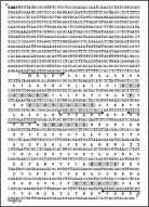
Fig. 2. Nucleotide and predicted peptide sequences for TcRjl. GTP-binding and hydrolysis conserved motifs are shaded. The EcoRI cloning site is represented in bold. Arrows under the nucleotide sequence represent annealing sites of primers used for sequencing (Tcrjl-start, I; Tcrjl-sense, II; Tcrjl-antisense, III; Tcrjl-end, IV).

Fig. 3. Northern blot assay using the radio-isotope labelled RT–PCR fragment as probe. The single lane contains total RNA from 5×10⁷ exponentially growing epimastigotes. Numbers on the left indicate migration position of Trypanosoma cruzi rRNA, used as markers for approximate mRNA size determination.
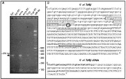
Fig. 4. Mapping the 5′ region of the TcRjl transcript. (A) Ethidium bromide-stained agarose gel of the RT–PCR reaction described in the text. Lane designations: Epi, reaction using epimastigote RNA; Epi RT−, negative control using epimastigote RNA not submitted to reverse transcription; Ama, reaction using amastigote RNA; Ama RT−, same as Epi RT−, but for amastigote mRNA; Tryp, reaction using trypomastigote RNA; Tryp RT−, same as in Epi RT−, but for trypomastigote RNA; 100 bp molecular weight marker. The arrow at the left indicates the 370 bp specific product generated. (B) Upper sequence segment: genomic sequence of the 5′ region of TcRjl. Pyrimidine stretches are boxed and the AG dinucleotide used as acceptor for trans-splicing is circled. The sequence below the black horizontal arrow corresponds to the annealing region of the Tcrjl-antisense primer. The putative 5′UTR poly-CA regulatory element is underlined. TcRjl coding sequence is represented in uppercase. Bottom sequence segment corresponds to the nucleotide sequence from cloned products from the Epi reaction in (A). Mini-exon sequences are represented in bold and the coding region in uppercase.
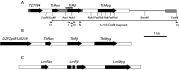
Fig. 5. TcRjl locus and synteny with loci from Trypanosoma brucei and Leishmania major. (A) Map of TcRjl locus: Grey boxes indicate the region covered by our sequencing, while white boxes correspond to sequences available in databases and thin lines represent regions uncovered by sequencing. Thick arrows represent identified genes and their coding orientation, with their names indicated above (TC7784 ORF is incomplete at its 5′). The striped box covers the region corresponding to the RT–PCR derived probe used for characterizing TcRjl. Relevant restriction sites are indicated underneath the map as well as the region corresponding to the 5·7 kb cloned EcoRI fragment. The sequencing strategy is also represented, thin arrows matching annealing sites of primers used for sequencing (T7; T3; Tcrjl-start, I; Tcrjl-sense, II; Tcrjl-antisense, III; Tcrjl-end, IV). (B and C) Rjl loci from T. brucei and L. major: white boxes and black thick arrows correspond to the same as for (A). Spotted boxes correspond to intronic-like elements in LmRjl. The distance bar corresponds to 1 kb for A, B and C.
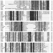
Fig. 6. Multiple sequence alignment of trypanosomatid Myg proteins with representative orthologues from other eukaryotes obtained by BlastP searches on non-redundant (nr) database of NCBI and aligned by ClustalX program (v 1.81). Black shading indicates identical residues, while grey shading represents conservative substitutions. First two letters are indicative of species (Tc, Trypanosoma cruzi; Tb, Trypanosoma brucei; Hs, Homo sapiens; Dm, Drosophila melanogaster; At, Arabidopsis thaliana; Sc, Saccharomyces cerevisae; Pf, Plasmodium falciparum; Gi, Giardia intestinalis). Accession numbers as follows: HsMyg1, AAG17847; Dm_CG11980, AAN13404; AtGAMM1, BAB08430; ScYer156, AAB64683; PfMyg1, CAD50537; GiMyg, EAA42308.
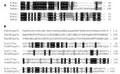
Fig. 7. Multiple sequence alignment of putative genes located at 5′ of TcRjl. Origin of sequences is detailed in the methodology section. Black shading indicates identical residues, while grey shading represents conservative substitutions. (A) Trypanosomatid Rjl associated Orfs (Rao) proteins. (B) Gene located at 5′ of TcRao and TbRao. Trypanosoma cruzi orf is incomplete at the 5′ terminal.
 You have
Access
- 6
- Cited by