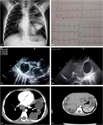
Cystic echinococcosis rarely presents with cardiac involvement, with a prevalence of only 0.5–2% of all patients.Reference Kahlfuß, Flieger, Roepke and Yilmaz 1 Cardiac impairment, such as rhythm disorders, may occur in these patients. A previously healthy 5-year-old boy was admitted with cough and intermittent chest pain during the previous month. Tachypnea (40/min) and hepatomegaly were determined during physical examination. Laboratory results revealed leucocytes 13,830/mm3, consisting of 38% neutrophils, 12% lymphocytes, 8% monocytes, and 42% eosinophils, C-reactive protein 0.6 (0–5) mg/dl, erythrocyte sedimentation rate 23 mm/hour, and immunoglobulin E at 674 (<50) mg/dl. Chest X-ray revealed multiple opacities in the left hemithorax (Fig 1a). Electrocardiography demonstrated negative T waves on V5, V6, and inferior leads (DII, DIII, and aVF) (Fig 1b). Echocardiography revealed a single, well-circumscribed cyst, 23×29 mm in diameter, inside the lateral wall of the left ventricle (Fig 1c and d and video 1). CT of the chest revealed multiple cysts, with the largest measuring 5.5 cm in diameter on the left lung and a cyst measuring 4.5 cm inside the mediastinum (Fig 1e). CT of the brain was normal, whereas abdominal CT revealed multiple cysts, with the largest measuring 3 cm in size on the liver (Fig 1f). Hydatid cyst serology was 1/640. The patient was started on peroral albendazole (15 mg/kg/day). After Holter monitoring, he was referred to a tertiary hospital specialising in cardiovascular surgery for simultaneous heart, bilateral-lung, and liver-cyst hydatid surgery in a single session. After cardiac and pulmonary lesions were removed, normal cardiac and ECG findings without any arrhythmia were observed on outpatient controls. Cystic echinococcosis is a multisystemic infection. Systemic workup including investigation of cardiac involvement is mandatory to prevent fatal complications.
( a ) Posteroanterior chest X-ray revealed multiple opacities on the left hemithorax. ( b ). Electrocardiography showed negative T waves on V5, V6, and inferior leads (DII, DIII, and aVF) ( c and ( d ). Modified subcostal (Fig 1c) and apical (Fig 1d) four-chamber images on the echocardiograph showed a well-bordered, encapsulated, hypoechoic lesion measuring 23×29 mm in diameter. The lesion is located far from the mitral inflow and left ventricular outflow tracts ( e ). Chest CT showed multiple cysts, of which the largest was 5.5 cm, on the left lung ( f ). Abdominal CT revealed multiple cysts, of which the largest was 3 cm, on the liver.

Acknowledgement
None.
Financial Support
This research received no specific grant from any funding agency, commercial, or not-for-profit sectors.
Conflicts of Interest
None.