INTRODUCTION
Chagas disease (CD), caused by the hemoflagellate Trypanosoma cruzi and also known as American Trypanosomiasis, is a parasitic infection endemically transmitted through blood-sucking triatomine bugs (i.e. reduviids) in 21 countries of Central and South America. According to the World Health Organization, in the endemic area CD currently provokes more than 7000 annual deaths and maintains over 25 million people at risk for the infection (WHO, 2015). Moreover, the international migratory movements together with the existence of alternative routes of transmission such as transfusion of infected blood or mother-to-child, has triggered the emergence of this parasitosis in non-endemic countries (e.g. Canada, Australia, Japan or Spain) (Gascón et al. Reference Gascón, Bern and Pinazo2010) contributing this way to the spreading of a disease that at this time affects an estimated 7 million people worldwide (WHO, 2015).
Focusing on the chemotherapy, the only two drugs up to now available for the specific treatment of CD − i.e. benznidazole (BZ) and nifurtimox − are not completely satisfactory. In this regard, both have limited efficacy especially at the later chronic stage and upon naturally-resistant parasite strains, also exhibiting wide range of side effects (Bahia et al. Reference Bahia, Figuereido Diniz and Mosqueira2014) and therefore, encouraging and supporting the search for alternative approaches potentially applicable to CD treatment.
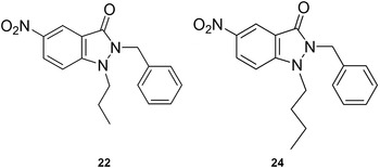
In previous studies, we have introduced the 1,2-disubstituted 5-nitroindazolinone system as a putative scaffold for the design of potential antichagasic drugs, according to the results achieved on several T. cruzi models (Vega et al. Reference Vega, Rolón, Montero-Torres, Fonseca-Berzal, Escario, Gómez-Barrio, Gálvez, Marrero-Ponce and Arán2012; Fonseca-Berzal et al. Reference Fonseca-Berzal, Escario, Arán and Gómez-Barrio2014, Reference Fonseca-Berzal, da Silva, da Silva, Vasconcelos, Batista, Escario, Arán, Gómez-Barrio and Soeiro2015). Concretely, the 2-benzyl-1-propyl (22) and the 2-benzyl-1-butyl (24) derivatives (Fig. 1) have demonstrated outstanding outcomes as inhibitors of intracellular amastigotes belonging to several T. cruzi strains (i.e. the drug-sensitive CL Brener and Tulahuen and the moderately drug-resistant Y strain), with no toxic effect on various mammalian cell lines (i.e. J774 macrophages and L929 fibroblasts) and primary cultures of cardiomyocytes (Vega et al. Reference Vega, Rolón, Montero-Torres, Fonseca-Berzal, Escario, Gómez-Barrio, Gálvez, Marrero-Ponce and Arán2012; Fonseca-Berzal et al. Reference Fonseca-Berzal, Escario, Arán and Gómez-Barrio2014, Reference Fonseca-Berzal, da Silva, da Silva, Vasconcelos, Batista, Escario, Arán, Gómez-Barrio and Soeiro2015). Consequently, in the present work we extended the biological evaluation of these two derivatives to further investigate their direct impact on bloodstream trypomastigotes (BT), due to the clinical relevance that this form possesses together with amastigotes. Additionally, to gain a deeper knowledge regarding the mechanisms involved in the trypanocidal activity of these compounds, studies of transmission electron microscopy (TEM), high-resolution respirometry and flow cytometry were also performed on BT in order to identify putative targets and achieve more information regarding the 5-nitroindazolinones mode of action on T. cruzi.
Chemical structures of prototypes 2-benzyl-5-nitro-1-propylindazolin-3-one (22) and 2-benzyl-1-butyl-5-nitroindazolin-3-one (24).

MATERIALS AND METHODS
Ethics
All procedures involving mice were carried out in accordance with the guidelines established by the FIOCRUZ Committee of Ethics for the Use of Animals (CEUA LW16/14).
Compounds
The synthesis of these two 5-nitroindazole derivatives was previously described (Vega et al. Reference Vega, Rolón, Montero-Torres, Fonseca-Berzal, Escario, Gómez-Barrio, Gálvez, Marrero-Ponce and Arán2012). The numbering of compounds used in this reference has been followed in the present work. Stock solutions of 22 and 24 were prepared in dimethyl sulfoxide (DMSO) and extemporaneously added to the cultures in a final concentration of the solvent non-toxic itself (up to 0·6% v/v). BZ (Laboratório Farmacêutico do Estado de Pernambuco – LAFEPE, Brazil) was used as reference drug.
Parasites
For all the assays, BT of T. cruzi from Y strain (Silva and Nussenzweig, Reference Silva and Nussenzweig1953) were harvested by heart puncture from infected Swiss mice at the parasitemia peak day. After their purification, BT were resuspended in RPMI-1640 medium supplemented with 5% heat-inactivated (56 °C, 30 min) foetal bovine serum (FBS).
BT susceptibility assay
The assay was performed in 96-well microtiter plates by seeding 200 µL well−1 of a suspension in RPMI containing 5 × 10⁶ BT mL−1. Parasites were incubated for 2 and 24 h at 37 °C in a humidified 5% CO2 atmosphere, within increasing concentrations of compounds 22 and 24 (0–250 µ m). In each assay, concentrations were assayed by duplicate and untreated BT included as control. After each time point, parasite death rates were determined by light microscopy through direct quantification of live BT in a Neubauer chamber. The assays were run in the same conditions three times separately (n = 3) and the EC50 (i.e. the concentration causing 50% of parasite death) estimated by plotting drug concentrations vs percentages of parasite death (%PD). The results are expressed as the mean value of EC50 ± standard deviation (s.d.).
TEM analysis
BT treated for 2 and 24 h with the corresponding EC50/24 h of each compound (37 °C in a humidified 5% CO2 atmosphere), were rinsed with phosphate buffered saline (PBS) and fixed for 60 min at 4 °C with 2·5% glutaraldehyde and 2·5 mm CaCl2 in 0·1 M cacodylate buffer (pH 7·2) and then post-fixed for 1 h at 4 °C with 1% OsO4, 0·8% potassium ferricyanide and 2·5 mm CaCl2 using the same buffer. Control samples were performed in parallel by using BT only incubated with the vehicle solution (DMSO, not exceeding 0·6% v/v). Then, samples were dehydrated in a graded series of acetone and finally embedded in Polybed 812 resin. Sections were stained with uranyl acetate and lead citrate and the analysis were performed in Jeol JEM1011 TEM (Tokyo, Japan) in Plataforma de Microscopia Eletrônica, Fundação Oswaldo Cruz (Da Silva et al. Reference Da Silva, Batista, Batista, Lionel, Ripoll Hammer, Brun and Soeiro2014).
Oxygen uptake analysis
As previously reported (Gonçalves et al. Reference Gonçalves, Menna-Barreto, Polycarpo, Gadelha, de Castro and Oliveira2011), the oxygen uptake of both treated and untreated BT were measured by high-resolution respirometry (Oxygraph 2 K; OROBOROS Instruments, Innsbruck, Austria) with a constant temperature of 37 °C throughout the experiment. In the chamber, 5 × 10⁷ BT were resuspended in 2 mL of the respiration buffer containing 125 mm sucrose, 65 mm KCl, 10 mm Tris-HCl pH 7·2, 1 mm Mg2Cl and 2·5 mm K2PO4 (Vercesi et al. Reference Vercesi, Bernardes, Hoffmann, Gadelha and Docampo1991, with adaptations). Both the oxygen concentration and flux were recorded in DatLab Software (OROBOROS Instruments, Innsbruck, Austria) for up to 2 h of incubation with the addition of increasing concentrations of derivatives 22 and 24, reaching the respective EC50/24 h as final concentration. Finally, oxygen consumption was blocked by the addition of 22 µ m antimycin A (AA). Control samples containing BT only treated with the vehicle (DMSO, not exceeding 0·6% v/v) were run in parallel. The results indicate the percentage of inhibition in oxygen consumption (pmol min−1 mL−1 10−7 parasites) experimented by treated BT in comparison with that of the control, expressed as mean ± s.e.m. of three different experiments (n = 3).
Flow cytometry analysis
A suspension of 5 × 10⁶ BT mL−1 in RPMI was treated for 2 and 24 h (37 °C in a humidified 5% CO2 atmosphere) with the respective 22 and 24 EC50/24 h, previously determined. After treatment, samples of either treated or untreated BT were incubated for 15 min at 37 °C with tetramethylrhodamine ethyl ester (TMRE, 50 nM). A suspension of BT exposed to heat shock during 2 h at 56 °C was run in parallel as a positive control of parasite death. Data acquisition and analysis were performed with a FACSCalibur flow cytometer (Becton Dickinson, San Jose, CA) equipped with Cell Quest Software (Joseph Trotter, Scripps Research Institute, San Diego, CA). A total of 10 000 events were acquired in the region previously established as that corresponding to BT. Alterations in TMRE fluorescence were quantified by calculating the mean percentages of treated and untreated parasite populations that displayed depolarization of the mitochondrial membrane ± s.d., from the results obtained in three independent experiments (n = 3).
Statistical analysis
SPSS software (version 20, IBM) was used for the statistical analysis. The non-parametric U Mann–Whitney test was applied to compare the oxygen uptake of treated and untreated parasites, as well as for the comparison of the parasite population that displayed depolarization of the mitochondrial membrane among treated and untreated samples. P < 0·05 was considered statistically significant.
RESULTS
Trypanocidal profile on BT
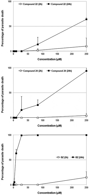
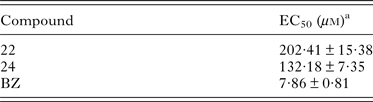
The direct impact of derivatives 22 and 24 over BT was evaluated after 2 and 24 h of incubation at 37 °C. As Fig. 2 reflects, after 2 h of drug treatment, neither compound 22 nor 24 exerted trypanocidal effect on BT, exhibiting EC50 > 250 µ m, likewise BZ. However, a time-dependent effect was observed for these compounds that improved their trypanocidal profiles after 24 h of drug exposition. By this time, both derivatives reached great percentages of parasite death (% PD) at the highest concentration tested, being also BT motility and morphology distorted, especially for 24 that was more potent than 22 (Fig. 2). However, after 24 h of incubation none of these compounds resulted as active as BZ, displaying both lower EC50 in comparison with the value achieved by the reference drug (Table 1).
Activity of compounds 22 (A) and 24 (B) on BT of T. cruzi after 2 and 24 h of incubation at 37 °C. BZ (C) was tested as reference drug. BT, bloodstream trypomastigotes; BZ, benznidazole.

Activity of derivatives 22 and 24 on BT after 24 h of incubation at 37 °C, expressed as EC50
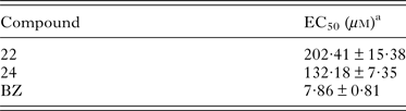
a Estimated by plotting drug concentrations vs percentages of parasite death (%PD). Results are expressed as mean ± s.d. of three independent experiments (n = 3).
TEM analysis
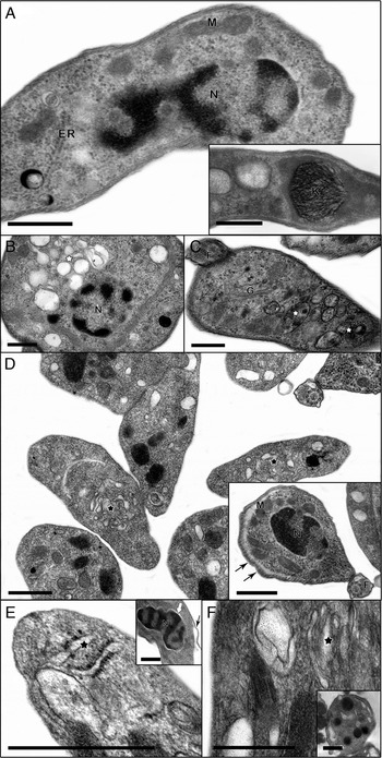
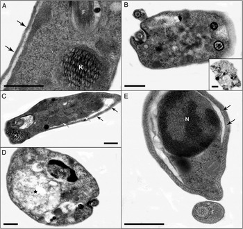
TEM analysis of BZ-, 22- and 24-treated BT was performed using the corresponding EC50/24 h, after 2 (Fig. 3) and 24 h (Fig. 4) of compound exposure at 37 °C. The shorter incubation time (i.e. 2 h) allowed the evaluation of the initial cellular alterations and the potential primary targets of the studied agents. After the period of 2 h, untreated BT (Fig. 3A) exhibited typical morphology, showing nuclei (N), endoplasmic reticulum (ER) and an unique mitochondrion (M) with its characteristic rounded kinetoplast (K). Otherwise, BZ-treated parasites showed an intense intracellular vacuolization (Fig. 3B and C, white stars) besides swollen mitochondrion (data not shown). When both 22- and 24-treated parasites were evaluated, most N and kDNA were unaltered (Fig. 3D and E) even after the longer period of 24 h of incubation (Fig. 4A, C and E). After 2 h of drug exposure, the most frequent and prominent ultrastructural modifications were dilated profiles (black stars) at the ER (Fig. 3E) and Golgi apparatus disorganization (Fig. 3F). Also, the derivatives lead to detachment of the plasma membrane (PM) (Figs. 3D–E and 4A, black arrows) and of the nuclear envelope (Fig. 3E, inset thick white arrow). Some treated parasites also presented swollen mitochondrion suggestive of loss of organelle integrity, especially those exposed to compound 24 (Fig. 4C, white arrows). In both treated samples, an intense shedding of cytoplasmic elements was found at the flagellar pocket (Fig. 4A and C). A large number of intracellular vesicles were early observed in both 22- (Fig. 3D) and 24- (Fig. 3E and F) treated parasites, as well as profiles of ER (double black arrow) surrounding cytoplasmic organelles such as mitochondrion (Fig. 4C) and nuclei (Fig. 4E) after longer periods of incubation.
Ultrastructural findings of T. cruzi BT after 2 h of treatment: (A) untreated parasites exhibited typical morphology, (B–C) parasites treated with BZ showed intense intracellular vacuolization (white stars), (D) 22-treated parasites revealed detachment of the PM (black arrows) and the appearance of intracellular vesicles, (E and F) 24-treated parasites showed dilated profiles at the ER and Golgi apparatus disorganization (black stars) as well as detachment of the PM (black arrow) and of the nuclear envelope (white thick arrow). BT, bloodstream trypomastigotes; BZ, benznidazole; M, mitochondrion; N, nuclei; ER, endoplasmic reticulum; K, kinetoplast; F, flagellum; G, Golgi apparatus; PM, plasma membrane. Scale bars = 0·5 µm.

Ultrastructural findings of T. cruzi BT after 24 h of treatment: (A and B) 22-treated parasites exhibited detachment of the PM (black arrows), (C–E) 24-treated parasites showed swollen mitochondrion (white arrows) and profiles of ER surrounding mitochondrion and nuclei (double black arrows). BT, bloodstream trypomastigotes; PM, plasma membrane; ER, endoplasmic reticulum; F, flagellum; K, kinetoplast; N, nuclei. Scale bars = 0·5 µm.

Oxygen consumption analysis
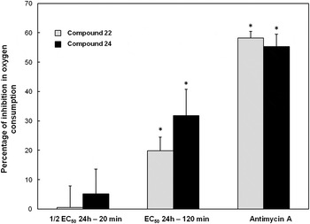
The inhibition in the oxygen uptake of BT treated up to 2 h with derivatives 22 and 24 is reflected in Fig. 5. It is noteworthy to point out that the concentrations administered throughout the experiment were those defined in the trypanocidal assay after 24 h of BT treatment and therefore, they did not induce a lytic effect on the parasites along the assay duration (as Fig. 1 shows, the EC50 values defined for both compounds after 2 h of treatment are higher than 250 µ m). According to this, reductions in the oxygen uptake of 22- and 24-treated BT were the consequence of mitochondrial damage and not caused by a reduction in BT population.
5-Nitroindazolinones reduce mitochondrial oxygen consumption of T. cruzi BT. Non-permeabilized parasites were incubated with 101 µ m of 22 and 66 µ m of 24 for 20 min at 37 °C and then added equal concentrations up to 2 h. Oxygen consumption was blocked by the addition of 22 µ m antimycin A. Asterisks indicate significant differences in relation to control group (P < 0·03). Bars represent mean ± s.e.m. and similar results were obtained with preparations from different experiments (n = 3). BT, bloodstream trypomastigotes.

As it can be seen, treatments of BT for 20 min with 1/2 EC50/24 h did not induce important reductions in oxygen consumption (0·50 and 5·20% for 22 and 24, respectively). Once stabilized the O2 flux in the chamber, equal concentrations of compounds were added and the treatments maintained up to 2 h, reaching this way the EC50/24 h values. After completing 2 h, oxygen consumption was inhibited up to 19·83 and 31·79% by compound 22 and 24, respectively (P < 0·03). Finally, oxygen consumption was strongly reduced by the addition of 22 µ m AA (i.e. 58·15 and 55·35% in 22- and 24-treated BT, respectively), indicating that uptake was due to mitochondrial respiration.
Flow cytometry analysis
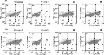
Results of the flow cytometry analysis are described in Table 2 and Fig. 6. As it can be seen, after 2 h of treatment with the EC50/24 h of derivatives 22 and 24, a loss in the mitochondrial membrane potential (MMP) of treated BT was accomplished when compared with untreated ones (P < 0·05). However, for both treated and untreated parasites, the population displaying such an effect increased in a time-dependent manner (P < 0·05). The values registered in untreated parasites correspond to the percentage of BT subpopulation that displayed it before incubation with drugs (Da Silva et al. Reference Da Silva, Batista, Batista, de Souza, da Silva, de Oliveira, Meuser, Shareef, Boykin and Soeiro2008). Nevertheless, after completing 24 h of compounds exposure, ca. 75% of the population treated with derivative 24 suffered depolarization of the mitochondrial membrane (P < 0·05), being this outcome observed in practically the 100% of the parasites treated with 22 (P < 0·05). Positive control (control +) led to mitochondrial membrane depolarization in nearly the total of BT population.
Flow cytometry analysis of TMRE-labelled BT. From left to right: untreated, positive control (heat shock, 2 h at 56 °C) and treated with the respective EC50/24 h of derivatives 22 and 24 for 2 (above) and 24 h (below) at 37 °C. BT, bloodstream trypomastigotes.

Percentages of parasite population that displayed depolarization of the mitochondrial membrane

Results are mean ± s.d. of three independent assays (n = 3).
a BT exposed to heat shock during 2 h at 56 °C (positive control of parasite death).
*Significant differences (P < 0·05) compared with untreated parasites.
#Significant differences (P < 0·05) compared with 2 h of incubation.
DISCUSSION
The complex life cycle of T. cruzi, maintained between both invertebrate (triatomines) and mammalian hosts, involves a successive differentiation of the parasite into epimastigotes, trypomastigotes and amastigotes, as well as other intermediate forms (Tyler and Engman, Reference Tyler and Engman2001). Regarding experimental chemotherapy research, the first T. cruzi form supposes a recurrent model to preliminary explore the trypanocidal activity of novel compounds (Vega et al. Reference Vega, Rolón, Montero-Torres, Fonseca-Berzal, Escario, Gómez-Barrio, Gálvez, Marrero-Ponce and Arán2012). In fact, an epimastigote-like form has been described as an intermediate form in the intracellular cycle of T. cruzi (Faucher et al. Reference Faucher, Baltz and Petry1995; Almeida-de-Faria et al. Reference Almeida-de-Faria, Freymüller, Colli and Alves1999). Otherwise, since epimastigotes are not directly involved in the pathology of the disease, a more specific assay against the relevant forms of T. cruzi for human infection (i.e. intracellular amastigotes and BT) is strictly required in preclinical studies (Miranda et al. Reference Miranda, Solana, Curto, Lammel, Schijman and Soto2015). In this context, we have previously introduced the potential of the 5-nitroindazolinones 22 and 24 to hamper the proliferation of intracellular amastigotes (Fonseca-Berzal et al. Reference Fonseca-Berzal, Escario, Arán and Gómez-Barrio2014, Reference Fonseca-Berzal, da Silva, da Silva, Vasconcelos, Batista, Escario, Arán, Gómez-Barrio and Soeiro2015). Both compounds were also evaluated in vivo and although they did not suppress the parasitemia when mouse models of T. cruzi acute infection were evaluated, these 5-nitroindazolinones considerably reduced the number of circulating BT after concluding an oral treatment of 5 consecutive doses of 100 mg kg−1 day−1, being derivative 24 the most active one (Fonseca-Berzal et al. Reference Fonseca-Berzal, Escario, Arán and Gómez-Barrio2014). Moreover, 22 and 24 did not induce toxic effects either in vitro on different cultures of mammalian cells, or in vivo as a consequence of this treatment scheme since no apparent signs of acute toxicity were observed (Fonseca-Berzal et al. Reference Fonseca-Berzal, Escario, Arán and Gómez-Barrio2014, Reference Fonseca-Berzal, da Silva, da Silva, Vasconcelos, Batista, Escario, Arán, Gómez-Barrio and Soeiro2015). Due to the promising biological effect of these novel compounds, presently our aim was to investigate the direct impact of both derivatives upon BT, exploring the putative targets and mechanisms involved in their anti-T. cruzi activity.
The trypanocidal profile achieved by derivatives 22 and 24 when tested in vitro directly on BT was not as good as those noticed when they were evaluated over both replicative stages of the Y strain (Fonseca-Berzal et al. Reference Fonseca-Berzal, da Silva, da Silva, Vasconcelos, Batista, Escario, Arán, Gómez-Barrio and Soeiro2015). As after longer periods of incubation the bloodstream forms of T. cruzi mostly lose their cellular integrity and viability in in vitro conditions lacking blood constituents, the maximum incubation time for the trypanocidal assay on BT was 24 h, being much shorter than those previously used for the analysis over intracellular amastigotes (i.e. 2, 4 or 7 days according to the strain of T. cruzi) and epimastigotes (up to 72 h according to the phenotypic assay performed) (Vega et al. Reference Vega, Rolón, Montero-Torres, Fonseca-Berzal, Escario, Gómez-Barrio, Gálvez, Marrero-Ponce and Arán2012; Fonseca-Berzal et al. Reference Fonseca-Berzal, Escario, Arán and Gómez-Barrio2014, Reference Fonseca-Berzal, da Silva, da Silva, Vasconcelos, Batista, Escario, Arán, Gómez-Barrio and Soeiro2015). It is possible that short periods of incubation may be not enough for 22 and 24 to exert anti-parasitic effect (death), what differs from BZ capable to induce a more evident lytic effect on BT of the Y strain after 24 h of treatment (Table 1). Conversely, both compounds displayed an efficacy similar to that of the reference drug when assayed on intracellular amastigotes of parasite strains belonging to DTU TcII and TcVI, and also achieved better trypanocidal profiles than BZ on epimastigotes (Vega et al. Reference Vega, Rolón, Montero-Torres, Fonseca-Berzal, Escario, Gómez-Barrio, Gálvez, Marrero-Ponce and Arán2012; Fonseca-Berzal et al. Reference Fonseca-Berzal, Escario, Arán and Gómez-Barrio2014, Reference Fonseca-Berzal, da Silva, da Silva, Vasconcelos, Batista, Escario, Arán, Gómez-Barrio and Soeiro2015). According to it, another hypothesis is that these compounds may be acting differently upon distinct cellular targets towards proliferative forms (amastigotes and epimastigotes) as compared with the non-multiplicative stage (trypomastigotes). Otherwise, previous assays performed with other series of 5-nitroindazoles pointed out a remarkable activity on trypomastigotes (Boiani et al. Reference Boiani, Gerpe, Arán, Torres de Ortiz, Serna, Vera de Bilbao, Sanabria, Yaluff, Nakayama, Rojas de Arias, Maya, Morello, Cerecetto and González2009; Rodríguez et al. Reference Rodríguez, Gerpe, Aguirre, Kemmerling, Piro, Arán, Maya, Olea-Azar, González and Cerecetto2009a ) or a similar pattern on the three stages of the parasite (Muro et al. Reference Muro, Reviriego, Navarro, Marín, Ramírez-Macías, Rosales, Sánchez-Moreno and Arán2014). However, the differences noticed in the scaffold of these molecules (1,3-disubstituted nitroindazole vs 1,2-disubstituted nitroindazolinone), parasite strain or assay conditions could be involved in the different biological activity registered among derivatives, what deserves additional studies to deeper understand the susceptibility profile of T. cruzi to these 5-nitroindazolinones according to the parasite stage.
In this framework and to initiate studies of the mechanism of action triggered by these 5-nitroindazolinones, an ultrastructural analysis was performed by applying TEM. Presently, the incubation of BT with 22 and 24 mainly induced an intense cytoplasmic vacuolization, the occurrence of dilated ER profiles, Golgi apparatus disruption and the frequent appearance of multiple vesicle profiles (multivesicular bodies). Also, our ultrastructural findings showed cytoplasmic organelles such as nuclei and mitochondrion surrounded by ER, which is suggestive of autophagy (Figs 3 and 4), a type II programmed cell death (PCD) mechanism (Vannier-Santos and de Castro, Reference Vannier-Santos and de Castro2009). In fact, PCD mechanisms (i.e. apoptosis, autophagy and necrosis) are well-characterized in higher eukaryotes, actively participating in homeostasis. Concretely, autophagy is fundamental to maintain the metabolic balance and the recycling of cytoplasmic structures and therefore, a deregulation of these procedures is likely to start death pathways (Menna-Barreto et al. Reference Menna-Barreto, Salomão, Dantas, Santa-Rita, Soares, Barbosa and de Castro2009a ). Autophagy is present during the life cycles of major parasitic protists such as trypanosomatids (Brennand et al. Reference Brennand, Gualdrón-López, Coppens, Rigden, Ginger and Michels2011), but also has been described as a response triggered by antiparasitic drugs (Menna-Barreto et al. Reference Menna-Barreto, Salomão, Dantas, Santa-Rita, Soares, Barbosa and de Castro2009a , Reference Menna-Barreto, Corrêa, Cascabulho, Fernandes, Pinto, Soares and de Castro b ). In fact, this type of PCD has been reported during the treatment of T. cruzi with both natural (Luize et al. Reference Luize, Ueda-Nakamura, Dias Filho, García Cortez, Morgado-Díaz, de Souza and Nakamura2006; Menna-Barreto et al. Reference Menna-Barreto, Laranja, Silva, Coelho, Paes, Oliveira and de Castro2008; Da Silva et al. Reference Da Silva, Batista, de Araújo, Batista, Lionel, de Souza, Ripoll Hammer, da Silva, de Mieri, Adams, Zimmermann, Hamburger, Brun, Schühly and Soeiro2013; Veiga-Santos et al. Reference Veiga-Santos, Desoti, Miranda, Ueda-Nakamura, Dias-Filho, Silva, García Cortez, de Mello and Nakamura2013) and synthetic drugs (Fernandes et al. Reference Fernandes, Da Silva, Pinto, de Castro and Menna-Barreto2012, Da Silva et al. Reference Da Silva, Batista, Batista, Lionel, Ripoll Hammer, Brun and Soeiro2014). It is important to note that with both studied compounds, earlier events (as after 2 h of drug incubation) included the detachment of PM and nuclear envelope, high levels of shedding elements at flagellar pocket besides numerous cytoplasmic vacuoles and dilated Golgi apparatus and abnormal ER, leading to later incubation periods (24 h) to the engulfment of organelles by ER profiles that may be suggestive of parasite autophagy possibly triggered by an osmotic shock that deserves to be further explored in future studies.
According to its singularity, the mitochondrion of trypanosomatids has turned into one of the preferred targets for designing antiparasitic drugs. The alterations that they induce in this organelle are time-dependent and vary with the compound class. Mitochondrial damage can appear either as a direct effect of the drug, or as consequence due to the loss in organelle viability related to their primary interference with other cellular components or metabolic pathways (Silva Paes et al. Reference Silva Paes, Suarez Mantilla, Barisón, Wrenger and Silber2011; Menna-Barreto and de Castro, Reference Menna-Barreto and de Castro2014). The affectation of mitochondrion was further explored through respirometry assays and according to the results (Fig. 5), once reached compounds EC50/24 h, 22- and 24-treated BT suffered noteworthy inhibitions in oxygen consumption, greater for derivative 24. These outcomes correlate with the ultrastructural findings, since a loss in mitochondrial integrity was registered in a number of 24-treated parasites (Fig. 4).
Some trypanocidal compounds capable of redox cycling (e.g. quinone derivatives or nifurtimox) seem to act by selectively promoting the oxidative stress in T. cruzi (Pinto and de Castro, Reference Pinto and de Castro2009; Wilkinson and Kelly, Reference Wilkinson and Kelly2009). These potent oxygen redox cyclers induce the increase of reactive oxygen species (ROS) in the parasite and in combination with T. cruzi deficient antioxidant defences results in the selective killing of the parasite (Turrens, Reference Turrens2004). Focusing on 5-nitroindazoles, the potential of 2-benzyl derivatives to selectively induce oxidative stress in the parasite has been previously proposed (Folch-Cano et al. Reference Folch-Cano, Olea-Azar, Arán and Díaz-Urrutia2010). This study also showed their capacity to generate nitro-anion radical species and their immediately reoxidization to the original conformation in presence of oxidizing agents (redox cycling). A similar behaviour is usually accepted for nifurtimox, which undergoes intracellular reduction in presence of type II nitroreductases yielding nitro-anion radicals, followed by cycling in the presence of oxygen with the regeneration of the parent nitrocompound (Wilkinson and Kelly, Reference Wilkinson and Kelly2009). However, our results illustrate that oxygen uptake was not increased in T. cruzi by 22 and 24 treatments (Fig. 5), being these compounds suspected not to act through oxygen redox cycling at the concentrations tested and up to the incubation time assayed using BT (up to 2 h). Otherwise, they would raise parasite oxygen uptake even when added AA, similarly to the cyanide-insensitive oxygen consumption experimented by nifurtimox (Maya et al. Reference Maya, Bollo, Nuñez-Vergara, Squella, Repetto, Morello, Périe and Chauvière2003). The lesser capacity of 5-nitroindazole derivatives to be reduced compared with that of nifurtimox (Folch-Cano et al. Reference Folch-Cano, Olea-Azar, Arán and Díaz-Urrutia2010) or the requirement of higher concentrations of drug to induce oxygen redox cycling (Boiani et al. Reference Boiani, Piacenza, Hernández, Boiani, Cerecetto, González and Denicola2010) may explain the different pattern registered among our nitroderivatives and nifutimox. According to this, 22 and 24 are more probable to act by inhibiting the mitochondrial electron transport chain (ETC) and therefore, to reduce parasite mitochondrial respiration (Fig. 5), likewise other 5-nitroindazole derivatives previously assayed on T. cruzi (Rodríguez et al. Reference Rodríguez, Arán, Boiani, Olea-Azar, Lavaggi, González, Cerecetto, Maya, Carrasco-Pozo and Speisky Cosoy2009b ). However, further assays focussed to explore NADH-cytochrome c oxidoreductase (complex I–III) and cytochrome c oxidase activity (complex IV) are needed to assert the specific ETC alterations triggered by these compounds (Menna-Barreto et al. Reference Menna-Barreto, Goncalves, Costa, Silva, Pinto, Oliveira and de Castro2009c ).
According to the nitro-anion radical instability and the rapid dismutation to nitroso species it suffers in several compounds, it has been proposed that megazol and other 5-nitroimidazoles, poor oxygen cyclers and time-dependent respiration inhibitors, exert their anti-T. cruzi activity by binding to trypanothione [T(SH)2] and glutathione (GSH) (Maya et al. Reference Maya, Bollo, Nuñez-Vergara, Squella, Repetto, Morello, Périe and Chauvière2003). The profile both 22 and 24 displayed along the respirometry assays, together with the reactivity of GSH with the nitro-anion radical showed in some 5-nitroindazole derivatives (Folch-Cano et al. Reference Folch-Cano, Olea-Azar, Arán and Díaz-Urrutia2010), resembles a similar behaviour between these two groups of molecules. Nevertheless, the higher stability of nitro-anion radical in these kind of 5-nitroindazoles (Folch-Cano et al. Reference Folch-Cano, Olea-Azar, Arán and Díaz-Urrutia2010) compared with that of the aforementioned 5-nitroimidazoles (Maya et al. Reference Maya, Bollo, Nuñez-Vergara, Squella, Repetto, Morello, Périe and Chauvière2003), suggests different mechanisms of interaction with thioles. The referred 5-nitroimidazoles are proposed to interact with thioles through the formation of electrophilic metabolites (Maya et al. Reference Maya, Bollo, Nuñez-Vergara, Squella, Repetto, Morello, Périe and Chauvière2003). Otherwise, 5-nitroindazole derivatives are supposed to carry out this kind of interaction in a reaction mediated by the nitro-anion radical with GSH acting as oxidizing agent (Folch-Cano et al. Reference Folch-Cano, Olea-Azar, Arán and Díaz-Urrutia2010). At this point, it is noteworthy to highlight the importance of this thiol as precursor in T(SH)2 biosynthesis and therefore, in the antioxidant defences of T. cruzi (Irigoín et al. Reference Irigoín, Cibils, Comini, Wilkinson, Flohe and Radi2008).
Finally, due to the findings assessing some mitochondrial changes in both morphology and functionality, TMRE was used to monitor the MMP of treated and untreated BT, since this kind of fluorescent rhodamine derivatives is accumulated within energized mitochondrion (Menna-Barreto et al. Reference Menna-Barreto, Goncalves, Costa, Silva, Pinto, Oliveira and de Castro2009c ; Fernandes et al. Reference Fernandes, Da Silva, Pinto, de Castro and Menna-Barreto2012). Consistent with TEM and high-resolution respirometry, flow cytometry analysis pointed out the affectation of mitochondrion triggered by these 5-nitroindazolinones, which was very pronounced after longer periods of incubation (24 h of drug exposure). In fact, both compounds led to a time-dependent decrease in TMRE fluorescence, reflecting this way the parasite population with depolarizated mitochondrial membrane (Table 2). The use of this marker suggested the interference of derivatives 22 and 24 with the proton electrochemical potential gradient of the T. cruzi mitochondrial membrane (Da Silva et al. Reference Da Silva, Batista, Batista, de Souza, da Silva, de Oliveira, Meuser, Shareef, Boykin and Soeiro2008).
In physiological conditions mitochondrial respiration generates ROS, such as H2O2 and O2 −. However, any alteration like the inhibition of this metabolic procedure conduces to an increase in ROS steady-state concentrations (Turrens, Reference Turrens2003). In addition to being the main source of these molecules, the mitochondrion is particularly susceptible to ROS-induced damage and therefore, dysfunctional mitochondrion is able to hydrolyze ATP and accelerate ROS production. The removal of this defected organelle results critical for the cell protection and mainly occurs through a selective autophagic process known as mitophagy (Kim et al. Reference Kim, Rodríguez-Enríquez and Lemasters2007). According to the results achieved here, this kind of autophagy may be implicated as a T. cruzi response against the mitochondrial damage induced by these 5-nitroindazolinone prototypes, whose deregulation could conduce to the cell death (Kim et al. Reference Kim, Rodríguez-Enríquez and Lemasters2007).
Concluding remarks
The results compiled in the present work suggest that T. cruzi undergoes an autophagic pathway as a consequence of the treatment with 5-nitroindazolinones 22 and 24. Besides, the different cell death features were detected including mitochondrial collapse. In this regard, further biochemical and molecular studies are needed in order to fully elucidate the mechanism of action of these 5-nitroindazole derivatives.
ACKNOWLEDGEMENTS
The authors thank the Program for Technological Development in Tools for Health (PDTIS-Fiocruz) for the facilities. C. F. -B., J. A. E., V. J. A. and A. G. -B. acknowledge M. N. C. S. for kindly accepting C. F. -B. predoctoral stay in LBC/IOC/Fiocruz. The Spanish Ministry of Economy and Competitiveness (MINECO; ref. SAF2015-66690-R) and the 911120 UCM-CEI Moncloa Research Group are also acknowledged.
FINANCIAL SUPPORT
The present study was supported by grants from FAPERJ, CNPq, PAEF, PDTIS and Fiocruz. M. N. C. S. is CNPq and CNE (FAPERJ) researcher. Research by C. F. -B. was supported by a PICATA predoctoral fellowship and a PICATA predoctoral mobility grant of the Moncloa Campus of International Excellence (UCM-UPM & CSIC).