I. INTRODUCTION
Hydrogels are macromolecular network materials, which has important medical applications. As we know, hydrogel is a polymer that can swell and is insoluble in water; and, which has a three-dimensional structure. These properties make it widely used to control release of drugs, immobilization of enzyme, and control of protease activity, etc. Reference Khor1 Early developments in hydrogels focused mainly on synthetic materials such as poly(ethylene glycol)/poly(butylene glycol) block copolymers, poly(ethylene glycol)/poly(lactic acid) block copolymers, and copolymers of N-isopropylacrylamide and so on. These materials are basically suitable for drug delivery, but they are nonbiodegradable and some of them have slight toxicity which limits their application. Reference Peppas, Huang and Torreslugo2 In recent years, to solve some drawbacks of synthetic materials, many people are concerned about the natural polymers, such as hydroxyethylacryl CS/alginate hydrogel films, alginate/polyoxyethylene hydrogels, alginate/gelatin hydrogels, and hyaluronate/polygalacturonic acid hydrogels and so on. Reference Khor1–Reference Shoichet3 Hydrogels prepared with natural materials not only have above-mentioned excellent properties, but also advantages like biodegradable, nontoxic, etc. Reference Shoichet3–Reference Alvarez, Blanco, Puga and Concheiro5 So we prepared hydrogels with natural materials in this work.
CS is a kind of natural polysaccharide obtained from the deacetylation of chitin. Reference Chen, Tang, Ning, Wang and Zhang6 It was often used to make hydrogels for its biocompatibility, nontoxicity, biodegradability, and function of antihypertensive, antibacterial, and antineoplastic. Reference Honarkar and Barikani7,Reference Yeng, Salmah and Sam8 However, CS is not water-soluble, acetic acid or other acids can facilitate dissolution of CS in water, which can generate cytotoxicity. Reference Aziz, Cabral and Moratti9,Reference Yin, Zhao and Ding10 Therefore, we utilize the covalent attachment of reactive on the –NH3 and C6–OH to produce hydrophilic groups and enhance the water-solubility of CS. Reference Lim and Hudson11–Reference Fu and Sun13
Safflower/ligusticum wallichii polysaccharide-II are certain biological active ingredient from safflower and ligusticum wallichii, respectively. They have similar functions of antitumor, and antioxidant, etc. by viewed in recent literature. Reference Wang, Liu, Yang and Sun14–Reference Zhao, Huang and Liu17 Moreover, they have good biocompatibility and biodegradability. Reference Huang18 So the mixing polysaccharide of SLWP-II was selected to prepare hydrogel in this study. Research showed that safflower polysaccharide-II is comprised of D-glucose, D-rhamnose, L-arabinose, and D-galactose. The ligusticum wallichii polysaccharide-II is comprised of D-glucose, D-seminose, L-arabinose, and D-galactose. Reference Zhang, Zhang and Zhang19,Reference Sun, Yan and He20
While, up to now, it was not reported whether hydrogels can be made from CS and mixing polysaccharide of SLWP-II, and whether they can be used in drug delivery system and tissue engineering. In present work, SLWP-II could be functionalized with periodate to produce functional aldehyde groups that could serve as a novel macromolecular cross-linker for those polymers bearing free amino groups to form hydrogels. Reference Liao and Zhang21 So a novel hydrogel based on oxidized SLWP-II and N,O-carboxymethyl CS was prepared.
II. EXPERIMENTAL
A. Materials
CS (deacetylation degree (DD) of 95%, Mw = ∼210 kDa) was purchased from Qingdao Dive Light Biological Engineering Co., Ltd. (Qindao, China). Safflower and ligusticum wallichii polysaccharide-II (
 $M_\eta ^{\rm{s}}$
= 4.2 × 10⁴,
 $M_\eta ^{\rm{L}}$
= 3.8 × 10⁴) were purchased from West Anrui Bo Biological Technology Co., Ltd. (Xi'an, China). Chloroacetic acid and sodium periodate were purchased from Tianjin Evergreen Chemical Reagent Manufacturing Co., Ltd. (Tainjin, China). CBB (Coomassie brilliant blue) and BSA (bovine serum albumin) supplied by Biotopped (Beijing, China). All other chemicals used in this paper were analytical grade, without further purification. Ultrapure water (Milli-Q Gradient A10 Water Purification System, Millipore Corporation, Billerica, Massachusetts), it was used to prepare all solutions.
B. Synthesis and characterization of N,O-carboxymethyl CS and oxidized SLWP
N,O-carboxymethyl CS was prepared by electrophilic substitution reaction. Reference Lin, Xu and Liu22 Specifically, CS (5 g) was added into 25 mL of NaOH (40%, w/w) solution at −20 °C overnight. Subsequently, chloroacetic acid (20 g) dissolved into 100 mL of isopropanol was added into the frozen CS suspension guttatim in 30 min. The mixture was stirred by magnetic force at 25 °C for 2 h, and then at 60 °C for additional 2 h. In the end, the resultant was filtered and washed three times with 95% ethanol. After drying by oven at 40 °C, the final product of N,O-carboxymethyl CS was obtained and characterized by 1H NMR (D2O as solvent; Agilent NMR inova600 [Agilent Technologies, Santa Clara, California]; Relax delay 1.000 s; width 9598.1 Hz) and IR (v: 4000–400 cm−1) analysis. The substitution degree of CS was measured by potentiometric titration method. Reference Fu and Sun13
Oxidized SLWP-II was prepared by methods reported previously. Reference Cabral, Roxburgh, Shi and McConnell23 Specifically, a weight of SLWP-II was first added into an ethanol/water (1:1, v/v) solution (100 mL) with continuous stirring by magnetic force at 25 °C for 2 h. Then, a weight of NaIO4 (20, 40, and 60% molar equivalent with SLWP-II monomer) dissolved into 20 mL of distilled water was added to the SLWP-II solution for another 8 h at dark. After that, an equimolar amount of diethylene glycol is added to quench the unreacted NaIO4. The resultant was filtered and the residue was dissolved into distilled water, dialyzed 3 days in a dialyzer, then freeze-dried by lyophilizer to obtain the final product. SLWP-II with theoretical oxidization degrees of 20, 40, and 60% were prepared and named as OSLWP-1, OSLWP-2, and OSLWP-3, respectively. In addition, the OSLWP-2 was characterized by 1H NMR (D2O as solvent; Agilent NMR inova600; Relax delay 1.000 s; width 9598.1 Hz) and IR (v: 4000–400 cm−1) analysis. The oxidization degree of OSLWP-1 to OSLWP-3 was measured by a potentiometric titration method. Reference Wang, Liao and Chen24,Reference Li, Weng and Kong25
C. Preparation and characterization of CSLH
OSLWP-2 was used to prepare the CSLH for further analysis and characterization. A calculated weight of OSLWP-2 and N,O-carboxymethyl CS were dissolved into 10 mL distilled water to form 5 and 14% (w/w) solutions, respectively. Then, 4 mL of OSLWP-2 and N,O-carboxymethyl CS solutions, regardless of the weight ratio (1:1; 1:2; 1:3, w/w), were mixed at room temperature with gentle stirring, and then placed at 37 °C for 10 min to form the hydrogels (CSLHs). The obtained CSLHs with weight ratios (1:1; 1:2; 1:3, w/w) were named CSLH-1, CSLH-2, and CSLH-3, respectively. In addition, the CSLH-2 was prepared with D2O as solvent and then characterized by 1H NMR (Agilent NMR inova600; Relax delay 1.000 s; width 9598.1 Hz) and IR (v: 4000–400 cm−1) analysis. Reference Li, Weng and Kong25
D. Appearance morphologic characterization
The morphology of the hydrogels (CSLH-1, CSLH-2, and CSLH-3) were studied with scanning electron microscopy (SEM; JSM-6010LV, JEOL Ltd., Dongguan, Japan) at 20 kV. The lyophilized rupture pieces of the CSLHs were sputtered with gold, and placed in a cabinet drier for 24 h before observation. Reference Li, Weng and Kong25
E. Swelling ratio of CSLHs
The CSLHs degree of swelling was determined by gravimetrical method. Hydrogels (CSLH-1, CSLH-2, and CSLH-3) dried to constant quality were placed in acidic aqueous solution (hydrochloric acid, pH2), phosphate buffered saline (PBS, pH7.4), and alkaline aqueous solution (sodium hydrate, pH12) at 37 °C respectively. Then, samples were removed from the medium and weighed at 24 h. Reference Li, Weng and Kong25 The degree of swelling was calculated as follows:
 $${\rm{Swelling}}\;{\rm{ratio}}\;\left( {{{\rm{g}} \mathord{\left/ {\vphantom {{\rm{g}} {\rm{g}}}} \right. \kern-\nulldelimiterspace} {\rm{g}}}} \right) = {{{M_{24}} - {M_0}} \over {{M_0}}}\quad .$$
M 24 and M 0 are the weights of the swollen wet hydrogel and initial dry hydrogel, respectively. Measurements were performed in triplicate.
F. In vitro protein release
Bovine serum albumin (BSA) was selected as a model protein to evaluate the release of biomacromolecules from the hydrogels. Briefly, 0.01 g of BSA was dissolved into N,O-carboxymethyl CS aqueous solution in a 15 mL Eppendorf centrifuge tube (EP tube), soon after added into various amounts of oxidized SLWP-II, then stirred vigorously. The resulting solution was placed at 37 °C for 10 min to form the BSA-loaded CSLHs. The control samples are hydrogels without BSA. Thenceforward, 5 mL of PBS as a release medium, was added to the CSLHs and the EP tube was placed in a shaker (100 rpm/min) at 37 °C. Every 12 h, medium was removed for analysis and was replaced with fresh PBS. Reference Li, Weng and Kong25 Protein concentrations were measured from 1 mL of each sample by the Coomassie Brilliant Blue assay, measured for 156 h in total. Reference Yang and Wang26 Measurements were performed in triplicate.
G. In vitro degradation
The CSLHs samples (CSLH-1, CSLH-2, and CSLH-3) were prepared and cylindrical samples were completely immersed into 5 mL PBS solution in three small beaker respectively, incubated at 37 °C. Then, samples were removed from the medium and weighed at predetermined time-points (1, 3, 7, 14, 21, and 28 days). Reference Li, Weng and Kong25 The degree of in vitro degradation was expressed by the weight loss:
 $${\rm{Weight}}\;{\rm{loss}}\;(\% ) = {{{M_t} - {M_0}} \over {{M_0}}} \times 100\quad .$$
M 0 is the fully swollen weight of the hydrogel before degradation and M t is the weight of the hydrogel after t h. Measurements were performed in triplicate.
H. Rheological analysis
Rheological measurement was performed using a RS6000 rheometer (TA Instruments, Wetzlar, Germany). Mixing N,O-carboxymethyl CS with oxidized SLWP-II solutions to form CSLH-1, CSLH-2, and CSLH-3, respectively. Then, 1 mL of the CSLHs was immediately loaded onto a lower stationary thermostated rheometer plate (35 mm diameter) and the upper plate was adjusted to the predefined gap size (1 mm gap) at 25 °C. Ten minutes later, the oscillation measurements were carried out in a controlled deformation mode at a constant frequency of 1 Hz with 10 Pa shear stress in 5 min. Reference Tamburic and Craig27
I. In vitro cytotoxicity of the hydrogel
The in vitro cytotoxicity of the hydrogels was examined according to the reported method. Reference Han, Yang and Zhao28 Each hydrogel was incubated in a-MEM medium (5 mL) maintained at 37 °C for 48 h to get the hydrogel extracts. The WI-38 cells were placed in a 96-well plate at a seeding density of 1 × 10⁴ cells per well. Thereafter, 0.1 mL of fresh growth medium containing either a series of extractions of the hydrogel was added. Untreated cells in growth medium were used as a control. After another 7 days of incubation, MTT assay was performed to determine the in vitro cytotoxicity at predetermined time-point. After a further incubation of 2 h, the MTT solution (0.5 mg/mL) was carefully removed from each well, and 150 μL of DMSO was added to dissolve the MTT formazan crystals. The absorbance was recorded at 570 nm using an ELISA microplate reader (Bio-Rad, Hercules, California). Cell viability (%) was calculated according to the following equation: cell viability (%) = absorption test/absorption control × 100%, where absorption control is the absorbance from control wells. All data are presented as the mean of six measurements (±SD).
J. Statistical analysis
Statistical analyses were performed by t test. P value less than or equal to 0.05 means significant difference (Origin Pro 7.5). Values of results are shown as the mean and standard deviation.
III. RESULTS AND DISCUSSION
A. Characterization of N,O-carboxymethyl CS and oxidized SLWP-II
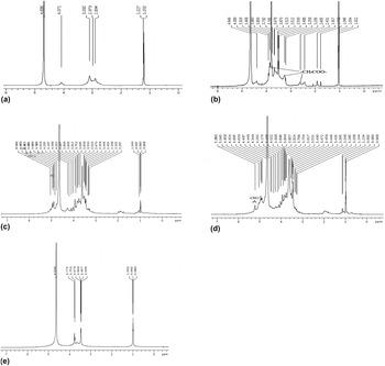
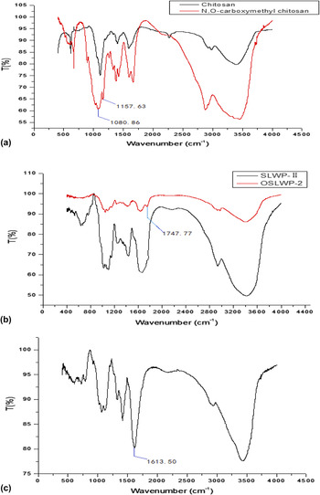
CS is insoluble in neutral water, but dissolved in acetic acid solution. This property limits its scope of application. Reference Yeng, Salmah and Sam8 To improve its water-solubility, chloroacetic acid was used to graft carboxymethyl groups onto the CS skeleton in an alkaline medium. Reference Lin, Xu and Liu22,Reference Li, Weng and Kong25 The resultant N,O-carboxymethyl CS was characterized by 1H NMR. The 1H-NMR spectra of carboxymethyl CS showed the successful synthesis of N,O-carboxymethyl CS [Figs. 1(a) and 1(b)]. The methylene protons of O- and N-substituted carboxymethyl CS were found at chemical shift 3.91 and 3.26 ppm, respectively. The methine proton at position-C2 of carboxymethyl CS was showed at chemical shift 2.61 ppm [Fig. 1(b)], which was in accordance with previous study. Reference Jaidee, Rachtanapun and Luangkamin29 As can be seen from Fig. 2(a), the stretching vibration absorption peak 1158.65 and 1089.17 cm−1 were found on carboxymethyl chitosan, which indicates that the –OH on chitosan has a carboxyl methylation reaction. The substitution degree of carboxymethyl was measured by titration method, which was 32% on N,O-carboxymethyl chitosan. Reference Fu and Sun13
1H NMR spectrum of chitosan (a), N,O-carboxymethyl chitosan (b), SLWP-II (c), oxidized SLWP-II (d) in D2O and N,O-carboxymethyl chitosan/oxidized SLWP-II hydrogel (e), in C2D4O2 at 25 °C.

IR spectrum of chitosan, N,O-carboxymethyl chitosan (a); SLWP-II, oxidized SLWP-II (b); and N,O-carboxymethyl chitosan/oxidized SLWP-II hydrogel (c); at 25 °C.

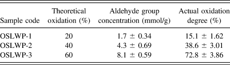
Based on the oxidation method, the SLWP-II was oxidized into their dialdehyde derivatives by NaIO4. Reference Cabral, Roxburgh, Shi and McConnell23 Compared with the 1H NMR spectrum of SLWP-II, oxidized SLWP-II has a new peak of aldehyde proton at 5.2 ppm [Figs. 1(c) and 1(d)], which was in accordance with previous study. Reference Li, Weng and Kong25 As can be seen from Fig. 2(b), the stretching vibration absorption peak 1747.77 cm−1 of C=O in the aldehyde group was found on OSLWP-2, which indicates that part of the –OH on CS was oxidized to aldehyde groups. In addition, the actual oxidization degrees of SLWP-II were varied from 15.1 to 72.8% at the different NaIO4 concentrations (Table I). However, SLWP-II was becoming more disaggregate with the increase of the NaIO4 concentration, which influenced its inherent properties greatly, for example, reducing its water-solubility and viscosity. Reference Li, Weng and Kong25 Therefore, the amount of NaIO4 used for oxidization of SLWP-II should be strictly controlled. In addition, the appropriate amount of aldehyde groups on oxidized SLWP-II has yet to be investigated. In this study, OSLWP-2 with aldehyde group concentration of 4.1 mmol/g was selected to prepare the following hydrogel.
Aldehyde group concentration and oxidation degree of oxidized SLWP-II at different oxidant concentration.

B. Characterization of CSLH formation
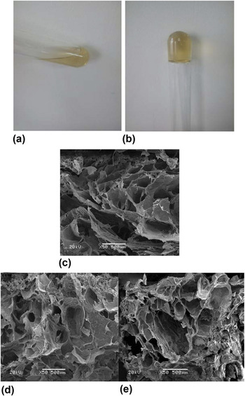
After the CSLH was prepared, it was detected by 1H NMR. Theoretically, amino groups on N,O-carboxymethyl CS and aldehyde groups on oxidized SLWP-II could react into a Schiff base in hydrogel. Reference Shoichet3,Reference Ma, Yang, Zhou, Xiao, Kennedy and Nie12 In Fig. 1(e), the disappeared peak at 5.4 ppm of the oxidized SLWP-II. As can be seen from Fig. 2(c), the stretching vibration absorption peak 1747.77 cm−1 of C=O in the aldehyde group disappeared, and the stretching vibration absorption peak of –C=N– was found at 1613.50 cm−1. This indicate that Schiff base reaction between OSLWP-2 and N,O-carboxymethyl CS did occur. Figures 3(a) and 3(b) showed the appearance of N,O-carboxymethyl CS/oxidized SLWP-II system before and after formation of hydrogel. As can be seen, mixture solution showed a flowing state before hydrogel formation, but the mixture turned into hydrogel by crosslinking soon.
Appearance of N,O-carboxymethyl chitosan/oxidized SLWP-II system at before (a) and after (b) the formation of the hydrogel and SEM images of the CSLHs (CSLH-1 c, CSLH-2 d, CSLH-3 e).

C. Morphological analysis
Morphology of the hydrogels (CSLH-1, CSLH-2, and CSLH-3) were observed with scanning electron microscope (SEM). As can be seen in Fig. 3, the CSLHs had porous interior structures with interconnecting pores, pores diameter ranging from several tens to several thousands of micrometers. Moreover, with the proportion of oxidized SLWP-II to N,O-carboxymethyl CS from 1:1 to 1:3, the more crosslinking between amino group and aldehyde group, the diameter of the pores are decreased and the compactness of pores increased. The CSLH-1, with the lowest contents of N,O-carboxymethyl CS, had the largest pores (1080 μm). Additionally, the walls of this gel had a loose sheet-like appearance implying their weak mechanical strength. Conversely, CSLH-3, with the highest contents of N,O-carboxymethyl CS, had smaller, more compact pores (140 μm) and thicker walls. That is to say, when there are sufficient number of aldehyde groups in oxidized SLWP-II, with the amount of N,O-carboxymethyl CS increasing, the amount of amino group is increasing accordingly. Therefore, with the proportion of oxidized PP-II to N,O-carboxymethyl CS from 1:1 to 1:3, intramolecular and intermolecular crosslinking of N,O-carboxymethyl CS will be increased, and the compactness of pores will be also enhanced. Reference Shoichet3,Reference Yin, Zhao and Ding10–Reference Ma, Yang, Zhou, Xiao, Kennedy and Nie12 This porous interior structures showed that the CSLH can be used in drug delivery and cell culture system.
D. Swelling behavior analysis
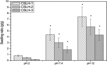
As we can see from Fig. 4, with the proportion of oxidized SLWP-II to N,O-carboxymethyl CS from 1:1 to 1:3, the swelling ratio of the CSLHs is decreased. This might be expected because CSLH-3 has higher crosslinking activity and thus a decrease in its swelling ratio. Reference Jayakumar30 The swelling ratio of CSLH is relatively low at pH 2. This might be attributed to the protonation of –COO− to –COOH in acid environment lead to the formation of hydrogen bonds between N,O-carboxymethyl CS and oxidized SLWP-II, which prevent the entry of water molecules. Furthermore, the protonation of –NH2– to –NH3 + and unprotonation of –COO− result in net charge reducing to zero, which lead to free ion concentration close to zero, and outside osmotic pressure make the hydrogel dehydrate and shrank. Reference Muzzarelli, Jari and Petrarulo31,Reference Ostrowska and Gierszewska32 Finally, these two reasons induce the swelling ratio of CSLHs significantly to decrease at pH 2.0. The swelling ratio is relatively high at pH 12. This was caused by a large swelling force created by the electrostatic repulsion of the ionized carboxylic acidic groups (–COO−). Reference Han, Yang and Zhao28,Reference Muzzarelli, Jari and Petrarulo31 Therefore, the gelation of these hydrogels can be regulated not only by the oxidization degree of SLWP-II, but also by the pH conditions. This might have potential applications in oral drug delivery where the acidic environment of the stomach could decrease the swelling of the hydrogel and prevent biomacromolecule release.
Swelling ratio of CSLHs (CSLH-1, CSLH-2, CSLH-3) in various pH condition (pH = 2, 7.4, and 12) at 37 °C. *P < 0.05 compared with pH = 2.

E. In vitro release behavior
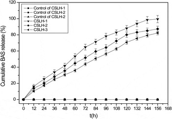
We measured the release behavior of BSA over 156 h from CSLHs at 37 °C in PBS. We can see from Fig. 5, the degradation products of control samples doesn’t react with Coomassie Blue. From Fig. 5, the results showed that the BSA released from CSLH-1 reach 88% in 120 h, while 80% and 70% from CSLH-2 and CSLH-3, respectively, and the cumulative release of BSA were approximately 99, 87, and 82% in 204 h. This is probably a result of the larger pore sizes in CSLH-1 (1080 μm) that we observed by SEM. However, CSLH-2 and CSLH-3, with a greater degree of crosslinking and more compact microstructure, would decrease the diffusion of BSA. This result is similar to cumulative BSA release of 80% from N,O-carboxymethyl CS/oxidized alginate hydrogel reported previously. Reference Li, Weng and Kong25 If it is a small molecule drug in the hydrogel, the release rate may be faster than BSA. Therefore, we can control the release speed by adjusting the components of the gels or loaded with different molecular weight drugs in the hydrogel, and make it better suited to different application requirements.
In vitro release behavior of BSA from CSLHs in PBS solutions at 37 °C.

F. In vitro degradation
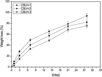
We performed in vitro degradation behavior of the hydrogels (CSLH-1, CSLH-2, and CSLH-3) at pH 7.4 in PBS. The ions in the PBS solution can break the intramolecular and intermolecular hydrogen bonds of polysaccharides. Therefore, the structural stability of hydrogels is decreased, and with the increasing of time, the hydrogels were degraded gradually. Reference Ostrowska and Gierszewska32,Reference Fan, Du, Zhang, Yang, Zhou and Kennedy33 As shown in Fig. 6, CSLH-1 exhibits a relatively rapid degradation rate, which is approximately 49% in 7 days, faster than 40% of CSLH-2 and 33% of CSLH-3. Samples lost 94%, 82%, and 76% of their weight at 28 days, respectively. This could be attributed to increased stability from CSLH-1 to CSLH-3 for chang to be with more crosslinking between N,O-carboxymethyl CS and oxidized SLWP-II, and the effect of ions on hydrogels decreased gradually. Hence, we have demonstrated that the physiological degradation of the CSLHs can be regulated by adjusting the components of the gels, and make it better suited to different application requirements.
In vitro degradation profile of CSLH-1, CSLH-2, and CSLH-3 in PBS solution at 37 °C.

G. Rheological analysis
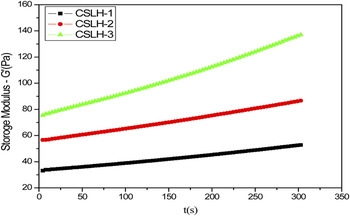
In rheology analysis, storage modulus (G′) also called elasticity modulus, reflects the stiffness of material. Reference Chambon, Petrovic, Macknight and Winter34 As shown in Fig. 7, storage modulus (G′) of CSLH-1, CSLH-2, and CSLH-3 increased in turn. This might be due to CSLHs becoming more and more compact structure from CSLH-1 to CSLH-3. The storage modulus (G′) of CSLH-1 to CSLH-3 increased gradually. This could be attributed to the crosslinking and gel formation process that still continues after 10 min. From Fig. 6, the results showed that the hydrogels have suitable stiffness (33–136 Pa). Moreover, there is an increasing trend in the CSLH-1 to CSLH-3. Reference Li, Weng and Kong25,Reference Kreilgaaed35 Therefore, it has suitable stiffness strength for a few applications in the drug delivery system and tissue engineering.
Storage modulus (G) of CSLH-1, CSLH-2, and CSLH-3 in 5 min at 25 °C.

H. In vitro cytotoxicity
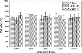
The biocompatibility of biomaterials is crucial for their clinical and therapeutic applications. To evaluate this novel hydrogel as a potential delivery system for biomacromolecules, we measured the cytotoxicity of these hydrogels and their components by a MTT test during one week. Reference Shin, Olsen and Khademhosseini36 For N,O-carboxymethyl CS/oxidized SLWP-II hydrogel is a solid system, which prevented the direct use of MTT method for the determination of its cytotoxicity. Therefore, extractions from the hydrogels were used to evaluate the cytotoxicity of the hydrogels. As shown in Fig. 8, the crosslinked hydrogel exhibited no toxicity toward WI-38 cells at predetermined time-point, and they have a role in promoting cell proliferation. This is likely to be caused by the formation of Schiff bases between the amino groups on N,O-carboxymethyl CS and the aldehyde groups on oxidized SLWP-II during the gelation process that decreases the toxic free aldehyde groups in the hydrogel. In addition, it may be because in the process of the degradation of the hydrogel, only the glycosidic bond can be broken, and the structure of the Schiff bases were not destroyed. Reference Li, Yang, Ma, Xu and Chen37 The results showed that the hydrogel has exhibited good cell compatibility.
In vitro cytotoxicity of hydrogels against WI-38 cells was confirmed by MTT assay in function with different concentrations of hydrogel extract of 7 days.

IV. CONCLUSIONS
A series of N,O-carboxymethyl CS/oxidized SLWP-II (CSLHs) was prepared. With amounts of N,O-carboxymethyl CS increasing, crosslinking degree of the CSLHs was increased. Meanwhile, the swelling ratio, degradation rate, and the cumulative release of BSA were reduced. The CSLHs exhibit porous three-dimensional structure, which is very important in the drug delivery and cell culture. The CSLHs showed suitable stiffness and can withstand several tens to several hundreds Pa pressure. Importantly, the CSLH is noncytotoxic and cytocompatibility. Thus, we have developed a novel hydrogel that might have important clinical applications, serving as a carrier gelling system suitable for local drug delivery as well as for tissue engineering. In future, we will use CSLH to make some pharmaceutical formulations, and then examine their practical application.
ACKNOWLEDGMENT
This work was supported by the National Natural Science Foundation of China (No. 81260070).