INTRODUCTION
Morphological abnormalities or deformities in fishes have received the attention of several researchers since the 16th Century (Berra & Au, Reference Berra and Au1981) and since then a number of studies have been documented for the appearance of various types of abnormalities in wild fishes (Boglione et al., Reference Boglione, Costa, Giganti, Cecchetti, Di Dato, Scardi and Cataudella2006; Jawad & Hosie, Reference Jawad and Hosie2007; Jawad & Oktoner, Reference Jawad and Oktoner2007; Koumoundouros, Reference Koumoundouros2008; Al-Mamry et al., Reference Al-Mamry, Jawad, Al-Rasady and Al-Habsi2010; Jawad & Al-Mamry, Reference Jawad and Al-Mamry2012). Abnormalities involve the absence of the tail (Honma, Reference Honma1994) or compression (Lemly, Reference Lemly1993). Partial tail incidence is very rare in wild fish populations (Divanach et al., Reference Divanach, Boglione, Menu, Koumoudouros, Kentouri, Cataudella, Chantain, Saroglia, Sweetman and Lavens1996). A lethal effect of severe skeletal deformities has been observed in fishes living under natural conditions by several authors (Matsuoka Reference Matsuoka1987; Boglione et al., Reference Boglione, Costa, Giganti, Cecchetti, Di Dato, Scardi and Cataudella2006). The development of skeletal deformities can occur during a fish's early life stages due to unfavourable environmental conditions (Sfakianakis et al., Reference Sfakianakis, Koumoundouros, Divanach and Kentouri2004; Sfakianakis et al., Reference Sfakianakis, Georgakopoulou, Papadakis, Divanach, Kentouri and Koumoundouros2006), pollutants (Bengtsson, Reference Bengtsson1988; Lemly, Reference Lemly1993) and nutrition, and can also be caused by inbreeding and mutations (Tave et al., Reference Tave, Bartels and Smitherman1983; Gjerde et al., Reference Gjerde, Pante and Baeverfjord2005). In wild fishes, visible anomalies are usually encountered through fishing activities, angling and scientific studies. Abnormal fishes can be used as indicators of water pollution because of their high incidence in polluted areas (Bengtsson, Reference Bengtsson1979).
Epinephelus coioides (Hamilton, 1822), belonging to the family Serranidae under the order Perciformes, is found in the Western Pacific, the Indian Ocean and the Red Sea (Lieske & Myers, Reference Lieske and Myers1994). It has 11 dorsal spines; 13–16 dorsal soft rays; three anal spines and eight anal soft rays. The colour of E. coioides is tan dorsally and whitish ventrally. Numerous small brownish-orange or reddish-brown spots are present on the head, body and median fins, and they are further characterized by four tan-coloured, irregular H-shaped dark bars; back with three to four blackish saddles; head, body and fins with numerous small brownish-orange or reddish-brown spots (Craig et al., Reference Craig, de Mitcheson and Heemestra2011). They inhabit turbid coastal reefs and are often found in brackish water (Randall et al., Reference Randall, Allen and Steene1997). They are also found offshore in depths of up to 100 m (Pogonoski et al., Reference Pogonoski, Pollard and Paxton2002). Juveniles are commonly found in estuaries and mangroves.
Cynoglossus cynoglossus (Hamilton, 1822), belonging to the family Cynoglossidae under the order Pleuronectiformes, is found in Indo-West Pacific regions. It has 95–102 dorsal soft rays and 72–78 anal soft rays. The dorsolateral line is usually undulating. There are no dark blotches or irregular cross bands on the body. It has an eyed-side lower jaw with a crenulated fleshy ridge comprising several folds, or a jaw with a single, broadly triangular-shaped, fleshy ridge (Munroe, Reference Munroe, Carpenter and Niem2001). They inhabit muddy and sandy bottoms, often in shallow areas, including coastal waters and estuaries. These species have a high commercial value within these areas. In India there is no record of abnormal wild fishes of this particular species and this may be the first record of abnormalities in such commercially important fishes.
MATERIALS AND METHOD
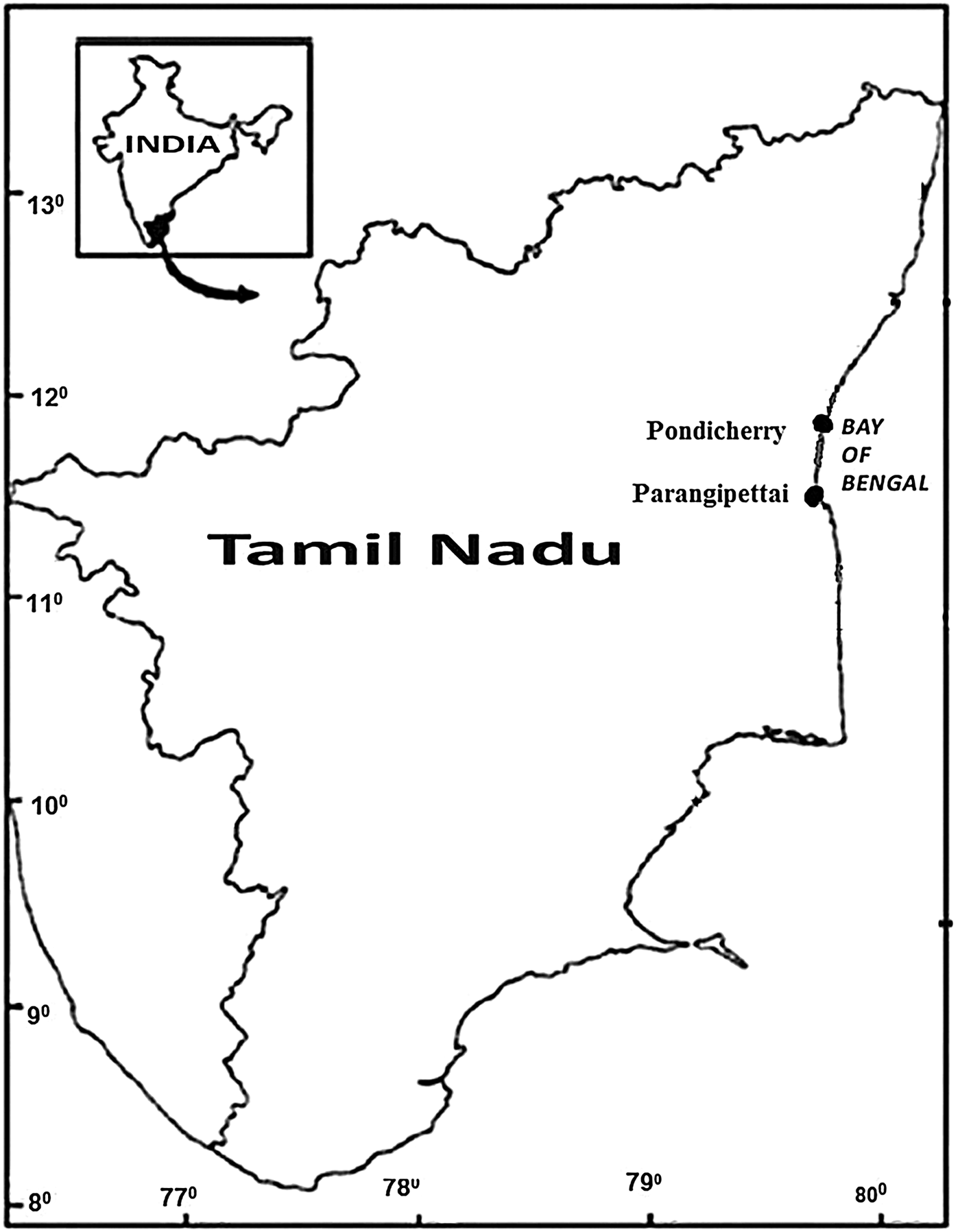
One specimen of each species of the abnormal fishes Epinephelus coioides and Cynoglossus cynoglossus was collected from the Parangipettai (11°29′N 79°47′E) and Nallavadu (11°46′03′′N 79°49′45′′E) landing centres, respectively, along the south-east coast of India (Figure 1). They were identified using a standard fish identification manual (Fischer & Whitehead, Reference Fischer and Whitehead1974; Fischer & Bianchi, Reference Fischer and Bianchi1984). The total length and weight of the fishes were recorded and the abnormalities were reported.
Study area map.

RESULTS AND DISCUSSION
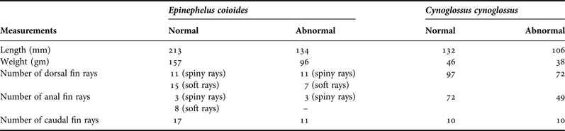
The total length and weight of the normal and abnormal species of Epinephelus coioides (Figure 2A, B) and Cynoglossus cynoglossus (Figure 3A, B) are given in Table 1.
(A) Normal and (B) abnormal Epinephelus coioides collected from the Parangipettai coast.
(A) Normal and (B) abnormal Cynoglossus cynoglossus collected from the Nallavadu coast.
Morphometric measurements of the normal and abnormal fishes collected from the Parangipettai and Nallavadu coasts.
SYSTEMATICS
Order PERCIFORMES
Family SERRANIDAE (Innamura & Yabe, 2002)
Genus Epinephelus (Bloch, 1793)
Epinephelus coioides (Hamilton, 1822)
Order PLEURONECTIFORMES
Family CYNOGLOSSIDAE
Genus Cynoglossus (Hamilton, 1822)
Cynoglossus cynoglossus (Hamilton, 1822)
Skeletal deformities are visible on the fish's body when compared to the normal fishes of the same species (Figures 2A & 3A). In both fishes, the caudal regions are completely deformed and fused. In general, the dorsal, anal and caudal fins of Epinephelus coioides are separated from one another, but in this abnormal E. coioides (Figure 2B) the dorsal fin, anal fin and caudal fins are joined together. The caudal region behind the eighth soft ray of the dorsal fin is fused and nearly one fourth of the total length of the body is completely deformed. In the abnormal Cynoglossus cynoglossus (Figure 3B), one fourth of the total length of the vertebral region is fused and approximately 3 cm of the caudal region is deformed. Nearly 25 dorsal rays and 23 anal rays are absent. This indicates that the caudal vertebrae may be completely deformed in these fishes.
Similar types of abnormalities have been reported by several authors. These include: the occurrence of various spinal deformations in a captive-bred wild line of Poecilia wingei, Poeser et al., 2005, described by Arbuatti et al., Reference Arbuatti, Salda and Romanucci2013; a partial tail in one specimen of the teleost fish Liza abu (Heckel, 1843) in Iran, reported by Jawad et al., Reference Jawad, Sadighzadeh and Valinassab2010; deformities in the last three thoracic and the first caudal vertebrae in Mugil cephalus Linnaeus, 1758, reported by Jawad, Reference Jawad2004.
The occurrence of compressed and fused vertebrae from the caudal to the dorsal fin, compression and ossification of the vertebral joints and excessive proliferation of collagen results in the increased thickness of the spinal column (Silverstone & Hammell, Reference Silverstone and Hammell2002). Spinal deformities may have a linkage with pollution (Couch et al., Reference Couch, Winstead and Goodman1977; Bengtsson, Reference Bengtsson1979). Abnormalities in fishes can have various causes, but are usually considered to originate from mutations and from the teratogenic effects of adverse environmental factors, such as mutagenic chemicals in the water (Longwell et al., Reference Longwell, Chang, Hebert, Hughes and Perry1992; Lien, Reference Lien1997). The coastal waters from Pondicherry to Parangipettai are also well known for being polluted by the presence of heavy metals and hydrocarbons in the water (Arockiamary et al., Reference Arockiamary, Vijayalakshmi and Balasubramanian2011; Prabhahar et al., Reference Prabhahar, Saleshraniand and Tharmaraj2011), in sediments (Veerasingam et al., Reference Veerasingam, Raja, Venkatachalapathy, Mohan and Sutharsan2010; Solai et al., Reference Solai, Suresh Gandhi and Sriram2010; Lyla et al., Reference Lyla, Manokaran and Khan2012) and in fishes and other marine organisms (Lakshmana Senthil et al., Reference Lakshmana Senthil, Ajith Kumar, Marudhu Pandi, Dhaneesh, Bala Murugan and Balasubramanian2012; Thiyagarajan et al., Reference Thiyagarajan, Dhaneesh, Ajith Kumar, Kumaresan and Balasubramanian2012; Jayaprabha et al., Reference Jayaprabha, Balakrishnan, Purusothaman, Indira, Srinivasan and Anantharaman2014). These pollutants could well affect the embryonic stage of these fishes and cause these types of abnormalities.
Other causes of fish deformities might include water temperature (Milton, Reference Milton1971), dissolved oxygen (Turner & Farley, Reference Turner and Farley1971), water surface tension (Setiadi & Tsumura, Reference Setiadi and Tsumura2007) or parasite infestations (Brown & Nunez, Reference Brown, Nunez, Leatherland and Woo1998). Vertebral deformities may affect the biology of the fish indirectly through inhibiting its free-swimming movements (Sadler, Reference Sadler1990; Jawad, Reference Jawad2004).
Any anomaly in the caudal fin will impair the flexibility of the tail, hindering the performance of the fish (including its capacity to get food and to avoid predators) (Jawad, Reference Jawad2004). However, it may also affect, to a certain limit, other aspects of performance such as reproduction success in species using fin display in courtship or intraspecific competition (De Girolamo et al., Reference De Giroloma, Lucini, Vega, Andreozzi, Coppola and Castaldo1999; Koumoundouros, Reference Koumoundouros2008). There are no previous reports of any abnormalities in these wild species off the south-east coast of India. An increased incidence of skeletal deformities among commercial fish species would suggest environmental deterioration and hence signal the need for prompt remedial action.
ACKNOWLEDGEMENTS
This work was supported by the DST-INSPIRE Fellowship (grant no. DST/INSPIRE Fellowship/2012/144) and the first author is grateful for this financial support.