Introduction
In birds, numerous oxidation reactions are continually taking place at the cellular and tissue level, resulting from catabolism and the activity of certain forms of immune cells. During normal physiological processes these are balanced out by a complex antioxidant mechanism aimed at minimising the generation of free radical species, thereby maintaining optimal redox balance. Partly responsible for these reactions are the antioxidant enzymes superoxide dismutase (SOD), glutathione peroxidase (PGX) and catalase (CAT). However, due to exposure to numerous stress-inducing and immunosuppressive factors and a high level of cellular metabolism resulting from intensive farming conditions (stocking density, changes in air temperature, epizootic agents and genetic potential), a very common phenomenon in large-scale poultry production is destabilisation of the redox balance towards excessive activity of reactive radical species, leading to oxidative stress (oxidation of lipids, proteins, DNA and sugars), which is an element in many diseases (Ognik and Wertelecki, Reference OGNIK and WERTELECKI2012). A significant role in the production of reactive oxygen species is attributed to the mitochondria. Functional damage to these organelles is linked to energy depletion and intensive production of reactive oxygen species, which leads to a vicious circle of cellular stress. Deterioration in mitochondrial metabolism can ultimately lead to necrosis of the liver cell and apoptosis, as well as impairment of heart function. The enzymes most commonly used in liver diagnostics are alanine aminotransferase (ALT), aspartate aminotransferase (AST), lactate dehydrogenase (LDH), gamma-glutamyl transferase (GGT), butyrylcholinesterase (BChE), acetylcholinesterase (AChE), alkaline phosphatase (ALP), sorbitol dehydrogenase (SORD), creatine kinase (CK), glucose-6-phosphate dehydrogenase (G6PD), hydroxybutyrate dehydrogenase (HBDH), acid phosphatase (AC) and ceruloplasmin. Certain enzymes included in standard liver profiles, such as ALT, AST, LDH, HBDH and CK, are used to evaluate heart function, in addition to Ca2+ATPase (Krauze, Reference KRAUZE2013).
Activity of antioxidant and liver profile enzymes in poultry can be affected by numerous factors, including age, sex, species, breed, nutrition, physiological state and farming techniques (Sembratowicz et al., Reference SEMBRATOWICZ, OGNIK, TRUCHLIŃSKI and MODZELEWSKA-BANACHIEWICZ2004; Brodacki et al., Reference BRODACKI, BATKOWSKA and ZADURA2006). There is high individual variation among birds, which means that results of enzyme activity analyses fall within broad ranges. This poses significant difficulties in establishing physiological norms for enzyme activity in this group of animals. It should be emphasised, however, that enzyme assays that are useful in the diagnosis of diseases and evaluating the effect of other factors on human and other mammalian organisms can also be used in poultry.
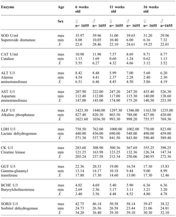
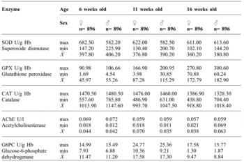
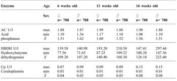
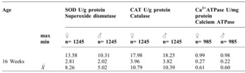
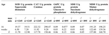
In order to properly interpret the results of enzymatic assays, they should be compared with the results of the same tests in birds from a healthy control group. The values presented in this paper are based on many years of research on healthy turkeys raised for slaughter (Tables 1-5) and other data presented in the literature (Ognik and Sembratowicz, Reference OGNIK and SEMBRATOWICZ2007; Ognik and Czech, Reference OGNIK and CZECH2010; Ognik and Sembratowicz, Reference OGNIK and SEMBRATOWICZ2011; Ognik and Krauze, Reference OGNIK and KRAUZE2012; Ognik and Wertelecki, Reference OGNIK and WERTELECKI2012; Czech et al., Reference CZECH, OGNIK and GRELA2012; Krauze, Reference KRAUZE2013; Ognik, Reference OGNIK2013; Ognik et al., Reference OGNIK, CZECH and STACHYRA2013; Czech et al., Reference CZECH, OGNIK, SEMBRATOWICZ and CHAŁABIS-MAZUREK2014; Ognik and Czech, Reference OGNIK and CZECH2014; Ognik et al., Reference OGNIK, SEMBRATOWICZ, CZECH, KULAK and MERSKA2015).
Activity of SOD, CAT, ALT, AST, AP, LDH, CK, GGT, BChE and SORD in the blood plasma of turkeys (Czech et al., Reference CZECH, OGNIK and GRELA2012; Ognik and Krauze, Reference OGNIK and KRAUZE2012; Krauze, Reference KRAUZE2013; Ognik, Reference OGNIK2013; Ognik et al., Reference OGNIK, CZECH and STACHYRA2013; Ognik and Czech, Reference OGNIK and CZECH2014).

Activity of SOD, PGX, CAT, AChE and G6PD in turkey erythrocytes (Ognik and Czech, Reference OGNIK and CZECH2010; Ognik and Sembratowicz, Reference OGNIK and SEMBRATOWICZ2011; Krauze, Reference KRAUZE2013; Ognik et al., Reference OGNIK, SEMBRATOWICZ, CZECH, KULAK and MERSKA2015).

Activity of AC, HBDH, and ceruloplasmins in the blood serum of turkeys (Krauze, Reference KRAUZE2013).

Activity of SOD, CAT and Ca2+ATPase in the pectoral muscle of turkeys (Ognik and Wertelecki, Reference OGNIK and WERTELECKI2012; Krauze, Reference KRAUZE2013; Ognik, Reference OGNIK2013; Czech et al., Reference CZECH, OGNIK, SEMBRATOWICZ and CHAŁABIS-MAZUREK2014).

Activity of SOD, CAT, G6PC, SDH and MDH in the liver of turkeys (Ognik and Wertelecki Reference OGNIK and WERTELECKI2012, Krauze Reference KRAUZE2013, Ognik Reference OGNIK2013, Czech et al., Reference CZECH, OGNIK, SEMBRATOWICZ and CHAŁABIS-MAZUREK2014).

Antioxidant enzymes profile
The antioxidant enzyme system in poultry is composed of specialised enzymes that catalyse reactions removing free radicals and preventing their generation. These enzymes include superoxide dismutase, (SOD), glutathione peroxidase (GPx) and catalase (CAT).
SOD is an enzyme which catalyses reactions that break down the superoxide anion radical to hydrogen peroxide and molecular oxygen (the dismutation reaction). SOD is a metalloenzyme and forms the main defence mechanism against the toxic effect of peroxides in cells. Apart from catalysing the dismutation reaction, SOD exhibits enzymatic activity in the case of other substrates. CuZnSOD has been shown to exhibit catalytic activity in the removal of singlet oxygen. This is highly significant because, as yet, no other enzyme has been detected that is able to eliminate singlet oxygen. The reaction is based on oxidation of histidine, tyrosine and methionine residues present in the enzyme molecule. SOD occurs in two forms: intracellular and extracellular. It is possible to distinguish three isoforms of superoxide dismutase. The intracellular form is located in the cytoplasm and its active centre contains copper and zinc (Cu/ZnSOD; SOD-1), as well as in the mitochondrial matrix, with manganese as the active centre (Mn-SOD; SOD-2). Extracellular superoxide dismutase SOD-3, located outside of the cells, also contains copper and zinc (Skrzycki and Czeczot, 2004).
Glutathione peroxidase (GPx) is a selenoenzyme that breaks down hydrogen peroxide, generated in the dismutation reaction, into water, at the same time transforming reduced glutathione (GSH) to its oxidized form (GSSG). GPx reduces organic peroxides (e.g. lipid peroxides) to alcohols. To prevent inactivation of proteins by GSSG, GPx interacts with glutathione reductase, an enzyme restoring the reduced from of glutathione at the expense of oxidation of NADPH, or GSSG is actively removed to the outside of the cell at the expense of energy accumulated as ATP. It is possible to distinguish four forms of glutathione peroxidase. The classical form, cGPx, occurs mainly in the erythrocytes and provides protection against oxidative stress, particularly hydrogen peroxide. GI-GPx is present in the digestive tract and the liver. Its task is providing protection against peroxides and xenobiotics. Plasma glutathione peroxidase (pGPx) is located mainly in the kidneys, and its function is defence of the extracellular space. The fourth form is phospholipid-hydroperoxide glutathione peroxidase (PHGPx), which protects cell membranes against lipid peroxidation (Łukaszewicz-Hussain, Reference ŁUKASZEWICZ-HUSSAIN2003).
Catalase (CAT) is an enzyme with two kinds of activity: catalase and peroxidase. The main function of CAT is participation in disproportionation of hydrogen peroxide if its concentration in the organism is very high (10-8 mol/l) and GPx is unable to break down the excess. The first stage of the reaction involves oxidation by hydrogen peroxide of catalase haem iron, causing the generation of porphyrin radical (compound A). Next, compound A is reduced by the next H2O2 molecule, generating molecular oxygen and water. Compound A returns to its initial state, in which iron has an oxidation state of 3. Catalase has limited peroxidase activity, where active compound A can oxidize substrates such as ethanol, methanol, phenol and formate. There are two types of catalase in mammalian cells: cytoplasmic (erythrocytes) and peroxisomal (Ścibior and Czeczot, Reference ŚCIBIOR and CZECZOT2006).
Many years of measurements of SOD activity in the blood, pectoral muscles and livers of turkeys show that SOD and CAT activity decrease slightly as turkeys age (Ognik et al., Reference OGNIK, CZECH and STACHYRA2013). In contrast, measurements of GPx activity in the blood of turkeys show that the activity of this enzyme increases with the age of turkeys. The increase in GPx activity with age can probably be explained by mobilisation of this enzyme to break down endogenous hydrogen peroxide, which increases with age (Drobek-Słowik et al., Reference DROBEK-SŁOWIK, KARCZEWICZ and SAFRANOW2007).
In order to properly interpret the effect of a factor (e.g. viral or bacterial infection, changes in diet or increased stress) on the antioxidant response in an organism, all of the attendant enzymes (SOD, GPx and CAT) should be determined, as they form a set of proteins interacting in successive reactions breaking down radicals and peroxides. It should be emphasised that antioxidant enzyme activity varies under the influence of the same factor action on an organism. This probably depends in part on the initial redox status of the organism.
There are reports indicating that factors inducing oxidation reactions in the organism, such as viral infections or diseases, can reduce the activity of antioxidant enzymes. In the liver tissue and brain of chickens infected with NDV (Newcastle disease), a decrease in SOD, GPx and CAT activity was observed in contrast with healthy control chickens. Experiments on chickens infected with NDV showed that a vitamin E feed supplementation reduced oxidative stress induced by NDV infection, which was manifested as a smaller decrease in the activity of antioxidant enzymes than in the NDV-infected group that did not receive vitamin E (Kadiam et al., Reference KADIAM, SUBBAIAH and RANIPRAMEEL2011). In the blood of chickens infected with Eimeria acervulina oocysts, a decrease in SOD activity and an increase in CAT activity were noted (Koinarski et al., Reference KOINARSKI, GEORGIEVA, GADJEVA and PETKOV2005). In sick animals, oxidative stress and phagocytosis are continually induced, during which production of radicals and peroxides increases. In pathological states there is an increase in the mobility of antioxidant enzymes, whereby their activity may increase in the initial period of infection. As antioxidant enzymes exploit low-molecular-weight antioxidants (glutathione) and trace elements (Zn, Cu, Fe, Mn, Se) in reactions breaking down radicals and peroxides (Kędzierska et al., Reference KĘDZIERSKA, BOBER, KWIATKOWSKA, STACHOWSKA, OLSZEWSKA, CHLUBEK and CIECHANOWSKI2008), a decrease in antioxidant enzyme activity is observed when reserves of low-molecular-weight antioxidants that are cofactors of these enzymes are depleted, which may be manifested in a later period of a disease. There are reports indicating that short-term stress induced in turkeys (crowding, decrease in ambient temperature or changes in light) caused an increase in SOD and CAT activity (Ognik et al., Reference OGNIK, SEMBRATOWICZ, CZECH, KULAK and MERSKA2015). Probably the duration of the stress was not long enough to cause a very large quantity of radicals to be generated in the bird. The increase in SOD and CAT activity may have been the effect of rapid mobilisation of enzymes to break down the radicals and peroxides generated. The short-term stress probably did not cause a decrease in the content of the low-molecular-weight antioxidants necessary for SOD, GPx and CAT reactions, and so no decrease in their activity was observed.
Analysis of antioxidant enzyme activity is very often used to evaluate the effect of various feed supplements (including antioxidants) on the redox status of birds. The response of the first line of antioxidant defence, i.e. antioxidant enzymes, to a dietary factor depends on the initial redox state of the organism, the amount of supplement (antioxidant) introduced, and the duration and frequency of administration of the supplement (Ognik et al., Reference OGNIK, CZECH and STACHYRA2013). An increase in SOD activity accompanied by no change or a reduction in PGX and CAT activity may indicate positive stimulation of SOD activity, which means stimulation of enzymatic defences. An increase in SOD activity in turkey blood with no effect on PGX and CAT activity was noted when the birds’ drinking water was supplemented with an aloe vera preparation containing natural antioxidants (Ognik and Czech, Reference OGNIK and CZECH2010) or the synthetic derivative 5-oxo-1,2,4-triazine (Ognik and Sembratowicz, Reference OGNIK and SEMBRATOWICZ2011). In a study on turkeys receiving Bioaron C (aloe vera and chokeberry extract) in drinking water, an increase was noted in SOD, PGX and CAT activity in the blood (Ognik and Sembratowicz, Reference OGNIK and SEMBRATOWICZ2007). An increase in SOD and CAT activity in the blood plasma and pectoral muscle of turkeys was noted when linseed oil was added to their feed mixture (Czech et al., Reference CZECH, OGNIK, SEMBRATOWICZ and CHAŁABIS-MAZUREK2014). A study on turkeys receiving natural antioxidants in their feed found decreased SOD and CAT activity in the blood plasma, which indicated that the birds were free of oxidation reactions (Ognik et al., Reference OGNIK, CZECH and STACHYRA2013). Similar positive results, i.e. a reduction in SOD and CAT activity in the pectoral muscle and liver of turkeys, were noted as a result of administration of natural RRR-d-alpha-tocopherol in their feed (Ognik and Wertelecki, Reference OGNIK and WERTELECKI2012). A reduction in SOD and CAT activity in the blood of turkeys was also noted following dietary supplementation with natural plant antioxidants (Ognik et al., Reference OGNIK, CZECH and STACHYRA2013), mannan-oligosaccharides (Ognik and Krauze, Reference OGNIK and KRAUZE2012), and protein-xanthophyll concentrate (PX) from alfalfa (Czech et al., Reference CZECH, OGNIK and GRELA2012). A reduction in CAT activity was observed in the blood of turkey hens receiving feed with linseed oil and natural RRR-d-alpha-tocopherol (Ognik and Czech, Reference OGNIK and CZECH2014).
It should be emphasised that dietary enhancement of antioxidant potential can only achieve the desired effect when a suitable amount of the antioxidant is selected. Excessive doses of an antioxidant or dietary supplement susceptible to oxidation processes can have the reverse effect - instead of acting as antioxidants, they become pro-oxidants (Gryszczyńska and Iskra, Reference GRYSZCZYŃSKA and ISKRA2008). A lack of effect in in vivo conditions with antioxidants with confirmed activity in vitro may be explained by the biotransformation mechanism of these compounds in the bird, during which hydroxyl groups (responsible for antioxidant activity) become conjugated with endogenous compounds, forming conjugates with glucuronic acid, glutathione, and sulphates. For this reason the in vivo activity of antioxidants may be only partly preserved. What effect of a dietary supplement will have (as an antioxidant or pro-oxidant) and for how long will depend on the activity of antioxidant enzymes (Gryszczyńska and Iskra, Reference GRYSZCZYŃSKA and ISKRA2008). To properly evaluate the cause of an increase or decrease in antioxidant enzyme activity, it is necessary to determine indicators of lipid oxidation processes (LOOH - lipid peroxides - and MDA - malondialdehyde).
Enzymes of the hepatic and cardiac profile
Sensitive indicators of liver and heart function in poultry are intracellular enzymes that enter the blood due to cell damage or death. The following liver profile enzymes are used in diagnostics.
Indicator enzymes are not present in the blood or their level of activity is low. They enter the blood following cell damage. These enzymes may be organ-specific - i.e. sorbitol dehydrogenase (SORD) and creatine kinase (CK) - or non-specific, e.g. glycolysis enzymes - lactate dehydrogenase (LDH) and its isoenzyme hydroxybutyrate dehydrogenase (HBDH). They also include enzymes of the pentose phosphate cycle - glucose-6-phosphate dehydrogenase (G6PD); Krebs cycle enzymes - succinate dehydrogenase (SDH) and malate dehydrogenase (MDH); and enzymes of protein metabolism - alanine aminotransferase (ALT), aspartate aminotransferase (AST) and gamma-glutamyl transferase (GGT). Cytoplasmic indicator enzymes such as alanine aminotransferase (ALT), aspartate aminotransferase (AST), gamma-glutamyl transferase (GGT), sorbitol dehydrogenase (SORD) and lactate dehydrogenase (LDH) appear in the blood after even slight damage to cellular membranes. In contrast, enzymes associated with the endoplasmic reticulum, mitochondria, ribosomes and lysosomes, such as glutamine dehydrogenase (GLDH) and acid phosphatase (AC), appear in the bloodstream only in the case of very severe liver damage.
Secreted enzymes are synthesized in the ribosomes of cells, mainly of the liver, and then secreted into the blood. Their activity in the blood serum decreases as a result of hepatocyte damage: e.g. cholinesterase (BChE and AChE).
Excreted enzymes are present in small quantities in the blood. They are excreted by the liver into the bile, so in the case of liver damage or impaired bile flow their concentration in the blood increases: e.g. alkaline phosphatase (ALP), acid phosphatase (AC) and ceruloplasmin (Cp).
The following cardiac profile enzymes are used in diagnostics, and include the indicator enzymes creatine kinase (CK), beta-hydroxybutyrate dehydrogenase (HBDH), lactate dehydrogenase (LDH), Ca2+ATPase, alanine aminotransferase (ALT) and aspartate aminotransferase (AST).
Enzymes commonly used in poultry diagnostics
Aspartate aminotransferase (AST) is an enzyme present mainly in skeletal muscles, heart muscle, the liver and brain. Many years of measurements of AST activity in turkey blood have shown that the activity of this enzyme increases with the age of the birds.
Alanine aminotransferase (ALT) is mainly present in the liver and, in smaller amounts, in the kidneys and muscles.
Alkaline phosphatase (ALP) is found in blood cells, the epithelium of bile ducts, the intestinal mucous membrane and the kidneys. Many years of measurements of ALP activity in turkey blood have shown that the activity of this enzyme decreases with the age of the birds.
Lactate dehydrogenase (LDH) is located in the liver, heart, skeletal muscles, kidneys and lungs. Many years of measurements of LDH activity in turkey blood have shown that the activity of this enzyme increases up to the 11th week of life and then declines, attaining a level similar to that seen in the early rearing period (6 weeks of age).
Determination of the activity of ALT, AST and LDH in the blood (serum or plasma) shows the degree of permeability of the cell membrane, e.g. in hepatocytes, in the disease process. ALT, AST and LDH, as cytoplasmic enzymes, are sensitive indicators of disease processes because they appear in the blood even when damage to the cell membrane is slight. Increased activity of enzymes in the plasma/serum indicates cell damage rather than impaired organ function. In cases of acute liver damage aminotransferase activity may increase 50- or 100-fold. The De Ritis ratio (AST/ALT) is used in diagnostics whereby in acute cases the De Ritis ratio is more than 1, while in chronic cases it does not exceed 1. Cytosolic AST appears in the plasma during the course of cardiac pathology and liver damage, after which the mitochondrial form of the enzyme appears.
Penetration of the mitochondrial form of AST through the membrane is associated with the loss of oxidative phosphorylation capacity and leads to cell death. The cell membrane is believed to be more susceptible to long-term damage than the mitochondrial membrane (James et al., Reference JAMES, MAYEUX and HINSON2003). In the case of monensin poisoning (ionophore coccidiostats) in turkeys, AST activity may increase even 20-30 times, while ALT activity can increase as much as 11 times. In the case of E. coli infection in seven week-old turkeys AST may increase by 30% (Koncicki and Krasnodębska-Depta, Reference KONCICKI and KRASNODĘBSKA-DEPTA2005). LDH activity increases in all disease states with tissue necrosis, during histomoniasis in turkeys, and in infectious diseases - IBD, HHS and E. coli infection (Koncicki et al., Reference KONCICKI, BUKOWSKA, MAZUR-GONKOWSKA, KRASNODEBSKA-DEPTA and STENZEL2006). In the case of E. coli infection in seven week-old turkeys, LDH activity can increase even two-fold. In the case of histomoniasis in nine week-old turkeys, LDH activity can increase as much as eight times (Koncicki et al., Reference KONCICKI, BUKOWSKA, MAZUR-GONKOWSKA, KRASNODEBSKA-DEPTA and STENZEL2006).
Analysis of AST, ALT and LDH activity is very often used in experiments conducted on turkeys and chickens in order to assess the effect of various feed supplements on the liver profile of the birds. A lack of effect on AST, ALT and LDH activity or a decrease in their activity indicates that the supplement does not interfere with liver function (Czech and Ognik, Reference CZECH and OGNIK2014).
Determination of ALP activity in the blood makes it possible to diagnose cholestatic liver disease, which is usually linked to bile duct damage. An increase in ALP activity has been observed during the course of rickets and osteomalacia, aflatoxin poisoning in chickens and ducks and bone cancer. On the other hand, in seven week-old turkeys infected with E. coli there is an approximately two-fold decrease in ALP (Koncicki and Krasnodębska-Depta, Reference KONCICKI and KRASNODĘBSKA-DEPTA2005).
Enzymes not commonly used in poultry diagnostics
Gamma-glutamyl transferase (GGT) is an enzyme present in the cells of many organs, including the liver, kidneys, heart and other muscles. The highest concentration of this protein is seen in the cell membranes of hepatocytes and nephrocytes and in cells of the epithelium of the bile duct, from where it is released in the case of damage (SriRajaskanthan and Preedy, Reference SRIRAJASKANTHAN and PREEDY2006). In birds, high activity of GGT may indicate liver disease or impaired bile flow (Kramer and Hoffmann, 1997). According to Alonso-Alvarez et al. (Reference ALONSO-ALVAREZ, PEREZ and VELANDO2007), activity of this enzyme increases in female birds during laying time, clearly reflecting an increase in metabolism in the liver during laying. Handler et al. (Reference HANDLER, GENELL and GOLDSTEIN1994) report that in advanced age bile flow decreases, which is manifested as decreased GGT activity in the blood. According to Mokondjimobe et al. (Reference MOKONDJIMOBE, LONGO-MBENZA, AKIANA, NDALLA, DOSSOU-YOVO, MBOUSSA and PARRA2012), the enzyme's activity decreases in stress conditions, which is correlated with an increase in the aminotransferases AST and ALT (Won et al., Reference WON, KIM, ANNAMALAI, SHUNMUGAVEL, SINGHM and SINGH2013).
Acetylcholinesterase (AChE) is present in the serum, erythrocytes, liver, pancreas, and other tissues, but in erythrocytes it is the enzyme with the highest esterolytic activity. Catalysing hydrolysis of acetylcholine, AChE controls metabolic pathways associated with the immune and inflammatory response (Kawashima and Fujii, Reference KAWASHIMA and FUJII2003; Inkson et al., Reference INKSON, BRABBS, GREWAL, SKERRY and GENEVER2004). A decrease in AChE activity may indicate exposure to a toxic agent, and according to Pal et al. (Reference PAL, KUSHWAH and KUSHWAH1990) AChE activity may therefore be a useful biochemical marker for diagnosis of toxic conditions in poultry. Decreased activity of this enzyme has been observed in chickens intoxicated with chloro-organic compounds (Garg et al., Reference GARG, PAL, JHA and JADHAO2004). Neeraj et al. (2012) demonstrated that AChE activity decreased after stress. Bukowska et al. (Reference BUKOWSKA, PIENIĄŻEK, HUTNIK and DUDA2007) report that a decrease in AChE activity may result from direct binding of the enzyme with a xenobiotic, modification of erythrocyte membranes, changes in the electric charge of cell membranes or an increase in the concentration of reactive oxygen species. At the same time, environmental toxins can block AChE nicotinic receptors, thereby inhibiting the flow of calcium through ion channels. Large quantities of generated H2O2 act as AChE inhibitors (Schallreuter et al., Reference SCHALLREUTER, ELWARY, GIBBONS, ROKOS and WOOD2004).
Butyrylcholinesterase (BChE) plays a significant role in lipoprotein metabolism, maintaining myelin structure and cell adhesion, and neurogenesis. Multiple measurements of BChE activity in the blood plasma of turkeys shows that activity of this enzyme increases slightly with the age of the birds. It is considered a detoxifying enzyme, which, owing to its esterase activity, hydrolyses hydrophilic and hydrophobic compounds containing an ester of carboxylic or phosphoric acid (Çokuğraş, Reference ÇOKUĞRAŞ2003). Estimation of BChE activity in the blood plasma is useful in diagnosis of hepatobiliary diseases (Ogunkeye et al., 2006). According to Meslam et al. (Reference MESLAM, GUILLOZET, SHAW and QUINN2002), AChE activity decreases with age, while BChE activity increases. A decrease in BChE activity is observed in cases of liver dysfunction (Ogunkeye et al., 2006), while an increase in the activity of this enzyme may occur in the case of fatty liver disease (Koda et al., Reference KODA, KAWAKAMI, MURAWAKI and SENDA2007). In a study with turkeys, caffeine alkaloid was observed to cause a harmful reduction in AChE activity and an increase in that of BChE. At the same time it was observed that such changes in activity can be eliminated by adding L-carnitine and choline, either together or separately (Krauze, Reference KRAUZE2013). Garg et al. (Reference GARG, PAL, JHA and JADHAO2004) observed a decrease in AChE activity in chickens following administration of a xenobiotic in the form of a pesticide. These authors also noted similar values for activity of this enzymatic protein to those obtained during our own long-term research.
Sorbitol dehydrogenase (SORD) is present mainly in the cytoplasm of hepatocytes, and in smaller quantities in the cells of the kidneys, spleen and muscles. SORD transforms sorbitol to fructose in the cell (Witmer et al., Reference WITMER, VRENSEN, VAN NOORDEN and SCHLINGEMANN2003). This enzyme is a highly selective indicator of hepatocyte damage and can be a useful marker in evaluating the function of the liver, for which it is specific. The activity of this enzyme increases in the case of toxic liver damage and may indicate liver dysfunction, and also increases in the initial stage of viral hepatitis; therefore it can be used in diagnosing toxic conditions in poultry. A flaw of this parameter is the rapid decrease in SORD activity in comparison with ALT and AST. Activity decreases as the clinical state improves. Glutathione may be an inhibitor of the enzyme (Divisi et al., Reference DIVISI, DI TOMMASO and SALVEMINI2006; Grucka-Mamczar et al., Reference GRUCKA-MAMCZAR, BIRKNER, ZALEJSKA-FIOLKA, KASPERCZYK, KASPERCZYK, STAWIARSKA-PIETA, BIRKNER and POLONIAK2007). In a study with turkeys the addition of a caffeine alkaloid caused a decrease in SORD activity. The alkaloid may have induced oxidative stress reactions in the liver cells of the turkeys (Krauze, Reference KRAUZE2013).
Malate dehydrogenase (MDH) in the Krebs cycle catalyses oxidation of L-malate to oxaloacetate, which can acquire the next acetyl-CoA residue. MDH transfers hydrogen protons to nicotinamide adenine dinucleotide NAD+, creating its reduced form NADH. Some authors claim that the activity of this enzyme increases under the influence of oxidative stress, so that analysis of MDH activity can be used to estimate stress levels in poultry (Neeraj et al., 2012). It is possible that stress is an inductor of this enzyme, which is linked to intensification of Krebs cycle metabolism due to increased demand for energy during stress.
Acid phosphatase (AC) occurs mainly in the lysosomes, but also in the erythrocytes, blood serum, platelets, marrow, bones, kidneys and liver. It is an indicator enzyme of lysosomes, where proteolysis of protein taken in with food occurs. Analysis of AC activity can be used to evaluate poisoning with xenobiotics in poultry, when acid phosphatase activity increases. Furthermore, analysis of AC activity can be used to estimate the rate of protein metabolism. AC protects the structure and functioning of the cell within the lysosome in conditions of protein deficiency (Pillay et al., Reference PILLAY, ELLIOT and DENNISON2002).
Glucose-6-phosphate dehydrogenase (G6PC) is widely distributed in animal tissues, mainly in the liver and kidneys, where it catalyses dephosphorylation of glucose 6-phosphate to glucose in glycogenolysis, making the energy contained in glycogen available (Ghosh et al., Reference GHOSH, SHIEH, PAN and CHOU2004). G6PC is responsible for detoxification, and glucose released into the bloodstream is an inhibitor of this enzyme. Measurements of G6PC activity in the blood of turkeys show that the activity of this enzyme increases up to about the 11th week of life, after which it decreases, and in the 16th week attains a level similar to that noted in the early rearing period (six weeks of age). A significant influence of age on the reduction in this enzyme's activity was clearly demonstrated by Plewka et al. (Reference PLEWKA, KAMIŃSKI, PLEWKA and NOWACZYK2000), who suggested that this can be explained by ageing of the endoplasmic reticulum, as evidenced by lipid peroxidation and increased synthesis of free radicals. The highest G6PC activity is observed during the middle of the rearing period (Łukaszewicz-Hussain et al., 2004; Plewka et al., Reference PLEWKA, PLEWKA and IHNATOWICZ2006), after which it decreases (Foye et al., Reference FOYE, UNI, MCMURTRY and FERKET2006). G6PC activity increases with age in rapidly growing birds, which can be explained by increased oxidative stress. Oxidative stress may cause a decrease in the level of glucose, which is an inhibitor of this enzyme (Dziewulska-Szwajkowska et al., Reference DZIEWULSKA-SZWAJKOWSKA, ŁOZIŃSKA-GABSKA, ADAMOWICZ, WOJTASZEK and DZUGAJ2003, Sangiao-Alvarellos et al., Reference SANGIAO-ALVARELLOS, GUZMAN, LAIZ-CARRION, MÍGUEZ, DEL RÍO, MANCERA and SOENGAS2005). Moreover, as birds age a decrease is observed in the blood glucose level, which contributes to a decrease in the activity of this enzyme (Krauze Reference KRAUZE2013). Glucose-6-phosphate dehydrogenase (G6PD) is an enzyme which during the pentose cycle catalyses oxidation of glucose 6-phosphate to 6-phosphogluconolactone. The results of studies by Ordonez et al. (Reference ORDONEZ, ROSETY-PLAZA and ROSETY-RODRIGUEZ2006) and Egesie et al. (Reference EGESIE, GESIE, ISIGUZORO and EGESIE2008) show that G6PD activity can vary widely, depending on breed, age and sex. According to many authors it is the only enzyme supplying NADPH that is activated during stress (Tripathi et al., 2004; Kotaka et al., Reference KOTAKA, GOVER, VANDEPUTTE-RUTTEN, AU, LAM and ADAMS2005; Neeraj et al., 2012). Łukaszewicz-Hussain et al. (2004) suggested that this enzyme plays a key role in the body's antioxidant defence, and its activity increases during enhanced phagocytosis. In their opinion, increased G6PD activity is a defence mechanism induced by stress, and a return of G6PD to normal activity is observed when the glutathione level increases. The activity of this enzyme increases considerably during stress, accompanied by a decrease in concentration of malondialdehyde (Hongu and Sachan, Reference HONGU and SACHAN2000). The activity of this biocatalyst can increase during increased demand for NADPH, which was observed during increased lipid anabolism (synthesis of fatty acids, cholesterol and other steroids, and sphingolipids). NADPH deficiency in the erythrocytes shortens the survival time of these blood cells. According to Divisi et al. (Reference DIVISI, DI TOMMASO and SALVEMINI2006), a state of reduced G6PD activity means a certain degree of deficiency of this enzyme appearing with age. Decreased activity of this enzyme causes NADPH oxidase activity to increase in order to produce peroxide anions, which, in the dismutase reaction, are transformed to hydrogen peroxide, necessary for phagocytosis (Das et al., Reference DAS, PAL, CHAKRABORTY, MANUSH, CHATTERJEE and APTE2006). Decreased activity of enzymes associated with generation of NADPH, i.e. G6PD and MDH, can directly lead to synthesis of fatty acids in subcutaneous fat tissue and cause carbon chain elongation, limiting the process of synthesis of these compounds (Xu et al., Reference XU, WANG, MAO, ZHAN and HU2003).
Measurement of the activity of succinate dehydrogenase (SDH) in the liver can be used to evaluate mitochondrial function. SDH activity may be modulated by several activators and inhibitors. Xenobiotics that disrupt the tricarboxylic acid cycle may inhibit succinate dehydrogenase, and impairment of the cycle initiates a reduction in energy production in the cell. This may result in the generation of reactive oxygen species and disrupt the bioenergetic balance of the cell, which can lead to its apoptosis or necrotic death. In older birds activity of this enzyme is lower than in younger individuals. Stress, e.g. in the form of low ambient temperature, does not cause changes in the activity of this biocatalyst (Schelling et al., Reference SCHELLING, MURRAY, YOO, ROW, CUSACK, CAPALDI and GIBSON2006).
Ceruloplasmin (Cp), also called ferroxidase or multicopper oxidase, is produced mainly by hepatocytes of the liver. Cp oxidizes toxic Fe2+ ions to non-toxic Fe3+ ions. Cp is an acute phase protein, which means that its concentration increases in response to inflammation, thereby restoring homeostasis, i.e. systemic balance. An increase in Cp is generally correlated with an increase in copper in the blood serum (Bento et al., Reference BENTO, PEIXOTO, ZAITSEV and LINDLEY2007). A study by Starcher and Hill (Reference STARCHER and HILL1965) showed that Cp activity in chickens does not exceed 1 U/l. Analysis of Cp in the blood serum of turkeys has confirmed that Cp activity in poultry (in this case turkeys) was lower than 1 U/l, and its activity increases with the age of the birds. According to Starcher and Hill (Reference STARCHER and HILL1965), Cp activity in poultry increases under the influence of stress factors inducing an increase in steroid hormones (e.g. corticosterone), as well as in pathological states, as indicated by an increase in Cp in chickens infected with S. gallinorum.
Calcium ATPase (Ca2+ATP) is located in the endoplasmic reticulum membrane of the heart. It is responsible for removing excess Ca2+ ions from the cytosol to the outside of the cell against the concentration gradient. This process takes place at the expense of energy acquired from ATP hydrolysis. ATPase activity is inhibited in stress conditions. A high concentration of amphiphilic compounds, such as lipids, can disrupt cell membrane structure, ion transport and ATPase activity (Ma et al., Reference MA, SUMBILLA, FARRANCE, KLEIN and INESI2004). According to Gryzińska et al. (Reference GRYZIŃSKA, KRAUZE and STRACHECKA2012), activity of Ca+2ATPase is inhibited as birds age. Calcium ions serve as a transmitter of an intracellular signal, and a reduction in Ca2+-ATPase activity and a pathological increase in the concentration of these ions in the cytoplasm may induce a non-specific response of the target cell, e.g. disruption of heart function (Ma et al., Reference MA, SUMBILLA, FARRANCE, KLEIN and INESI2004).
Creatine kinase (CK) is an enzyme occurring in the muscles. CK plays a key role in cellular bioenergetics and homeostasis of cellular ATP, where it catalyses a reversible transfer of a phosphate group, thereby regulating ATP synthesis. Huff et al. (Reference HUFF, HUFF, RATH, ANTHONY and NESTOR2008) claim that CK can be used as a marker indicating the degree of susceptibility to stress, and suggest that resistance to this factor in some genetic lines is manifested as high permeability of muscle cell membranes (Huff et al., Reference HUFF, HUFF, RATH, ANTHONY and NESTOR2008). Nicholson et al. (Reference NICHOLSON, LOCHMILLER, STEWART, MASTERS and LESLIE2000) demonstrated that CK activity in turkeys increases in stress situations, e.g. when the birds are handled, and suggested that sensitive individuals in the flock can be identified by measuring the activity of this enzyme. In studies on chickens, Sandercock et al. (Reference SANDERCOCK, HUNTER, NUTE, MITCHELL and HOCKING2001) and Sandercock and Mitchell (Reference SANDERCOCK and MITCHELL2003) showed that stress causes an increase in CK activity, which was correlated with disturbance of intracellular Ca2+ homeostasis in the muscles and with acid-alkaline imbalance. This may lead to damage to the skeletal muscles or myopathy, resulting from disturbances in the function of muscle cell membranes and their permeability (Sandercock et al., Reference SANDERCOCK, HUNTER, NUTE, MITCHELL and HOCKING2001, Sandercock and Mitchell Reference SANDERCOCK and MITCHELL2003). In the case of monensin poisoning in seven week-old turkeys, CK activity can increase four-fold (Koncicki and Krasnodębska-Depta, Reference KONCICKI and KRASNODĘBSKA-DEPTA2005).
Beta-hydroxybutyrate dehydrogenase (HBDH) is a considerably more specific indicator enzyme of damage to the cardiomyocytes than LDH. Muscle cell damage may appear when products of energy metabolism are accumulated and there is a local drop in pH in the myocytes. This leads to inactivation of mitochondria and cell membrane damage, as well as a sharp increase in the activity of HBDH, CK and LDH (Dembińska-Kieć et al., 2009). Disruption of biochemical transformations in the muscle cells of birds can have a negative effect on meat quality after slaughter.
Conclusions
The results presented in this review concerning the activity of selected enzymes are the effect of many years of research on healthy turkeys. Despite the high level of individual variation in turkeys, studies have made it possible to establish ranges of activity for these enzymes that can be considered physiologically normal for these birds. Measuring the activity of these enzymes may be very useful in determining the direction of redox reactions and in evaluating the health of turkeys. Analysis of the activity of these enzymes may be of great importance in determining whether oxidative stress reactions are induced in cells and which cells or organs have undergone damage.
During oxidative stress in cells, which leads to cell damage or organ dysfunction (of the liver or heart), there is generally an increase in the activity of AST, ALT, ALP, LDH, GGT, SORD, MDH, AC, G6PC, G6PD, Cp, CK and HBDH and a decrease in the activity of SOD, GPx, CAT, AChE, BChE, SDH and Ca2+ATPase.