Introduction
Dogs harbour a variety of intestinal parasites, some of which can also infect humans. In view of this, some of the dog parasites, such as Toxocara canis and Ancylostoma sp., are reported to be a significant public health problem, especially in developing countries and communities that are socio-economically disadvantaged (Craig & Macpherson, Reference Craig, Macpherson, Macpherson, Meslin and Wnadeler2000). In these communities, poor levels of hygiene and overcrowding, together with lack of veterinary attention and zoonotic awareness, exacerbate the risk of disease transmission (Hinz, Reference Hinz1980; Malgor et al., Reference Malgor, Oku, Gallardo and Yarzabal1996).
It is common to observe intestinal parasites in canines of all ages, but the prevalence of infection is usually high in puppies, mainly due to the fact that certain modes of transmission are exclusive to the newly whelped or neonates, and also because young dogs have not yet acquired immunity to parasites (Bowman, Reference Bowman1999; Ramirez-Barrios et al., Reference Ramirez-Barrios, Barboza-Mena, Munoz, Angulo-Cubillan, Henandez, Gonzalez and Escalona2004). The clinical signs of parasitic infection vary and occasionally some infected animals will be asymptomatic. However, severe clinical cases in young dogs will lead to diarrhoea, anaemia and sometimes death (Georgi & Georgi, Reference Georgi and Georgi1994; Reinemeyer, Reference Reinemeyer, Kirk and Bonagura1997; Bowman, Reference Bowman1999).
Information on the epidemiology of the parasite infections in a specific canine population is a useful tool for the veterinarian practitioner when he/she has to provide a clinical diagnosis (Smith, Reference Smith1991; Nolan & Smith, Reference Nolan and Smith1995). Several epidemiological studies of intestinal parasites in dogs have been carried out in Nigeria and other parts of the world. However, information on the intensity of infections is particularly scarce. The purpose of this study was to determine the prevalence and intensity of gastrointestinal helminth parasites of dogs brought for care at two veterinary clinics at Ibadan.
Materials and methods
Faecal samples were collected from a total of 959 dogs comprising 493 males and 466 females, from two veterinary clinics in Ibadan between January 2001 and December 2002. All faecal samples were obtained from privately owned dogs taken to the two veterinary clinics at Ibadan, for either a medical problem or for routine examination. Faecal samples were collected directly from the dog's rectum and placed in previously labelled, sterile 30 ml bottles. Information was collected on the dog's approximate age, sex, breed and mode of life. Each faecal sample collected was mixed thoroughly with 10% aqueous formaldehyde for preservation.
Faecal samples were processed in the laboratory by means of the modified Kato–Katz procedure (Forrester & Scott, Reference Forrester and Scott1990) and examined for helminth eggs using an optical microscope. Each observed helminth egg was identified using established structural and morphometric criteria (Bowman, Reference Bowman1999). In addition to qualitative diagnosis, an indirect measure of helminth intensity was obtained by counting eggs, expressed as eggs/gram of faeces (epg).
Statistical analysis
Statistical tests were performed using SPSS 11.0 (SPSS Inc., Chicago, Illinois, USA). Chi-squared tests were used to study the relationship between parasite prevalence and host age, sex and breed. Mann–Whitney U tests were used to explore the relationship between the egg intensity and sex, while Kruskal–Wallis tests were used for variables with more than two levels, i.e. the relationship between the egg intensity and host age and breed.
Results
The eggs of six helminth species were observed in this study. The prevalence and intensity of the different worms are shown in tables 1 and 2. The overall prevalence of infection with intestinal helminth parasites was 24.7%. The most frequently observed intestinal helminth parasite in this study was Ancylostoma spp. (17.9%) followed by T. canis (9.0%). The prevalences for the other four worms (Toxascaris leonina, Trichuris vulpis, Dipylidium caninum and Uncinaria stenocephala) were low and varied from 0.6% to 0.2% (table 1). The overall mean intensities (epg) for the various helminth species were 462.0 ± 100.5 for T. canis, 54.1 ± 8.6 for Ancylostoma spp., 0.8 ± 0.4 for T. leonina, 0.1 ± 0.0 for T. vulpis, 0.2 ± 0.1 for D. caninum and 1.0 ± 0.7 for U. stenocephala (table 2). The prevalence of intestinal parasites was significantly higher in Mokola Clinic (39.8%) than in Bodija Clinic (12.8%) (P < 0.05).
Prevalence (%) of helminth eggs in faeces of dogs in Mokola and Bodija clinics.

Intensity (I) of helminth eggs in faeces of dogs in Mokola and Bodija clinics.

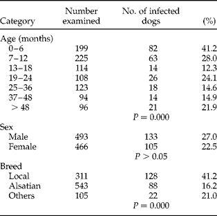
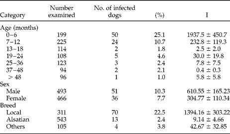
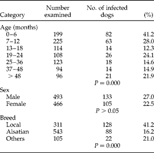
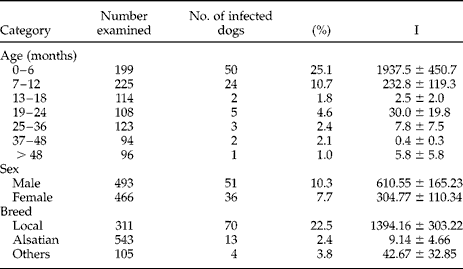
When the prevalence of intestinal helminth parasites was analysed by age, results showed that young dogs ( < 12 months old) were more infected than older ones. The highest prevalence of intestinal parasites was observed in the 0–6 months age group and was significantly higher than other, older ones. There was no significant difference observed in the overall prevalence of intestinal parasites between the male (27.0%) and female dogs (22.5%) (P>0.05) (table 3). Similarly, for T. canis infection, the prevalence was significantly higher (P < 0.05) in dogs < 6 months old when compared with older dogs (table 4). No significant difference in the prevalence of T. canis was observed between male (10.3%) and female (7.7%) (χ2 = 1.993, df = 1, P>0.05) dogs (table 4). The intensity of T. canis infection in young dogs (0–6 months) was significantly higher (P < 0.05) than in older age groups; however, there was no significance difference in the intensity between male and female dogs (U = 732.5, df = 1, P>0.05) in males (610.55 ± 165.23) than in female dogs (304.77 ± 110.34) (table 4).
Prevalence (%) of intestinal parasites of dogs relative to host age, sex and breed.
Prevalence (%) and intensity (I) of Toxocara canis in dogs relative to host age, sex and breed.
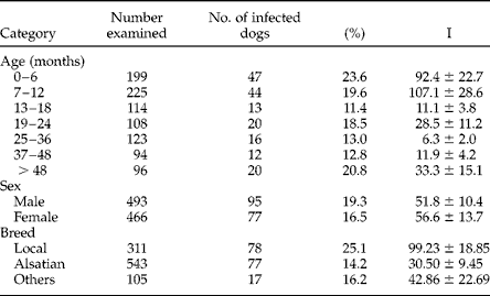
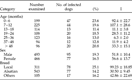
The prevalence of Ancylostoma spp. was observed to be highest in dogs of age group 0–6 months. The prevalence of infection was higher in male dogs (19.3%) than in female dogs (16.5%); however, there was no significant difference (χ2 = 1.227, df = 1, P>0.05) (table 5). The intensity of Ancylostoma spp. infection showed that young dogs ( < 12 months) were more heavily infected than the older ones. There was no significant difference observed in the intensity of Ancylostoma sp. infection between the male dogs (51.85 ± 10.41) and female dogs (56.57 ± 13.72) (U = 111955.50, df = 1, P>0.05) (table 5). The prevalence and intensity of other helminth parasites observed in this study were similar in both male and female dogs (table 6). Multiple infections with two or more helminth parasites (3.5%) were far less common than were infections with a single parasite type (85.7%).
Prevalence (%) and intensity (I) of Ancylostoma spp. in dogs relative to host age, sex and breed.
Prevalence (%), intensity (I) and range (R) of helminth eggs in the faeces of dogs of different sexes.

The prevalence of helminth parasites in dogs of different breeds is presented in table 3. The highest prevalence of helminth parasites (41.2%) was found in the local breed (African shepherd) and significantly higher (P < 0.05) than in Alsatian or other exotic breeds (Labrador, Ridgeback, Doberman and mongrel). Similarly, T. canis and Ancylostoma spp. were more prevalent in the local breed (African shepherd) than in Alsatian or other exotic breeds (tables 4 and 5).
Discussion
The overall prevalence of gastrointestinal helminth parasites of dogs observed in this study (24.7%) was less than the value of 77.4% reported in a similar study involving dogs from veterinary clinics, University of Ibadan and Oyo State Veterinary Clinic, over two and a half decades ago (Olufemi & Bobade, Reference Olufemi and Bobade1979). This may be attributed to the increasing number of veterinary outfits established in Ibadan and also the awareness among the dog owners of the need to take their dogs to veterinary clinics for deworming. Similar results were obtained by Onyenwe & Ikpegbu (Reference Onyenwe and Ikpegbu2004), who reported a prevalence of 24.12% among 908 dogs presented to the University of Nigeria Veterinary Teaching Hospital (UNVTH), Nsukka Nigeria. This may be due to the similarity between the dog populations studied, since most of the faecal samples examined were also obtained from the dogs presented for veterinary care. The overall prevalence obtained in this study differs from that reported by Ramirez-Barrios et al. (Reference Ramirez-Barrios, Barboza-Mena, Munoz, Angulo-Cubillan, Henandez, Gonzalez and Escalona2004), who obtained a prevalence of 35.5% among 614 dogs presented to the veterinary clinic of the University of Zulia, Venezuela.
The prevalence of helminth infections of dogs observed in Mokola Clinic was higher than that of Bodija Clinic. This may be due to the fact that most dogs attending Mokola Clinic were local dogs which freely roam the streets and may acquire helminth infections through the ingestion of paratenic hosts as a result of their scavenging habits. On the other hand, dogs attending Bodija Clinic were mostly exotic breeds, whose movements are restricted by their owners. In addition, dogs taken to Bodija Clinic, which is privately owned, might be receiving more care from their wealthy owners through regular administration of anthelminthic drugs, compared to dogs taken to Mokola Clinic, a public clinic where effective parasiticides may be less available.
The most commonly encountered parasites in this study were Ancylostoma spp. (17.9%) and T. canis (9.0%). This is in agreement with other research findings in Nigeria and other parts of the world (Ugochukwu & Ejimadu, Reference Ugochukwu and Ejimadu1985; Jordan et al., Reference Jordan, Mullins and Stebbins1993; Blagburn, Reference Blagburn2001; Onyenwe & Ikpegbu, Reference Onyenwe and Ikpegbu2004; Ramirez-Barrios et al., Reference Ramirez-Barrios, Barboza-Mena, Munoz, Angulo-Cubillan, Henandez, Gonzalez and Escalona2004). This is of great importance since T. canis and Ancylostoma spp. are well-recognized zoonotic agents which may constitute a significant public health risk due to the frequent contact between humans and their pets (Chiejina & Ekwe, Reference Chiejina and Ekwe1986; Ramirez-Barrios et al., Reference Ramirez-Barrios, Barboza-Mena, Munoz, Angulo-Cubillan, Henandez, Gonzalez and Escalona2004).
Dog infections with single worm species were encountered more frequently in this study while multiple infections were less commonly detected. These results are in agreement with the findings of other authors (Kirkpatrick, Reference Kirkpatrick1988; Vanparijs et al., Reference Vanparijs, Hermans and Van Dar Flaes1991; Ramirez-Barrios et al., Reference Ramirez-Barrios, Barboza-Mena, Munoz, Angulo-Cubillan, Henandez, Gonzalez and Escalona2004). Uncinaria stenocephala, another hookworm observed in this study, occurs with a low prevalence; this may be due to the fact that this parasite is best adapted to colder temperature climates (Gualazzi et al., Reference Gualazzi, Embil and Pereira1986). Therefore, the low prevalence observed was expected in this study. A similar observation was reported from another tropical country, Kenya, where a lower prevalence was reported for the worm (Kagira & Kanyari, Reference Kagira and Kanyari2000).
The prevalence of the tapeworm D. caninum (0.2%) observed in this study was lower than the prevalence (4.0%) reported from a study investigated in the same study area about two and a half decades ago (Olufemi & Bobade, Reference Olufemi and Bobade1979). This may be due to an increased awareness of environmental hygiene in most houses, coupled with modern housing structures that create a unsuitable environment for the survival of the intermediate host (the flea Ctenocephalides canis). The closed endemic focus of infection is thus broken in the transmission cycle.
In this study, the prevalences of T. canis and Ancylostoma spp. were higher in younger dogs aged 0–6 months than in older ones. This finding is in agreement with the observation of previous workers in Nigeria and other parts of the world (Idowu et al., Reference Idowu, Okon and Dipeolu1977; Chiejina & Ekwe, Reference Chiejina and Ekwe1986; Kirkpatrick, Reference Kirkpatrick1988; Blagburn, Reference Blagburn2001; Ramirez-Barrios et al., Reference Ramirez-Barrios, Barboza-Mena, Munoz, Angulo-Cubillan, Henandez, Gonzalez and Escalona2004). The higher prevalence of T. canis in young dogs was expected because of transplacental infection of the fetus, and documented evidence of age-associated immunity in adult dogs, as reported by previous workers (Bowman, Reference Bowman1999; Blagburn, Reference Blagburn2001). Similarly, Ancylostoma sp. was observed more in young puppies aged 0–6 months, probably due to transmammary passage (Lindsay & Blagburn, Reference Lindsay and Blagburn1995; Ramirez-Barrios et al., Reference Ramirez-Barrios, Barboza-Mena, Munoz, Angulo-Cubillan, Henandez, Gonzalez and Escalona2004).
Acknowledgements
The authors wish to thank the staff of the Magma Veterinary Clinic, Bodija and Oyo State Veterinary Clinic, Mokola, both at Ibadan, for their help and co-operation during sample collection. This research study was funded partly by the Obafemi Awolowo University Postgraduate Research Grant.