Introduction
The class Kutorginata Williams et al., Reference Williams, Carlson, Brunton, Holmer and Popov1996 includes both the kutorginids and the nisusiids, which are some of the oldest known Cambrian rhynchonelliform brachiopods with a shell composed of calcium carbonate and primitive types of articulation (Popov et al., Reference Popov, Holmer and Bassett1996; Williams et al., Reference Williams, Carlson, Brunton, Holmer and Popov1996; Popov and Williams, Reference Popov and Williams2000; Bassett et al., Reference Bassett, Popov and Holmer2001). The Kutorginata first appeared in the Atdabanian (Unnamed Series 2), rapidly diversified, and attained a cosmopolitan distribution before becoming extinct by the end of the Unnamed Cambrian Series 3.
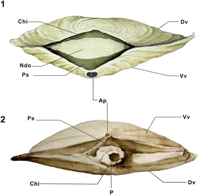
Perhaps the most unusual morphological feature of the kutorginates is that they are provided with both a ventral apical perforation and a widely triangular posterior notothyrial and delthyrial opening between the outer margins of the notothyrium and pseudodeltidium (Fig. 1), and both openings have at some time been interpreted as accommodating the pedicle (see discussions and reviews in Williams and Carlson, Reference Williams and Carlson2007 and Popov et al., Reference Popov, Egerquist and Holmer2007, Reference Popov, Bassett, Holmer, Skovsted and Zuykov2010). To date, the best exceptionally preserved record of a kutorginate with soft anatomy, including the attachment structures, is Kutorgina chengjiangensis Zhang et al., Reference Zhang, Shu, Emig, Zhang, Han, Liu, Li and Guo2007 from the early Cambrian Chengjiang Lagerstätte of southern China, and the original report (Zhang et al., Reference Zhang, Shu, Emig, Zhang, Han, Liu, Li and Guo2007, Reference Zhang, Robson, Emig and Shu2008) described the pedicle as emerging from the posterior notothyrial and delthyrial opening (Fig. 1.1).
Diagrammatic drawings of the shell valves of kutorginides. (1) Posterior view of shell valves of Kutorgina chengjiangensis, showing the wide notothyrial and delthyrial opening between the outer margins of the notothyrium and pseudodeltidium (based on ELI BK-064A); (2) posterior view of Nisusia sulcata (based on Rowell and Caruso, Reference Rowell and Caruso1985, fig. 9.7, KUMIP 163499), showing two apertures of the apical opening and the notothyrial and delthyrial opening plugged with silicified cylindroid pedicle discussed in this paper. Ap=apical perforation; Chi=chilidium; Dv=dorsal valve; Ndo=notothyrial and delthyrial opening between the outer margins of the notothyrium and pseudodeltidium; Ps=pseudodeltidium; P=pedicle; Vv=ventral valve.

Here we show that additional better-preserved material clearly illustrates that the pedicle emerges from the apical perforation of Kutorgina chengjiangensis, while the notothyrial and delthyrial opening lacks any evidence for having been associated with any adult attachment structure. This new information has important phylogenetic connotations for our understanding of brachiopod evolution. The known record of early Paleozoic rhynchonelliform brachiopods with exceptionally preserved attachment structures (variously termed) emerging from apical perforations have increased recently, mainly recorded from the enigmatic chileates, but also from the obolellates (Zhang et al., Reference Zhang, Holmer, Ou, Han and Shu2011a, Reference Zhang, Holmer, Popov and Shu2011b; Holmer et al., Reference Holmer, Popov and Bassett2014). This type of attachment may have been widespread in early rhynchonelliforms, representing a potential plesiomorphic feature that possibly was lost repeatedly. However, critical exceptionally preserved silicified material of another kutorginate—Nisusia sulcata Rowell and Caruso, Reference Rowell and Caruso1985—shows that the complete shells in this species have preserved cylindroid protrusions, which are here interpreted as the adult attachment (Popov et al., Reference Popov, Egerquist and Holmer2007, Reference Popov, Bassett, Holmer and Pour2009; Williams and Carlson, Reference Williams and Carlson2007; Zhang et al., Reference Zhang, Shu, Emig, Zhang, Han, Liu, Li and Guo2007), and in this kutorginate, the pedicle emerged posteriorly from the notothyrial and delthyrial opening, rather than from the apical perforation (Fig. 1.2). As noted by, for example, Williams and Carlson (Reference Williams and Carlson2007), pedicles emerging from the apical foramen (such as in Kutorgina chengjiangensis) and from the posterior gap between the valves (such as in Nisusia sulcata) cannot be homologous structures, indicating that the kutorginates had a dual system of attachment, but the detailed distributions and polarity of character transitions that are important for understanding the phylogeny are still far from clear. However, the apical foramen may represent an early larval type of attachment in the stem of the Brachiopoda as suggested by numerous previous authors (e.g., Holmer, Reference Holmer2001; Popov et al., Reference Popov, Egerquist and Holmer2007, Reference Popov, Bassett, Holmer, Skovsted and Zuykov2010; Williams and Carlson, Reference Williams and Carlson2007; Holmer et al., Reference Holmer, Stolk, Skovsted, Balthasar and Popov2009).
Materials and methods
The discussed and illustrated rhynchonelliform brachiopods are derived from localities in China, Canada, and the United States. The age range is from the early Cambrian to the Late Ordovician.
China
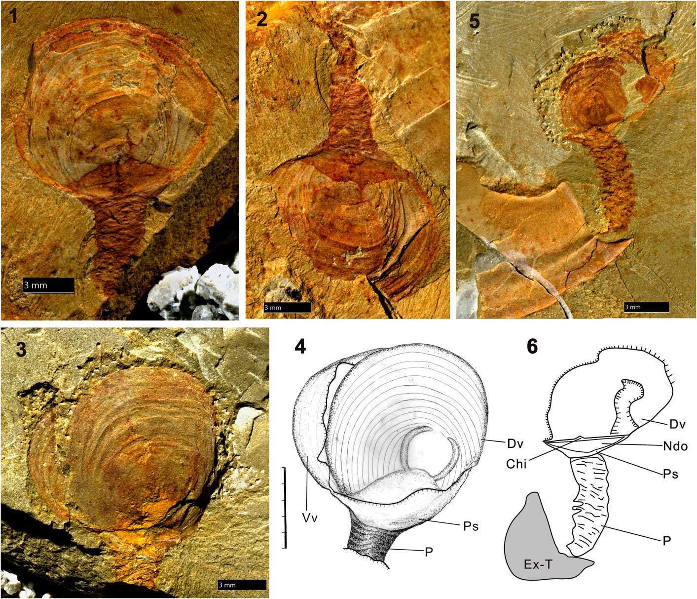
More than 150 specimens of Kutorgina chengjiangensis have been collected by the work team from Northwest University, Xi’an, China. The early collected 60 specimens for the original study (Zhang et al., Reference Zhang, Shu, Emig, Zhang, Han, Liu, Li and Guo2007) and the additional (over 90) specimens for the current study (Fig. 2) were collected at the sections exposed around the south of Erjie town. The early Cambrian Chengjiang fauna from which the new specimens of Kutorgina were collected belong to the Yu’anshan Member (the Eoredlichia-Wudingaspis trilobite Zone) of upper Heilinpu Formation (Qiongzhusian stage). More detailed account of the localities and stratigraphy can be found in Zhang et al. (Reference Zhang, Shu, Emig, Zhang, Han, Liu, Li and Guo2007).
The Burgess Shale-type preservation of the rhynchonelliform brachiopod Kutorgina chengjiangensis from the Chengjiang fauna of China, showing the laminar tabular pedicle just aligned with ventral valve. (1) ELI BK-064A, dorsoventrally compressed internal mold of shell valves with well-developed pedicle, noting the widely triangular posterior opening between the posterior margin of two valves; (2) posterior view of the counterpart of (1), ELI BK-064A; (3) ELI BK-038A, rephotographed specimen from Zhang et al. (Reference Zhang, Shu, Emig, Zhang, Han, Liu, Li and Guo2007, fig. 2a) , showing the displaced and conjoined shell valves with the ventral below, aligned with stout pedicle proximal; (4) interpretative drawing of (3); (5) ELI BK-066, a dorsoventrally crushed specimen showing posterior view of shell valves with wide notothyrium and delthyrium, rephotographed specimen from Zhang et al. (Reference Zhang, Robson, Emig and Shu2008, fig. 6h); (6) interpretative drawing of (5). Chi=chilidium; Dv=dorsal valve; Ex-T=exoskeletons of trilobite; Ndo=notothyrial and delthyrial opening between the outer margins of the notothyrium and pseudodeltidium; Ps=pseudodeltidium; P=pedicle; Vv=ventral valve.

United States
The silicified specimens of Nisusia sulcate come from the mid-Cambrian Marjum Limestone of western Utah (Rowell and Caruso, Reference Rowell and Caruso1985). A drawn interpretation of KUMIP 163499 is illustrated here (Fig. 1.2).
Canada
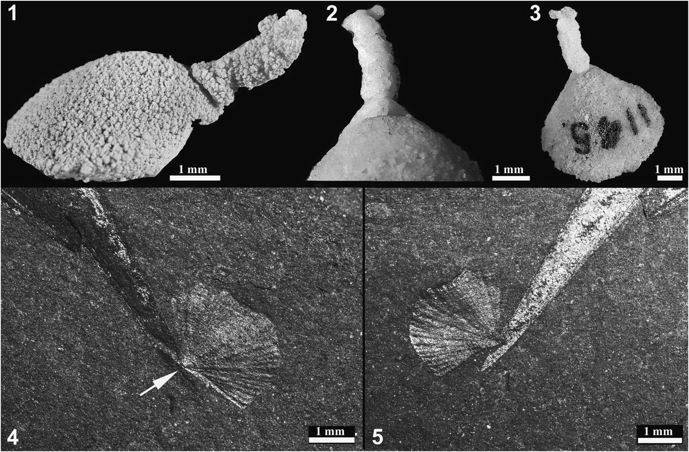
The illustrated attached specimens of Nisusia? burgessensis Walcott, Reference Walcott1924, discussed in the following, all come from the middle Cambrian Burgess Shale Lagerstätte in British Columbia, Canada (Fig. 3.4, 3.5). Many species of Nisusia are provided with spines, which seemingly are lacking in the Burgess species; pending further taxonomic revision, the generic assignation is conditional. Detailed accounts of the brachiopod faunas, localities, and stratigraphy can be found in Topper et al. (Reference Topper, Holmer and Caron2014, Reference Topper, Strotz, Holmer and Caron2015). A single silicified specimen of Eichwaldia subtrigonalis with preserved pedicle (Fig. 3.1–3.3) comes from the Late Ordovician Ottawa Formation, Allumette Island, Quebec, Canada (see Wright, Reference Wright1981, and references therein).
(1–3) GSC 1145, silicified internal of mold of Eichwaldia subtrigonalis; (1) lateral view of shell (whitened with ammonium chloride), showing silicified cylindroid pedicle, with closely spaced transverse grooves and emerging from the umbonal perforation; (2) detail of pedicle and umbo in dorsal view; (3) oblique ventral view; (4, 5) ROM 61116, two specimens of Nisusia? burgessensis attached to the dorsal spines of Wiwaxia (Series 3, Stage 5) Cambrian Burgess Shale, indicating that the pedicle probably emerged posteriorly between the valves (Topper et al., Reference Topper, Holmer and Caron2014, Reference Topper, Strotz, Holmer and Caron2015); (4) attached specimen with possible umbonal foramen lacking emerging pedicle marked by white arrow; (5) attached specimen with posterior commissure oriented parallel to the substrate.

Fossils were examined with a binocular Olympus zoom stereo microscope and photographed with a Nikon camera mounted on a photomicrographic system, with different illuminations for particular views when high-contrast images are required. The other specimens were taken to the Department of Palaeobiology, Uppsala University, and photographed with a camera lucida mounted on a Leica photomicrographic system. The artistic reconstructions and sketchy drawings of kutorginides were made by Zhang Z and Liu X in Northwest University of Xi’an.
Repositories and institutional abbreviations
All the materials of Kutorgina chengjiangensis from China are deposited in the Early Life Institute (ELI), Northwest University, Xi’an, China; the silicified specimens of Nisusia sulcate from the United States are housed in the Museum of Invertebrate Paleontology (KUMIP), Lawrence, Kansas. The material of Nisusia? burgessensis Walcott, Reference Walcott1924 is housed in the Royal Ontario Museum (ROM) in Toronto, Canada (ROM 61116), and the National Museum of Natural History, Smithsonian Institution (USNM) in Washington D.C., USA.
Redescription of attachment structures in Kutorgina chengjiangensis
The lamellose shell of Kutorgina chengjiangensis is strongly biconvex, and it appears to have an astrophic hinge and a pointed ventral valve, but due to the preservation as flattened molds, this is not as easy to discern (Fig. 2) as in those specimens derived from carbonate lithologies. The shell has a relatively large, posterior, widely triangular notothyrial and delthyrial opening, occupying somewhat more than half of the total width of the valve, and it is clearly not associated with any attachment structures in some well-preserved specimens with preserved pedicles (Fig. 2.1, 2.2). The ventral interarea is apsacline, and it is provided with a rather broadly convex pseudodeltidium that clearly separates the protruding apical pedicle from the large and empty notothyrial and delthyrial opening (Fig. 2.3, 2.4). The dorsal interarea has minute chilidium and dorsal hinge furrows well imprinted on the inner side of the interarea (Fig. 2.5, 2.6). In life, the relatively large posterior notothyrial and delthyrial opening of Kutorgina chengjiangensis would have accommodated the passage of the diductor muscle (see e.g., Rowell and Caruso, Reference Rowell and Caruso1985, fig. 7; Bassett et al., Reference Bassett, Popov and Holmer2001, fig. 18.2), and must have been closed by the posterior body wall, perhaps also protected by a flexible organic integument.
A total of 35 specimens of Kutorgina chengjiangensis have a preserved stout and annulated pedicle, and due to the flattened compressed molded preservation of the shells, it was previously described as protruding between the delthyrium and notothyrium of the shell valves (Zhang et al., Reference Zhang, Shu, Emig, Zhang, Han, Liu, Li and Guo2007, p. 1396); however, reexamination of existing specimens and newly collected better-preserved material clearly shows that the pedicle is separated from the posterior opening, is subtended between delthyrium and notothyrium, and emerges from the apical perforation, but the exact size and shape of the apical perforation is obscured by the emerging pedicle (Fig. 2.1–2.6). However, the foramen is clearly much smaller compared with the emerging pedicle; thus, the ventral apex is largely encased by the proximal part of the pedicle. The pedicle is remarkably thick, up to 3.1 mm in diameter, occupying up to 30%–40% the total valve width in some specimens, and it seems to be built up by series of regularly spaced, pronounced, stacked tabular discs (0.6–1.0 mm apart) (Fig. 2.1–2.5). The discs clearly have a pronounced three-dimensional preservation and may have been partly mineralized. A dark central lineation in some specimens suggests that it may have had a cavity running along its central axis (Zhang et al., Reference Zhang, Shu, Emig, Zhang, Han, Liu, Li and Guo2007). The distal termination of the pedicle attaches onto the exoskeletons of other animals, notably trilobites’ exuvae (Fig. 2.5, 2.6) (Zhang et al., Reference Zhang, Robson, Emig and Shu2008; Zhang and Holmer, Reference Zhang and Holmer2013).
Diversity of exceptionally preserved attachment structures in kutorginate and chileate brachiopods
Nisusia sulcata
Rowell and Caruso (Reference Rowell and Caruso1985) described exceptionally preserved silicified cylindroid protrusions emerging posteriorly from the notothyrial and delthyrial opening in 15 of 18 complete shells of Nisusia sulcata; the cylindroid has a maximum length of 6 mm and closely spaced transverse grooves (Fig. 1.2). The various interpretations of this structure were discussed in some detail by Williams and Carlson (Reference Williams and Carlson2007, p. 2848–2849), who noted that there are many problems associated with the original description by Rowell and Caruso (Reference Rowell and Caruso1985), where the cylindroid is interpreted as representing fecal matter rather than an exceptionally preserved pedicle. Most important, no other such large coprolites are known to be produced by any living or fossil brachiopods, and the minute amount of food consumed by living brachiopods (e.g., Peck et al., Reference Peck, Rhodes, Curry and Ansell1997) results at most in micrometer-sized fecal pellets (James et al., Reference James, Ansell and Collins1992). Moreover, as noted by Williams and Carlson (Reference Williams and Carlson2007, p. 2849), “It is unlikely that excreta could have retained a constant shape in such a high proportion of shells collected.” The main reason Rowell and Caruso (Reference Rowell and Caruso1985) rejected the pedicle nature of the cylindroid is the presence of the apical foramen, which was known to serve as a pedicle in comparable closely related strophomenate brachiopods, and they noted that “It is conceivable that the orthodox opinion is in error, but we find it difficult to ascribe any other role to the opening” (Rowell and Caruso, Reference Rowell and Caruso1985, p. 1234). However, as first suggested by Popov (cited in Williams and Carlson, Reference Williams and Carlson2007, p. 2848), the apical foramen in Nisusia may now be interpreted as the trace of a larval attachment, and the cylindroid most likely represents an adult type of pedicle; this was also discussed by Zhang et al. (Reference Zhang, Shu, Emig, Zhang, Han, Liu, Li and Guo2007, p. 1399).
Nisusia? burgessensis
Outside of the Chengjiang Lagerstätte, the only record of still-attached kutorginates is represented by Nisusia? burgessensis from the Burgess Shale. Topper et al. (Reference Topper, Holmer and Caron2014, Reference Topper, Strotz, Holmer and Caron2015) studied the life strategies of the attached brachiopod fauna, which includes only 25 specimens (of a total of about 250) of Nisusia? burgessensis that are preserved directly attached to a hard substrate. As noted by Topper et al. (Reference Topper, Strotz, Holmer and Caron2015), this may suggest that the pedicle attachment was relatively weak; however, the pedicle itself cannot be observed in any of the specimens. Presumably the pedicle of Nisusia? burgessensis was very short, as the majority of attached specimens are in direct contact with the substrate. The posterior margins of some attached specimens are separated from the substrate by approximately 0.1 mm (Fig. 3.4, 3.5), a distance that may represent the true length of the pedicle. The orientation of complete individuals appears to be parallel, or close to parallel, to the substrate (Fig. 3.4), and the short pedicle most likely emerged posteriorly between the valves. On one specimen, the pedicle foramen may be observed; however, with the valve’s orientation on the Wiwaxia sclerite, the foramen does not appear to be in a position conducive to attachment (Fig. 3.5).
Eichwaldia subtrigonalis
Wright (Reference Wright1981) made the most recent detailed study of the unique attachment structures of the problematic rhynchonelliform Ordovician–Permian brachiopods belonging to the Dictyonellida, which are now classified as the Chileata together with the equally problematic Cambrian–Silurian Chileida (Holmer et al., Reference Holmer, Popov and Bassett2014). The chileates most likely had posteriorly fused mantle lobes, and there is no delthyrium through which a posterior pedicle could pass in any of the known chileates (Popov and Holmer, Reference Popov and Holmer2007). Instead, the single chileate attachment structure consists of an umbonal perforation (Fig. 3.1–3.3), which is usually covered posteriorly by a plate—the colleplax in the sense of Wright (Reference Wright1981). A single articulated silicified juvenile ventral valve (8 mm long and wide) of the type species Eichwaldia subtrigonalis Billings, Reference Billings1858 has a 6 mm long and 2 mm wide silicified cylindroid emerging from the umbonal perforation, which is completely covered by the cylindroid. The cylindroid has closely spaced transverse grooves (Fig. 3.1, 3.2) and is more or less identical in morphology to the cylindroid in Nisusia sulcate; there is no indication that it represents “adventitious silica adhering to the silicified shell” as suggested by Wright (Reference Wright1981, p. 448).
Discussion and implications
The nature and origin of the brachiopod pedicle has for a long time been of critical importance for understanding the phylogeny and evolution within the Brachiopoda, and indeed some classificatory schemes, such as that of Beecher (Reference Beecher1892), were almost entirely based on the variation in the nature of the pedicle opening. Features of the brachiopod pedicle remain important characters in more broadly based phylogenetic analyses, such as that carried out by Williams et al. (Reference Williams, Carlson, Brunton, Holmer and Popov1996) and adopted in the revised brachiopod volumes of the Treatise on Invertebrate Paleontology (Williams et al., Reference Williams, Brunton and Carlson1997–2007), which still is the most up-to-date phylogenetic and classificatory framework of the Brachiopoda.
In the Treatise framework, the Kutorginata occupy an unstable and unresolved basal position in the Rhynchonelliformea (e.g., Popov et al., Reference Popov, Holmer and Bassett1996; Harper et al., Reference Harper, Popov and Holmer2017), associated with the Chileata and the Obolellata, but the phylogenetic positions of these groups remain problematic (Harper et al., Reference Harper, Popov and Holmer2017) and we still have a poor understanding of the stem and crown group relationships within the earliest Cambrian rhynchonelliforms. Consequently, the new soft anatomy information concerning the extinct kutorginates is of great phylogenetic significance, and the new type of apical pedicle described here from Kutorgina chengjiangensis is closely comparable in all major features with the pedicles emerging from the apical foramen of both the chileate Longtancunella chengjiangensis (Zhang et al., Reference Zhang, Holmer, Ou, Han and Shu2011a) and the obolellate Alisina sp. (Zhang et al., Reference Zhang, Holmer, Popov and Shu2011b), both also from the Chengjiang fauna. The pedicles of these species have an identical type of strong, three dimensionally preserved, distinctive annulations that consist of densely stacked tabular discs (Zhang et al., Reference Zhang, Holmer, Ou, Han and Shu2011a, fig. 3; 2011b, fig. 2). Moreover, exceptionally preserved similar pedicles are also known to emerge apically from the Silurian chileate dictyonellid Eichwaldia subtrigonalis (Fig. 3.1–3.3; briefly described in the preceding) as well as from the recently described Silurian chileate Trifissura rigida, where the pedicle is preserved as a short series of phosphatized pads (Holmer et al., Reference Holmer, Popov and Bassett2014, fig. 1).
The earliest ontogeny of the strophomenates also includes closely similar apical attachment structures, and in particular, the so-called pedicle sheath in, for example, Antigonambonites (Popov et al., Reference Popov, Egerquist and Holmer2007; Bassett et al., Reference Bassett, Popov and Egerquist2008) is long, hollow, and annulated and closely comparable to the pedicle of Kutorgina chengjiangensis; it is possible that the densely stacked tabular discs in Kutorgina were mineralized. The positions of the kutorginate, obolellate, chileate, and strophomenate pedicles indicate that they must have been secreted by a specialized modified region of the ventral mantle, rather than from a larval pedicle lobe as in all living rhynchonelliforms, and most likely represent homologous structures (Williams et al., Reference Williams, Carlson, Brunton, Holmer and Popov1996; Popov et al., Reference Popov, Egerquist and Holmer2007).
By contrast, the adult pedicle of the nisusiids emerged between the valves from the posterior gap as indicated by silicified pedicles in Nisusia (Rowell and Caruso, Reference Rowell and Caruso1985) as well as the apparent posterior attachment of the Burgess specimens of Nisusia? (Fig. 3.4, 3.5); this type of pedicle is most likely homologous with the pedicle of extant rhynchonelliform brachiopods. Thus, the kutorginates had a dual system of attachment, but the detailed distributions and polarity of character transitions that are important for understanding the phylogeny are still far from clear.
The fine details of the ontogeny in kutorginides unfortunately remain unknown, but the morphology of the umbonal region of the ventral valve, which clearly lacks a pedicle sheath, suggests that their ontogeny may have been closely similar to that of the orthotetidoids. The ontogeny of this strophomenate superfamily has been recently described in detail by Bassett and Popov (Reference Bassett and Popov2017). The ventral perforation in the orthotetidoid (Chilidiopsoidea) Coolinia shows that it has an apical plate that is closely similar to the chileate colleplax (Bassett and Popov, Reference Bassett and Popov2017), while at the same time the characters of the shell formation suggest that there is a substantial delay in the secretion of the ventral valve, possibly until the end of metamorphosis; moreover, a similar ontogeny was described earlier for the problematic stem rhynchonelliform chileate Salanygolina and the clitambonitoid Antigonambonites (Popov et al., Reference Popov, Egerquist and Holmer2007). In all these taxa, the delthyrial opening was completely covered by the pseudodeltidium at all growth stages, which is unlike the kutorginate ontogeny. The dorsal metamorphic shell (see Bassett and Popov, Reference Bassett and Popov2017) in Coolinia, Salanygolina, and Pelmanotreta (the last paterinate being the second-oldest brachiopod yet documented), in addition to the protegulum and two to three pairs of lobes in all these taxa, shows the position of larval setal sacs and, moreover, preserves a posterior flange, which indicates the presence of a larval pedicle emerged between valves, which presumably was used for the attachment on a settlement (Popov et al., Reference Popov, Egerquist and Holmer2007). This larval pedicle probably developed from the larval posterior pedicle lobe. If so, the larval pedicle of these brachiopods was homologous to the pedicle of the orthides and all crown group rhynchonellate brachiopods, as well as the pedicle of more derived paterinides, such as Dictyonina and Micromitra. The transition to the encrusting attachment by the surface of the ventral valve may have been due to the poorly developed mechanical properties of the body wall, which was lacking in dermal muscles. In the crown group rhynchonelliforms, there is invariably a cartilage-like tissue in the pedicle, which is clearly an apomorphic character that evolved later in brachiopod evolution.
Thus, the pedicle of the nisusiids was probably also developed from the larval posterior lobe, such as in the crown group rhynchonelliforms, and retained its function as a result of paedomorphic alteration; secretion of the ventral valve was delayed until the end of metamorphosis, which is indicated by the presence of the ventral umbonal perforation, although the ‘adult attachment’ by the surface of the ventral mantle in some groups became nonfunctional, as demonstrated by the specimens of Nisusia? burgessensis. Moreover, it is possible that connective-like tissue was present in the pedicle of nisusiids, which is supported by the fact that it is occasionally preserved.
Acknowledgments
Financial support from the National Natural Science Foundation of China (NSFC 41425008, 41720104002 and 41621003 to Z. Zhang) for the long-term collection of Chinese material is acknowledged. The work of L. Holmer was supported by a grant from the Swedish Research Council (VR 2012-1658), and his research visits in Xi’an for work with this paper were financially supported by the C.C. Yang Professor Scholarship from the Department of Geology, Northwest University, Xi’an. L. Popov acknowledges logistical and financial support from the National Museum of Wales. T. Topper was supported by a Marie Curie COFUND fellowship, Durham University. Fossil collection was partly financed by the National 973 Program (2013CB835002) and 111 Project of China (P201102007). Thanks are also extended to J.-P. Zhai for fossil preparation and to X. Liu for artistic reconstruction of fossils in this paper. We are also grateful to the editors J. Jin (Canada) and G. Brock (Australia) for assistance with the manuscript and to two referees for their helpful reviews, which greatly improved the paper.