Introduction
The endoscope is an effective and indispensable tool in otolaryngological clinics.Reference Woodson1, Reference Sharma, Price, Mierzwa, Montgomery, Qayyum and Bradnam2 Working in tandem with a charge-coupled device camera, the endoscope can be used for observation, presentation and documentation.Reference Yanagisawa and Carlson3 In addition, the video-endoscope offers superior image quality compared with conventional fibrescopy with a charge-coupled device camera.Reference Sato, Umeno and Nakashima4 However, dedicated charge-coupled device camera systems for endoscopy and video-endoscopy are expensive. Here, we report a new and economical method for recording extremely high quality laryngeal images, using a full digital, high definition video camera.
Methods
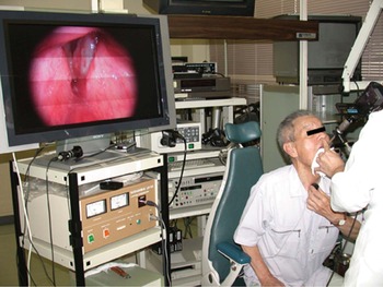
We used video cameras which had newly become available (i.e. the HDR-HC1 and HDR-HC3 models, Sony, Tokyo, Japan). An endoscopic adaptor (AT-N adaptor, Nagashima Medical Instrument Ltd, Tokyo, Japan) for a popular digital video camera (filter bore: 37.5 and 30.5 mm) was attached to the camera.Reference Tsunoda, Tsunoda and Hatanaka5, Reference Tsunoda, Tsunoda, Ozawa and Fujimine6 By screwing this adaptor to the lens top, either a flexible or rigid endoscope could then be attached to the camera. We mainly used a rigid endoscope (Karl Storz, Tuttlingen, Germany) because of its superior image quality (Figure 1).Reference Takwoingi, Kale and Morgan7 These video cameras could record a 1080i digital high definition video format. This system had 1080 effective scanning lines, and the image quality was much better than that obtained from ordinary digital video format (or from a different type of high definition video format, 720p).
A full digital, high definition video camera (HDR-HC3 model) and 80 cm high definition video monitor being used in the laryngeal examination of a 75-year-old man with laryngeal cancer. The patient can simultaneously watch a small camera monitor which is turned towards him.

Such cameras provided a high definition video output signal. Using a high definition multimedia interface cable or component video cable, the endoscopic images obtained via full digital, high definition video could be shown simultaneously on the monitor (Figure 1). Both focus and exposure were automatic. However, since glottic movement causes focal plane changes, we preferred to use a fixed focus and to bring the video into focus on the vocal folds. Date and time of recording, as well as the patient's voice, were automatically recorded.
The endoscopic image was recorded on a mini-digital videocassette, the same media as the previous models of digital video camera. The images were also displayed on the side viewer, and could be turned not only to the clinician but also to the patient, thereby allowing the patient to see the image simultaneously (Figure 1). Examination of the larynx and recording of the endoscopic image were performed in the same manner as normal video recording. One videocassette could record over 80 minutes of full digital, high definition video quality. The obtained data could be transferred to a personal computer via an Institute of Electrical and Electronic Engineers (IEEE) 1394 cable. The video image was easily dubbed and edited using popular software (iMovie HD, Apple Computer, Cupertino, California, USA).
Results
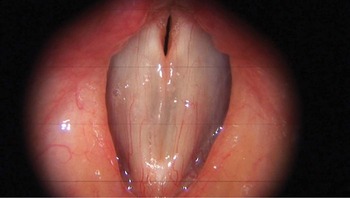
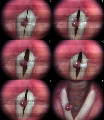
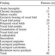
We began using a full digital, high definition video system in August 2005, and from March 2006 used a smaller device (the Sony HDR-HC3). Laryngostroboscopy was performed on 82 patients during an 18 month period (Table I). Compared with a proprietary charge-coupled device camera or a video-endoscope, the device itself was larger, so its use for surgery was somewhat more difficult (e.g. for biopsy of laryngeal lesions or removal of foreign bodies). Banding appeared only during stroboscopic examination; however, it did not cause problems during the observation (Figures 2 and 3). Since full high definition video generates a large amount of information, we mainly used desktop computers for capturing and editing the video (Power Mac G5, 1.8 GHz, dual central processing unit type, Apple Computer), as a notebook-type computer would have been insufficient for this purpose.
Full digital, high definition video stroboscopic image of a 52-year-old woman who complained of slight hoarseness. Slight oedema of Reike's space is clearly shown in the middle portion of the vocal fold; engorged vessels in the vocal fold may also be seen.

Videostroboscopic images (a–e) and laryngoscopic image (f) of a 59-year-old woman with a right vocal fold polyp. Tiny vessels and a slight sulcus of the left vocal fold are clearly visualised.

List of diseases of the patients

Except for the above-mentioned points, we experienced no difficulties with this system. Like a conventional video system, stroboscopic images could be monitored and checked frame by frame (Figure 3). Even tiny structures or lesions, such as small vessels, scars or oedema, could be clearly observed (Figure 2). Still images, which could easily be obtained from the video, were 1920 × 1080 pixels and comparable to those from a digital still camera. Since full digital, high definition video has a different pixel size and generates images which are fundamentally different, we could not compare the laryngeal findings from full digital, high definition video with those from conventional digital video and 720p high definition video. However, the findings obtained from full digital, high definition video were far superior to those obtained from our previous video-endoscope system.
Discussion
The endoscope is one of the most indispensable diagnostic tools in the laryngological field. In addition, motion recording of laryngeal movements or stroboscopic images is very important because of their clinical significance. Although it was originally designed for direct observation, use of a video camera has great advantages in laryngology.Reference Woodson1, Reference Sharma, Price, Mierzwa, Montgomery, Qayyum and Bradnam2 The video-endoscope provides clearer images, compared with a conventional flexible fibrescope with a charge-coupled device camera.Reference Sato, Umeno and Nakashima4 Recently, the 720p high definition video-endoscope, with 720 effective scanning lines, was made available and has proved useful.Reference Otto, Hapner, Baker and Johns8 However, such proprietary medical video systems are usually very expensive.Reference Tsunoda, Tsunoda and Hatanaka5, Reference Tsunoda, Tsunoda, Ozawa and Fujimine6
On the other hand, mainstream digital video systems have become much smaller and cheaper and have gained in popularity among consumers over the last 10 years.Reference Tsunoda, Tsunoda, Ozawa and Fujimine6 In 2005, a popular full digital, high definition video camera appeared on the market (the Sony HDR-HC1). This system had 1080 effective scanning lines, making video quality far better than that of conventional systems, and producing still images comparable to those of digital or conventional still cameras. In 2006, a much smaller full digital, high definition video camera, which weighed only 600 g, was introduced (the Sony HDR-HC3). We have been using these high definition video cameras and high definition video monitor systems for daily clinical work and to record patients' findings.
Using an adaptor and popular digital still or video cameras, endoscopic images can be monitored and recorded without special equipment.5.6 The endoscopic adaptor can also be applied to newly released full digital, high definition video cameras. These high definition video systems are substantially cheaper than proprietary medical imaging systems. Currently, prices for a full digital, high definition video camera start at ¥110 000 (approximately GB£460 or US$910). In addition, the image quality obtained is far superior to that of proprietary medical video systems.
In Japan, digital terrestrial broadcasting using a digital high vision system began in December 2003 in large cities, with a phased launch in other areas. Conventional analogue broadcasts are scheduled to cease in Japan in July 2015, with the rest of the world to follow. Even in the medical field, it is highly likely that the full digital, high definition video system will completely supersede conventional proprietary medical imaging systems in the near future. Full digital, high definition video products can be developed at less cost, with many more features. The endoscopic adaptor may be used for coming full digital, high definition video cameras which may have a filter attachment, which are much smaller and cheaper than current video cameras. In fact, in September 2006, the Canon company (Tokyo, Japan) released a new, compact, full digital, high definition video camera which weighed only 500 g, including the battery.
Conclusion
Using adaptors and popular, commercially available equipment, high quality imaging of the larynx can be obtained easily and economically. Although such products cannot be used directly in sterile areas, they stand to surpass proprietary medical video systems in many respects. These full digital, high definition video systems will be of great benefit, not only to doctors requiring improved methods of laryngeal observation, but also to doctors facing budgetary restraints.