Introduction
The family Ochetosomatidae Leão, Reference Leão1945 and in particular the genus Ochetosoma Braun, 1901 possess a rather complex taxonomic history. They were considered valid until Tkach (Reference Tkach2008) conducted an extensive review of this plagiorchioid group and did not consider Ochetosoma as a valid genus, proposing the resurrection of Reniferidae Pratt, 1902 and hence Renifer Pratt, 1902 (Leão Reference Leão1945). This nomenclatural change was based on the argument that the generic name Ochetosoma was pre-occupied by a genus of a marine echiuroid worm. However, as proven by Seo et al. (Reference Seo, Ansai, Sase, Saito, Takano, Kojima and Waki2024), the argument by Tkach (Reference Tkach2008) was mistaken because the proper echiuroid genus name is Ochetostoma Rüppell and Leuckart, Reference Rüppell, Leuckart and Rüppell1828 and not Ochetosoma (Rüppell and Leuckart, Reference Rüppell, Leuckart and Rüppell1828). Furthermore, Renifer (and Reniferidae) were recognised as valid, even though Ochetosoma was older since it was described by Braun (1901), whereas Renifer was described by Pratt (1902); according to the International Code of Zoological Nomenclature (ICZN), the first name has precedence. Based on the latter arguments.
Ochetosomatidae (Syn. Reniferidae) currently includes six genera – namely, Dasymetra Nicoll, 1911, Lechriorchis Stafford, 1905, Paralechriorchis Byrd & Denton, 1938, Pneumatophilus Odhner, 1910, Ochetosoma Braun, 1901, and Zeugorchis Stafford, 1905 (Tkach, Reference Tkach2008; Yamaguti Reference Yamaguti1971). In South America, only species of Dasymetra and Ochetosoma have been reported – the former with only one species, Dasymetra tupinambis Nasir & Diaz, 1971, and the latter with three species, O. chironius (Nasir & Diaz, 1971), O. heterocoelium (Travassos, Reference Travassos1921), and O. monstruosum (Braun, 1901) (Fernandes and Cohn Reference Fernandes and Kohn2014).
Ochetosoma heterocoelium is a unique species reported to infect at least 20 snake species across Brazil. This species was described by Travassos (1921) and posteriorly reported in other hosts and geographical locations by Corrêa et al. (Reference Corrêa, Paulino, Buononato and Federsoni1990), Silva et al. (Reference Silva, Rodrigues, Stein, Sipoli, Pinhão and Lopes1999), Pinto et al. (Reference Pinto, Mati and Melo2012), and Vicente-Ferreira et al (Reference Vicente-Ferreira, do Nascimento, Grezzana, Ávila and Garey2023). The species has also been found in Colombia; Lenis et al. (Reference Lenis, Arredondo and Calle2009) re-described O. heterocoelium based on specimens collected from seven snake species. In Brazil, O. heterocoelium is known to parasitise the mouth, oral cavity, oesophagus, or intestine of Bothrops cotiara (Gomes, 1913), B. jararaca (Wied, 1824), B. insularis (Amaral, 1922), B. moojeni Hoge, 1966, B. neuwiedi Wagler in Spix, 1824, Chironius bicarinatus (Wied, 1820), Ch. exoletus (Linnaeus, 1758), Ch. fuscus (Linnaeus, 1758), Drymarchon corais (Boie, 1827), Erythrolamprus aesculapii (Linnaeus, 1766), E. almadensis (Wagler in Spix, 1824), E. miliaris (Linnaeus, 1758), E. poecilogyrus (Wied, 1824), E. reginae (Amaral, 1935), E. typhlus (Linnaeus, 1758), Helicops modestus Gunther, 1861, Leptodeira annulata (Linnaeus, 1758), Palusophis bifossatus (Raddi, 1820), Philodryas patagoniensis (Girard, 1858), Pseudoboa coronata Schneider, 1801, and Xenodon merremii (Wagler in Spix, 1824) (Correa et al. Reference Corrêa, Paulino, Buononato and Federsoni1990; Pinto et al. Reference Pinto, Mati and Melo2012; Silva et al. Reference Silva, Rodrigues, Stein, Sipoli, Pinhão and Lopes1999; Travassos 1921; Vicente-Ferreira et al. Reference Vicente-Ferreira, do Nascimento, Grezzana, Ávila and Garey2023). Moreover, O. heterocoelium was reported from Atractus lasallei Amaral, 1931, Bothriechis schlegelli (Berthold, 1846), Bothrops asper (Garman, 1884), Chironius carinatus (Linnaeus, 1758), Leptodeira septentrionalis (Kennicott, 1859), Leptophis ahaetulla (Linnaeus, 1758), and Porthidium nasutum (Bocourt, 1868) in Colombia (Lenis et al. Reference Lenis, Arredondo and Calle2009).
The life cycle of O. heterocoelium possibly involves the physid snail Stenophysa marmorata (Guilding, 1928) as the first intermediate host which harbors a xiphidiocercariae (López-Hernández et al. Reference López-Hernández, de Melo and Pinto2023). The metacercariae of this digenean were reported from the intestinal serosa of Rhinella diptycha (Cope, 1862) (Syn. Rhinella schneideri (Werner, 1894)) tadpoles in Brazil (Pinto and Melo Reference Pinto and Melo2012). This basic information did not exclude the possibility that other snails and amphibians are involved in the life cycle of this digenean species.
Recently, an integrative taxonomy approach has been employed for species delimitation of digenean parasites of snakes in Brazil (Müller et al. Reference Müller, Morais and Silva2018, Reference Müller, Emmerich, Alcantara, Úngari, Ebert, Morais, O’Dwyer and Silva2021). Thus far, 40 species of digeneans have been reported, although only a small fraction of the snake diversity has been studied for parasites, and they remain understudied even though they are deeply threatened. During an investigation on the parasite diversity of snakes in Brazil, specimens of O. heterocoelium were sampled from the mouth and oesophagus of the Brazilian lancehead, B. moojeni, which is widely distributed across South American moist forests. In this study, we provide the first molecular characterization of O. heterocoelium using the 28S rDNA, and we assess their phylogenetic position within the Plagiorchoidea. Additionally, we add new morphological data of the species through light and scanning electron microscopy (SEM).
Material and methods
One specimen of B. moojeni (Figure 1) (male, adult, snout-vent length 78 cm and weight 185 g) was collected on December 3, 2019, at São Sebastião do Paraíso Farm (21°51’48.26" S, 48°26’56.78" W), municipality of Boa Esperança do Sul, São Paulo State, Brazil. The snake was killed using 50 mg/kg of sodium thiopental (Thiopentax®), administered intracerebrally. All procedures were submitted and authorized by the Ethics Committee on Animal Use of the Institute of Biosciences, Universidade Estadual Paulista ‘Júlio de Mesquita Filho’ (Authorization # 1061-CEUA) and by the Authorization and Information System in Biodiversity (SISBIO # 60640-1).
Specimen of Bothrops moojeni Hoge, 1966 (Viperidae) from São Sebastião do Paraíso Farm, municipality of Boa Esperança do Sul, São Paulo State, Brazil.

During the necropsy, digenean specimens were found in the mouth and oesophagus of the host. Some parasite specimens were fixed in an alcohol-formalin-acetic acid solution (AFA solution) under the slight pressure of a coverslip for 10 min. Then, specimens were transferred to 70% alcohol and stored for further processing. Other specimens were fixed using a hot AFA solution. Some specimens were directly transferred to vials with ethanol PA for molecular study. Digenean specimens were stained with alcoholic chloride carmine solution, cleared with eugenol, and analyzed with a computerized system for image analysis (V3 Leica Application Suite, Leica Microsystems, Wetzlar, Germany) in a microscope with Differential Interference Contrast (DIC).
Some specimens were also studied through scanning electron microscopy (SEM). For this, the specimens were post-fixed in 0.5% osmium tetroxide, dehydrated in a graded alcohol series, and critically point-dried. The specimens were mounted on an aluminum stub using conductive double-sided tape, coated with gold–palladium, and examined with the use of a Quanta 200 SEM (FEI Company, Eindhoven, the Netherlands) at the Electron Microscopy Center of the Institute of Biosciences, São Paulo State University, municipality of Botucatu, Brazil.
The morphometrical data of flattened and non-flattened specimens were compared using the t-test or Mann-Whitney test, according to the normality of the data. The significance level used was 5%.
Digenean vouchers were deposited in the Helminthological Collection of the Instituto de Biociências, Universidade Estadual Paulista (UNESP), in the municipality of Botucatu, São Paulo State, Brazil, under #CHIBB 10624. The snake hosts were deposited at the Herpetological Collection of the Universidade Regional do Cariri URCA-H, municipality of Crato, Ceará State, Brazil.
Photomicrographs of the holotype of O. heterocoelium from Travassos (1921) were examined for comparative purposes (CHIOC 973d) and were deposited at Coleção Helmintológica of the Instituto Oswaldo Cruz. Photomicrographs of the vouchers of O. Heterocoelium from Colombia after Lenis et al. (Reference Lenis, Arredondo and Calle2009) (CNHE 6459) were deposited in the Colección Nacional de Helmintos, Instituto de Biología, Universidad Nacional Autónoma de México.
Molecular procedures
DNA was extracted using the DNeasy Blood & Tissue Kit (Qiagen, Valencia, CA, USA), following the manufacturer’s protocol. Fragments of the 28S rRNA gene were amplified using the primers 28digl2 and 1500R (Curran et al. Reference Curran, Tkach and Overstreet2011). Amplifications were performed on a final volume of 25 μl containing 12.5 μl of 2× MyFi 176 TM Mix (Bioline, Taunton, MA, USA), 3.0 μl of extracted DNA, 7.5 μl of pure water, and 1.0 μl of each PCR primer. PCR products (2.0 μl) were run on an agarose gel (1%) using GelRedTM fluorescent nucleic acid dye and loading buffer to confirm amplicon size and yield. The mitochondrial gene COI was amplified using the primers JB3 and JB4 (Morgan and Blair Reference Morgan and Blair1998). Sequencing reactions were performed directly on the purified PCR products using a BigDye v3.1 Terminator Cycle Sequencing Ready Reaction kit (Applied Biosystems, Foster City, CA, USA), and sequences were run on an ABI 3500 DNA genetic analyzer (Applied Biosystems).
Contiguous sequences were assembled for phylogenetic analyses in Sequencher™ 5.2.4 (Gene Codes, Ann Arbor, MI, USA). Sequences of partial 28S rDNA were aligned with representatives of the Plagiorchiida available in GenBank, and the chosen outgroups were Maritrema neomi Tkach, 1998 (Microphallidae) (AF151927), Pleurogenes claviger (Rudolphi, 1819) (AF151925), and Prosthogonimus ovatus (Rudolphi, 1803) (AF151928). COI sequences were also aligned with representatives of the Plagiorchiida available in GenBank, and the chosen outgroup was P. ovatus. Newly obtained sequences were aligned using MUSCLE (Edgar Reference Edgar2004) implemented in Geneious version 11.1.4 (Kearse et al. Reference Kearse, Moir, Wilson, Stones-Havas, Cheung, Sturrock, Buxton, Cooper, Markowitz and Duran2012). Phylogenetic analyses were performed using Bayesian inference, and maximum likelihood (ML) using MrBayes and RaxML analyses were carried out using the computational resource CIPRES (Miller et al. Reference Miller, Pfeiffer and Schwartz2010). To search with the Markov chain Monte Carlo method, chains were run with 10,000,000 generations. On the burn-in, the first 25% of generations were discarded, and the consensus trees were estimated using the remaining trees. The nodal support for ML was estimated by performing 1,000 bootstrap replicates. The trees were visualized in FigTree v1.4.4 (Rambaut Reference Rambaut2009). Genetic divergence was calculated using the Kimura 2-parameter model in MEGA 11.0.13 software (Kimura Reference Kimura1980; Tamura et al. Reference Tamura, Stecher, Peterson, Filipski and Kumar2013).
The specimens were registered in SISGEN (Sistema Nacional de Gestão do Patrimônio Genético e do Conhecimento Tradicional Associado) according to Brazilian laws under the registration number (AB2C147), and the newly generated sequences were deposited in GenBank database. The accession numbers are provided in Figure 4.
Results
Four hundred and fifty-eight digenean specimens were found in the mouth and oesophagus of B. moojeni. The morphological analysis allowed us to identify these digeneans as O. heterocoelium (Figures 2 and 3; Table 1).
Specimen of Ochetosoma heterocoelium (Travassos, 1921) (Ochetosomatidae) parasite of Bothrops moojeni (Viperidae) from the São Sebastião do Paraíso Farm municipality of Boa Esperança do Sul, São Paulo State, Brazil. A. Whole body, ventral view; B. Detail of the anterior region, highlighting the anterior tegumental thickening; C. Detail of the female reproductive system, highlighting the ovary and Mehlis’ gland; and D. Detail of the seminal vesicle highlighting the insertion of each efferent duct in the cirrus sac. cs, cirrus sac; ed, efferent duct; eg, eggs; gp, genital pore; ic, intestinal caeca; lvd, left vitelline duct; m, metraterm; mg, Mehlis’ gland; oe, oesophagus; os, oral sucker; osm, oral sucker muscular projection; ov, ovary; p, pharynx; pp, pre-pharynx; rvd, right vitelline duct; sv, seminal vesicle; t, testis; vf, vitelline follicles; vs, ventral sucker; u, uterus.

Scanning Electron Microscopy (SEM) photomicrographs of Ochetosoma heterocoelium (Travassos, 1921) (Ochetosomatidae) parasite of Bothrops moojeni (Viperidae) from the São Sebastião do Paraíso Farm municipality of Boa Esperança do Sul, São Paulo State, Brazil. A. Whole body, ventral view; B. Oral sucker, highlighting the anterior tegumental thickening, ventral view; C. Detail of oral sucker, ventral view; D. Ventral sucker, ventral view; E. Genital pore, view on the anterior left lateral margin, ventral view; F. Laurer’s channel opening, located in the dorsal region, dorsal view; G. Spines in the dorsal region, dorsal view; H. Detail of the spines, posterior region, dorsal view; and I. Posterior region highlighting the excretory pore at the end.

Morphometry (in micrometres) of Ochetosoma heterocoelium (Travassos, 1920) with a comparison between measurements of flattened and non-flattened individuals

NI – not informed;
* not compared since few specimens were measured.
a data presented as range.
b data presented as mean followed by range in parentheses.
c data presented as mean followed by standard deviation and range in parentheses.
d data presented as mean followed by standard error and range in parentheses.
§ Comparison between measurements of flattened and non-flattened individuals.
Morphological characterization
Digeneans with elongated body, dorso-ventrally flattened. Spined tegument present. Oral sucker subterminal, with an anterior tegumental thickening in anterior end. Pre-pharynx short. Pharynx small, wider than longer. Oesophagus relatively long, and slightly sinuous in some specimens. Intestinal caeca broad, short and slightly unequal, ending at mid-level of ventral sucker region; largest cecal branch varies in each individual, sometimes is right one, sometimes left one. Ventral sucker larger than oral sucker, equatorial or pre-equatorial. Cirrus-sac broad, oblique, containing voluminous protractile cirrus and large terminal seminal vesicle; extends from ventral sucker level slightly transversal to open in genital pore. Genital pore lateral, sinistral, at caecal bifurcation level. Two symmetrical testes irregularly shaped, slightly lobed, post-equatorial, posterior to ventral sucker. Vas eferens of each testis connects directly to seminal vesicle at basis of cirrus sac. Ovary small, round to oval, located between right testis and ventral sucker. Vitelline follicles small, arranged in two clusters on each side of body, distributed from anterior region of ventral sucker to posterior end of testes. Uterus post-acetabular, starting in Mehlis’ gland, extends to posterior region where has many loops, and returns to anterior region to metraterm posterior to cirrus sac. Eggs small, ovoid, and operculate. Excretory vesicle Y-shaped (Figure 2). Scanning Electron Microscopy (SEM) revealed: Spined tegument present all over the body; dorsal spines reducing in size in anterior and posterior ends. Laurer’s channel located in the dorsal region. Excretory pore at the end of the body (Figure 3).
Comparison between flattened and non-flattened specimens
The comparison between the morphology of flattened and non-flattened specimens showed that, except for the pre-pharynx length and width, the ratio of oral sucker/ventral sucker length, ovary length and width, and eggs length and width, all other variables were enlarged in relation to the non-flattened specimens. Moreover, the cirrus sac of the non-flattened species could not be compared due to difficulties in observing the structure in stained specimens (Table 1).
Molecular analysis
Three partial sequences of the 28S rRNA gene of O. heterocoelium from different specimens were successfully sequenced and aligned with 35 digeneans species belonging to Plagiorchiida from 15 families available in GenBank. The final 28S rDNA alignment consisted of 1,208 bp, after trimming to the short sequences. Phylogenetic analysis of the 28S rRNA gene did not recover Ochetosoma as a monophyletic assemblage. Dasymetra nicolli Holl and Allison, 1935 and Lechriorchis tygarti were nested within Ochetosoma, with O. aniarum as the sister group of two clades, one formed by O. kansense and D. nicolli plus L. tygarti and Ochetosoma elongatum (Seo et al. Reference Seo, Ansai, Sase, Saito, Takano, Kojima and Waki2024), and one by O. heterocoelium and an unidentified ochetosomatid (Figure 4). Furthermore, the family Ochetosomatidae was yielded as the sister group of digeneans parasitising snakes and amphibians included in three families: Dicrocoeliidae, Cephalogonimidae, and Telorchidae (Figure 4).
Bayesian topology based on partial 28S ribosomal DNA sequences of Digenea. GenBank accession numbers are indicated next to species names. Numbers in the nodes represent supported nodes by posterior probabilities for Bayesian analyses (decimal numbers) and bootstrap for maximum likelihood analyses. The branch length scale bar indicates the number of substitutions per site.

The 28S intraspecific divergence among specimens of O. heterocoelium varied between 0 and 0.2%. Interspecific genetic divergence between O. heterocoelium and O. elongatum was 1%. Divergence between O. heterocoelium and O. aniarum was 1.3%, whereas between O. heterocoelium and O. kansense, it varied from 0.6 to 0.8%. Moreover, the divergence between O. heterocoelium and D. nicolli was 1.2% and 1% from L. tygarti (Figure 4).
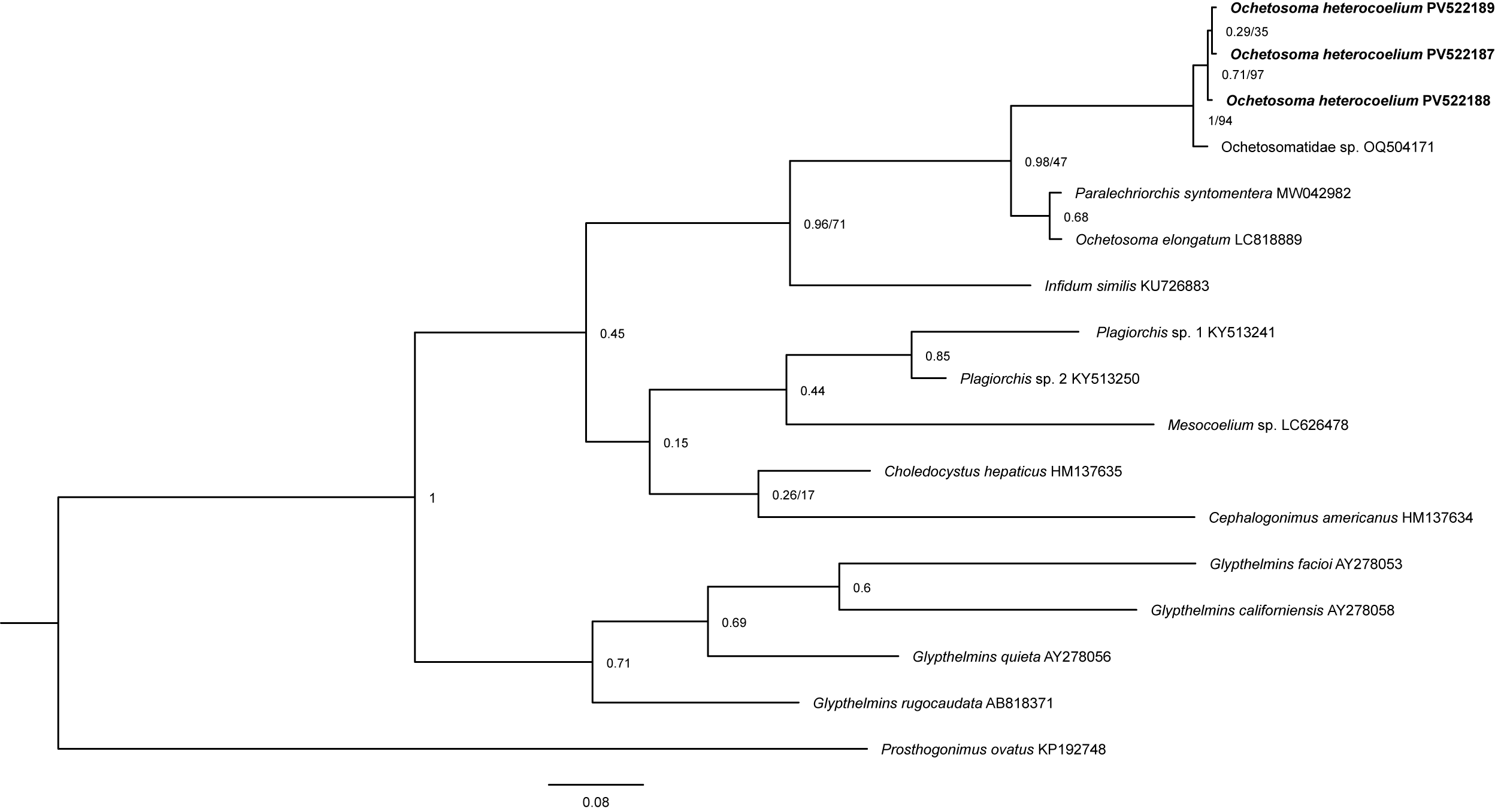
For the mitochondrial COI gene, three partial sequences from different specimens of O. heterocoelium were successfully sequenced and aligned with 14 digeneans belonging to the families Ochetosomatidae, Cephalogonimidae, Glypthelminthidae, Plagiorchiidae, Mesocoeliidae, and Prosthogonimidae; the latter was used as an outgroup with the taxon Prosthogonimus ovatus available in GenBank (Figure 5). The final COI alignment consisted of 195 bp long (Accession numbers will be given after manuscript acceptance). All sequence fragments of O. heterocoelium were identical. The genetically closest was Ochetosomatidae gen. sp., which is the only sequence available in GenBank for species of the genus with a divergence of 1% which corresponds to the sequence of a cercaria and is conspecific with O. heterocoelium. The sister taxon is Paralechriorchis syntomentera (Sumwalt, 1926) in which amphibians are intermediate hosts and snakes are definitive hosts which belongs to Ochetosomatidae with a divergence of 11%, this taxon was close to the other sequence of the genus, O. elongatum, with only 1.5% divergence between them.
The tree is a Bayesian topolgy based on partial mitochondrial COI gene DNA sequences of Digenea. GenBank accession numbers are indicated next to species names. Numbers in the nodes represent supported nodes by posterior probabilities for Bayesian analyses (decimal numbers) and bootstrap for maximum likelihood analyses. The branch length scale bar indicates the number of substitutions per site.

Discussion
The genus Ochetosoma includes about 36 species around the world (Yamaguti Reference Yamaguti1971). In South America, only three Ochetosoma species have been reported infecting snakes. Ochetosoma heterocoelium is the only species of this genus reported in Brazil (Fernandes and Cohn Reference Fernandes and Kohn2014). This species differs from the other congeners mainly in the extension of the intestinal caeca and the size of the cirrus-sac (Dubois and Mahon Reference Dubois and Mahon1959). After the original description by Travassos (Reference Travassos1921), new morphological data on the species as well as new hosts and new geographical records have been published (Correa et al. 1999; Pinto et al. Reference Pinto, Mati and Melo2012; Silva et al. Reference Silva, Rodrigues, Stein, Sipoli, Pinhão and Lopes1999; Vicente-Ferreira et al. Reference Vicente-Ferreira, do Nascimento, Grezzana, Ávila and Garey2023).
We describe for the first time the presence of an anterior tegumental thickening in the anterior end of O. heterocoelium which was clearly observed in stained specimens and through SEM. We confirmed the presence of the structure by studying the holotype of O. heterocoelium deposited in the Coleção Helmintológica of the Instituto Oswaldo Cruz (CHIOC 973d) (Figure 6A, B). Surprisingly, this structure was represented by Travassos (Reference Travassos1921) and Correa et al. (Reference Corrêa, Paulino, Buononato and Federsoni1990) in the illustrations of these publications, but it was not mentioned in the description in both studies.
Ochetosoma heterocoelium. A. Whole body, redrawn from Travassos (1921), highlighting the anterior tegumental thickening (att, full black arrow); B. Holotype deposited in the Coleção Helmintológica of the Instituto Oswaldo Cruz (CHIOC 973d) - detail of the oral sucker with anterior tegumental thickening (full black arrow); C. Voucher from Colombia after Lenis et al. (Reference Lenis, Arredondo and Calle2009) deposited in the Colección Nacional de Helmintos, Instituto de Biología, Universidad Nacional Autónoma de México (CNHE 6459), whole body; and D. Detail of the oral sucker showing the lack of the anterior tegumental thickening (dotted black arrow).

A redescription of O. heterocoelium based on specimens collected from several snake species in Colombia was conducted by Lenis et al. (Reference Lenis, Arredondo and Calle2009). However, when comparing the redescription provided by these authors with our specimens, we uncovered some important differences. For instance, anterior tegumental thickening was not described or illustrated in the study by Lenis et al. (Reference Lenis, Arredondo and Calle2009). We analyzed two voucher specimens deposited in the Colección Nacional de Helmintos, Instituto de Biología, Universidad Nacional Autónoma de México (CNHE 6459), and we confirmed that the specimens lacked such tegumental thickening associated with the oral sucker (Figure 6C, D). Furthermore, Travassos (Reference Travassos1921) described the intestinal caeca of O. heterocoelium as broad, short, slightly unequal, and ending in the ventral sucker level, with the longest being on the opposite side of the genital pore (i.e., the right cecum). Lenis et al. (Reference Lenis, Arredondo and Calle2009)’s redescription shows the same, but no further comments were provided about which cecum was longest. We conducted a detailed morphological study of 40 newly sampled specimens and found that this characteristic is variable in O. heterocoelium; 40% of the specimens exhibited the right cecum to be the longest. Lenis et al. (Reference Lenis, Arredondo and Calle2009) reported that O. heterocoelium possessed an efferent and defferent duct dorsal to the ventral sucker, although this is not observed in the illustrations. We observed, however, that O. heterocoelium lacks a defferent duct. Efferent ducts from each testis directly reach the seminal vesicle at the base of the cirrus sac (Figure 2D). Finally, Lenis et al. (Reference Lenis, Arredondo and Calle2009) described the position of the genital pore at the pharynx level which is more consistent with other Ochetosoma species, such as Ochetosoma ellipticus Pratt 1902 and Ochetosoma validum (Nicol, 1911) (Tkach Reference Tkach2008). Instead, we observed that in all 40 individuals analyzed, the genital pore always opens at the level of the oesophagus. Based on these divergent morphologies pointed out here, we concluded that O. heterocoelium found in Colombia and reported by Lenis et al. (Reference Lenis, Arredondo and Calle2009) must be considered as species inquirenda since it probably corresponds to an undescribed species of Ochetosoma; molecular data will be necessary to confirm this hypothesis.
In Brazil, digeneans frequently are fixed using a slight coverglass pressure that is sometimes useful for a better observation of internal anatomy. This methodology was commonly used by Brazilian helminthologists such as Travassos, and as a result, specimens deposited in parasite collections are mostly flattened; because of that, for an accurate morphological comparison with newly sampled individuals, it is convenient to have some specimens fixed in the same way. In addition, we compared the morphology of flattened and non-flattened specimens collected in this study. Most of the analysed variables were statistically different (Table 1). However, when comparing these data with other studies providing morphometrical data of O. heterocoelium (Lenis et al. Reference Lenis, Arredondo and Calle2009; Pinto et al. Reference Pinto, Mati and Melo2012; Travassos Reference Travassos1921), we found that irrespective of fixation procedure, the range of data obtained was consistent with the description of this digenean species.
For the first time, scanning electron microscopy was used to analyze O. heterocoelium (Figure 3). This method shows more accurately the structures not easily visible when analyzed through a light microscope. The spines that cover the entire body of the trematode are different in size, and the spines in the dorsal region near the anterior and posterior ends are reduced in size in relation to the spines in other areas of the parasite’s body. The anterior tegumental thickening associated with the anterior portion of the oral sucker was also observed through SEM, a feature never reported for the species; the opening of the Laurer’s canal was also observed using SEM.
Molecular analysis
The topology of the 28S phylogenetic tree is relatively congruent with that reported by Bespalaya et al. (Reference Bespalaya, Kondakov, Travina, Khrebtova, Kropotin, Aksenova, Gofarov, Lyubas, Tomilova and Vikhrev2022). These authors obtained similar results for the position of Opisthioglyphe ranae Frölich, 1791 within the family Telorchiidae.
Tkach et al. (Reference Tkach, Snyder and Swiderski2001) conducted a phylogenetic analysis of plagiorchiids showing Telorchiidae as a closely related group to Ochetosomatidae. Morphologically, species included in these families are morphologically similar and several traits of their life cycles are common (Macdonald and Brooks Reference Macdonald and Brooks1989). Still, in our analysis, Infidum similis Travassos, 1916, a dicrocoeliid, and Choledocystus hepaticus Lutz, 1928, a plagiorchiid, appear as the sister taxon of Ochetosomatidae. In a previous 28S phylogenetic analysis, at least C. hepaticus appeared as a sister taxon of Ochetosomatidae (Razo-Mendivil et al. Reference Razo-Mendivil, León-Règagnon and de Leon2006); however, for a more reliable taxonomic classification, taxa should be added for molecular studies and included for phylogenetic analysis (Tkach et al. Reference Tkach, Snyder and Swiderski2001).
In the present study, a difference in the phylogenetic position of Ochetosomatidae and Telorchiidae was found in comparison to previous studies; some taxa were added, such as Lechriorchis tygarti Talbot, 1933 and Infidum infidum (Faria, 1910).
The phylogenetic discrepancies uncovered in our study for members of the suborder Plagiorchiata were frequent before molecular studies, especially for some families belonging to this suborder, including the members of the family Ochetosomatidae. There were several taxonomic reassessments for the group due to studies on phylogeny being based exclusively on morphological characters, but they were not useful in resolving the classification of this group of digeneans. As pointed out by Tkack et al. (2010), these highly derived digenetic groups exhibit high levels of homoplasy, and their life cycles are poorly known.
Species in the genus Ochetosoma are distributed in the Nearctic, Neotropical, Palearctic, and Eastern biogeographical regions (Dyer Reference Dyer and Ballard1989; Santoro et al. Reference Santoro, Tkach, Mattiucci, Kinsella and Nascetti2011). Information on the life cycle of species of Ochetosoma is scarce, but it is known that their species exhibit a complex life cycle with three hosts – snails and amphibians as intermediate hosts, and snakes as definitive hosts. When these reptiles ingest an amphibian infected with the metacercariae, the adult parasites develop in the mouth and oesophagus and thus complete the parasite life cycle (Santoro et al. Reference Santoro, Tkach, Mattiucci, Kinsella and Nascetti2011). For O. heterocoelium, the information on the life cycle is fragmentary. The metacercariae of O. heterocoelium occurring on the external intestinal wall in amphibians was reported by Pinto and Melo (Reference Pinto and Melo2012) in Brazil. In addition, a xiphidiocercaria from the physid snail Stenophysa marmorata from Brazil was recently identified as a member of Ochetosomatidae and possibly of the genus Ochetosoma (López-Hernández et al. Reference López-Hernández, de Melo and Pinto2023). The present study confirms the hypothesis of these authors. Our three sequences of O. heterocoelium grouped together with the sequence of the xiphidiocercaria, and the divergence of the genes is very low (0.2%), allowing us to conclude that the S. marmorata is the first intermediate host for O. heterocoelium, and amphibians act as the second intermediate host in the life cycle of this parasite.
Another ochetosomatid, Dasymetra nicolli, grouped phylogenetically in the same clade as O. heterocoelium. The life cycle of Dasymetra villicaeca Byrd, Reference Byrd1935 is very similar to that of O. aniarum; both species infect the same definitive hosts, and the larval stages infect the same intermediate hosts (Byrd Reference Byrd1935). More sequence data is required to find a stable classification system for this group of plagiorchiid trematodes, and the study of the life cycle of more species will add relevant information to understand the phylogenetic interrelationships.
Supplementary material
The supplementary material for this article can be found at http://doi.org/10.1017/S0022149X25000343.
Acknowledgements
We sincerely thank researcher Julio Cesar Cenci de Aguiar for the support and intellectual insights that contributed to the preparation of this article.
Financial support
This study was supported by Fundação de Amparo à Pesquisa do Estado de São Paulo (FAPESP #2018/09623-4, #2023/07336-6, and #2018/00754-9) and Conselho Nacional de Desenvolvimento Científico e Tecnológico (CNPq) (R.J.S. #311635-2021-0, and E.E.P.C. #140903/2022-4). R.J.S. and G.P.P.L. were also supported by CAPES/PRINT (#88887.839573/2023-00 and #88887.839159/2023-00, respectively).
Competing interests
On behalf of all authors, the corresponding author states that there is no conflict of interest.
Ethical standard
The authors assert that all procedures contributing to this work comply with the ethical standards of the relevant national and institutional guides on the care and use of laboratory animals.