Introduction
Polyploidization occurs ubiquitously in nature and is known as an important factor in evolution that brings about various biological advantages. In yeast, the ability to adapt to environmental changes is improved by polyploidization (Selmecki et al., Reference Selmecki, Maruvka, Richmond, Guillet, Shoresh, Sorenson, De, Kishony, Michor, Dowell and Pellman2015). In plants, ancestral whole genome duplications have resulted in seed and flower improvements, which are driven by gene diversification (Jiao et al., Reference Jiao, Wichett, Ayyampalayam, Chanderbali, Landherr, Ralph, Tomsho, Hu, Liang, Soltis, Soltis, Clifton, Schlarbaum, Schuster, Ma, Leebens-Mack and dePamphilis2011). In vertebrates, multiple polyploidization, acquired by whole genome duplication, has promoted species evolution (Ohno, Reference Ohno1970; Holland et al., Reference Holland, Garcia-Fernandez, Williams and Sidow1994), and is supported by recent genomic sequence analysis, propounded as the 2R hypothesis (Panopoulou and Poustka, Reference Panopoulou and Poustka2005). Polyploidy exists in fish or amphibians, however polyploid mammals have exhibited various abnormalities during embryogenesis (Eakin et al., Reference Eakin, Hadjantonakis, Papaioannou and Behringer2005; Olzer et al., Reference Olzer, Ersoy, Oztas, Topcu, Celen and Danisman2015). Polyploidization triggers failures of cell division or defects in spindle assembly checkpoints (Eakin and Behringer, Reference Eakin and Behringer2003; Schmid et al., Reference Schmid, Steinlein, Tian, Hanlon Newell, Gessler, Olson, Rosenwald, Kneitz and Fedorov2014). Abnormal expression of cell division or cell cycle-related genes are also speculated as candidates for the lethality or abnormal development in mammalian tetraploid embryos (Kawaguchi et al., Reference Kawaguchi, Kano and Naito2009; Park et al., Reference Park, Hwang, Bui, Cho, Park, Oh and Kim2012). Other reports have shown that the more polyploidization, the less the number of the inner cell mass in blastocysts (Liu et al., Reference Liu, Czerwiec and Keefe2004; Lei et al., Reference Lei, Guan, Zu, Zhang, Shen and Jin2009). Tetraploid embryonic stem cells established from mouse tetraploid blastocysts have shown increment in apoptosis, and decrement of proliferation rate and meiosis-related gene expression (Horii et al., Reference Horii, Yamamoto, Morita, Kimura, Nagao and Hatada2015; Imai et al., Reference Imai, Kano, Fujii, Takasawa, Wakitani, Hiyama, Nishino, Kusakabe and Kiso2015; Wen et al., Reference Wen, Li, Cheng, Li, Zhang, Xiang, Wang and Han2017).
Most previous studies, focusing on tetraploid embryos or embryonic stem cells, have implied that various factors might be involved in abnormal development of mammalian polyploid embryos. However, how the extent to which polyploidization affects cell function has not yet been elucidated.
In our previous study, we demonstrate that tetraploid, octaploid, and even hexadecaploid, embryos had full capability to develop to the blastocyst stage with a full inner cell mass (Imai et al., Reference Imai, Fujii, Kusakabe, Kiso and Kano2019). Here we demonstrate for the first time that octaploid embryos might have the ability to implant and even develop in the uterus in mice.
Materials and methods
Recovery of mouse embryos and polyploidization
BDF1 female mice were ovulated by intraperitoneal (i.p.) injection of pregnant mare’s serum gonadotropin (Aska Pharmaceutical Co., Ltd) and human chorionic gonadotropin (Aska Pharmaceutical Co., Ltd), and then mated with BDF1 males. Two-cell-stage embryos were collected at 1.5 days post coitum (d.p.c.) and polyploidized using the electrofusion method. Embryos were cultured in M16 medium (Sigma).
Transfer of blastocysts
Blastocyst stage embryos were transferred into the uterine horn of pseudopregnant ICR female mice (at 2.5 d.p.c.; 5–8 embryos for each uterine horn) that had been anaesthetised with a combination of medetomidine, midazoram and butorphanol (Kawai et al., Reference Kawai, Takagi, Kaneko and Kunosawa2011). Following closure of the surgical incision, the mice were roused by i.p. injection of atipamezole. Carprofen was used for pain management. Mice were subjected to euthanasia by cervical dislocation at 7.5 d.p.c. The uterus was fixed with Bouin’s solution, embedded in paraffin, and serial sections were prepared and stained with haematoxylin and eosin.
Results and discussion
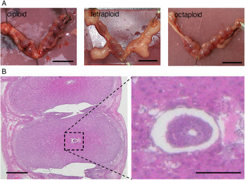
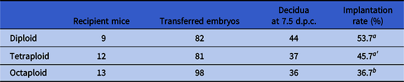
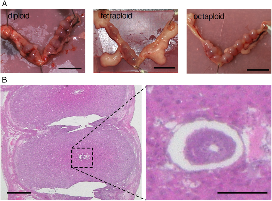
Decidua were formed at 7.5 d.p.c.in all uteri in which octaploid embryos were transferred, as for diploid and tetraploid embryos (Fig. 1A). However, the rate of implantation in octaploid embryos was significantly lower than that in diploid or tetraploid embryos (Table 1). As the total number of cells in polyploid embryos was lower than that of diploid embryos (Imai et al., Reference Imai, Fujii, Kusakabe, Kiso and Kano2019), we prepared serial sections that allowed comprehensive observation of whole uterine tissues, including in this study the whole concepti, rather than recovering the implanted embryos from decidua. Some octaploid embryos transferred into the uteri were observed in the decidua at the egg cylinder stage (Fig. 1B). Previous reports have indicated that octaploid embryos were absorbed and disappeared after implantation, leaving only empty decidua in mice (Wu et al., Reference Wu, Zhao, Zhu, Chen, Wei, Bao, Sun and Li2017). Wu and colleagues might have to failed to recover implanted octaploid embryos from the decidua by harvesting, because the polyploidized embryos were smaller than normal implanted embryos. Therefore, we adapted another approach to detect implanted octaploid embryos using whole uterine tissue analysis by serial sectioning. Our present study is the first to report that early octaploid embryos have the ability to implant and develop in the uterus after implantation, despite hyper-polyploidization in mammals.
Implantation and decidual formation of polyploid embryos. (A) Decidua-formed uteri after polyploid embryo transfer are shown. Uteri transferred with diploid, tetraploid and octaploid embryos are shown left to right. Bars, 1.0 cm. (B) Haematoxylin and eosin (H&E) staining of the implantation embryo after octaploid embryo transfer. A higher magnification image of the octaploid implantation embryo is shown on the right. Bar, 500 µm (left); 100 µm (right).
Implantation rate

Different letters a and b indicate significant differences at P < 0.05; a and a′ indicate approached significance at P < 0.1.
In haploid embryos, primitive streak formation is absent (Hu et al., Reference Hu, Zhao, TuanMu, Wei, Gao, Li, Ying and Zhang2015) while they develop to the egg cylinder stage (Graham, Reference Graham1970). In tetraploid embryos, lengthening of cell cycles and growth retardation have been reported in mice (Eakin et al., Reference Eakin, Hadjantonakis, Papaioannou and Behringer2005). In human embryos, chromosomal abnormality causes developmental defects through dysfunction of epithelial–mesenchymal transition (Kukharenko, Reference Kukharenko2018). The lower rate of implantation in polyploid embryos might reflect these abnormalities, in which the maternal immune and endocrine system is related to pregnancy maintenance and post-implantation development in the uterus (Robertson et al., Reference Robertson, Care and Moldenhauer2018). Although embryo morphology is correlated with chromosomal abnormalities and implantation rates (Gazzo et al., Reference Gazzo, Pena, Valdez, Chung, Velit, Ascenzo and Escudero2019), no report has shown a direct relationship between embryo chromosome number and implantation rates. Detailed mechanisms as to why whole genome duplication occurs and how the extent of polyploidization is involved in aberrational cell function in the early development of implanting embryo are still unknown. There is a need to study hyper-polyploid embryos, produced by repeated whole genome duplication, because most previous studies have only analyzed tetraploid embryos.
Here we describe for the first time that octaploid embryos have the potential to survive after implantation, and indicate that hyper-polyploid embryos have the ability to implant and even develop despite the harmful effect in mammals of larger genome content.
Our results could suggest a new model for analyzing the development of polyploid embryos that could unveil polyploidy development characteristics in mammals.
Financial support
This work was supported by Grant-in-Aid for Challenging Exploratory Research (K.K., JP25660254, JP15K14880) and Grant-in-Aid for JSPS Fellows (H.I., JP17J07902).
Conflicts of interest
All authors declare no conflicts of interest associated with this manuscript.
Ethical standards
All animal experiments were approved by the Experimental Animal Care and Use Committee of Yamaguchi University (protocol number: 267). Mice were housed in groups with ad libitum access to water and food.