Mucopolysaccharidosis are lysosomal storage diseases, characterised by deficient enzymatic degradation of glycosaminoglycanes: hyaluronic acid, chondroitin sulphate, dermatan sulphate, heparan sulphate, and keratan sulphate.Reference Aumailley and Gayraud1 The classification is based on the defective enzyme and seven types with heterogeneous clinical manifestations have been described: types I, II, III, IV, VI, VII, and IX.Reference Guertl, Noehammer and Hoefler2
Cardiovascular involvement is variable; however, cardiopulmonary failure contributes significantly towards morbidity and mortality in adults.Reference Muenzer3 Cytoplasmic vacuoles full of glycosaminoglycanes are observed in endothelial cells, myocyte and fibroblasts compromising the structure and function of endocardium, myocardium, valves, coronary arteries, conduction system, great vessels, and lung and systemic vasculature.Reference Robins, Kumar and Cotran4–Reference Hishitani, Wakita and Isoda6
It is speculated that cardiac lesions are more severe in patients whose enzymatic defect leads to the accumulation of dermatan sulphate (types I, II, VI, and VII), because this glycosaminoglycane prevails naturally in the valves and the blood vessels.Reference Dangel7, Reference Rigante and Segni8
On account of the perspective of the specific treatment for mucopolysaccharidosis through enzymatic replacement,Reference Braulin, Berry and Whitley9 it is essential to know the initial cardiovascular abnormalities to safely determine the impact of therapy during children development.
Materials and methods
The medical records and echocardiograms of 28 patients (15 male and 13 female) with mucopolysaccharidosis, aged 2–14 years, with a mean age of 9 years and a standard deviation of 3 years, seen at the Genetic Clinic between September, 2003 and November, 2005 were analysed retrospectively: six with type I, two with type II, six with type III, seven with type IV, five with type VI, and two with type VII. In all patients, the diagnosis was confirmed enzymatically. During the period of study no patient had enzymatic replacement, a therapy that is now available for types I, II, and VI at our hospital. The revision of the ambulatory medical records made it possible to identify alterations at cardiac auscultation (tachycardia, murmurs, second cardiac sound hyperphonesis, and presence of third cardiac sound) and cardiovascular symptoms (exercise dyspnoea and orthopnoea) detected by the paediatricians. Polysomnography test results were also recorded.
A single paediatric cardiologist executed 53 echocardiograms, as 17 patients underwent two or more examinations, with a mean interval of 10.3 months and a standard deviation of 5.6 months. None of the patients was receiving cardiovascular medications at the time of the echocardiograms.
Before each examination, weight and stature were recorded and the body surface area was calculated by the Dubois and DuboisReference Dubois and Dubois10 formula. A 12-lead electrocardiogram record was also obtained.
All the echocardiograms were performed at rest, without sedation, and included the M- and two-dimensional modes, besides Doppler examination with colour flow mapping. The ultrasound systems used were General Electric Logiq-500 and General Electric cardiovascular System Vivid-3 (Milwaukee, Wisconsin, United States of America), both equipped with electronic transducers from 2 to 7.5 megahertz.
The diastolic and systolic diameters were measured using the M-mode, as also the thickness of the septum and of the left ventricle posterior wall. The values obtained were compared to the expected average for the body surface,Reference Kampmann, Wiethoff and Wenzel11 allowing the calculation of the Z-score for each measure. The values of Z-score were considered normal between −2 and +2.
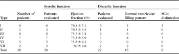
The systolic function of the left ventricle was evaluated through the ejection fraction obtained by the Teichholz method, and values equal to or above 55% were considered normal.
It was possible to characterise the diastolic function of the left ventricle through the ventricular filling pattern,Reference O’Leary, Durongpisitkul and Cordes12 when there was no mitral valve dysfunction worse than mild regurgitation.
The systolic pressure of the pulmonary artery was estimated through the tricuspid insufficiency and the mean pressure was estimated through pulmonary insufficiency. Pulmonary hypertension was diagnosed whenever the systolic pressure exceeded 35 millilitres of mercury and/or the mean pressure exceeded 25 millilitres of mercury.Reference Carvalho, Almeida and Lopes13
The morphological aspect of the valves was evaluated by the two-dimensional mode. The severity of mitral and aortic regurgitation, and of aortic stenosis, was determined according to the recommendations of the American Society of Echocardiography.Reference Zoghbi, Sarano and Foster14 The mitral valve orifice area was determined by planimetry at the parasternal short-axis view. The valve was considered stenotic whenever the area obtained at the tip of the leaflets was inferior to the third percentile expected for the patient’s body surface.Reference Riggs, Lapin, Paul, Muster and Berry15
Initially, all the patients were analysed according to both clinical and echocardiographic parameters, and then a comparison was made among the patients who accumulated (mucopolysaccharidosis types I, II, VI, and VII) and those who did not accumulate dermatan sulphate (mucopolysaccharidosis types III and IV).
The statistical program used was the Statistical Package for the Social Sciences and the applied tests were the Fisher’s exact test and the Spearman correlation, where a p-value < 0.05 was considered significant.
The study was approved by the Hospital’s Commission for Ethics in Research and was carried out without any subsidy.
Results
Global evaluation
Of the 28 patients that were studied, 26 (93%) showed some echocardiographic alteration at the final examination. However, abnormal auscultation was recorded in 16 patients (57%) and only 6 (21%) presented any cardiovascular complaint.
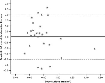
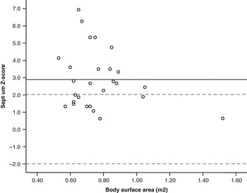
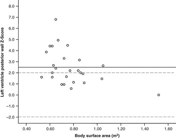
The Z-score values of left ventricle diastolic diameter, inter-ventricular septum, and posterior wall are shown in graphs 1, 2, and 3, respectively.
Z-score values for diastolic left ventricle diameter. Continuous line represents the mean Z-score of the studied population. The normal range is bounded by dashed lines.

Z-score values for inter-ventricular septum. Continuous line represents the mean Z-score of the studied population. The normal range is bounded by dashed lines.

Z-score values for left ventricle posterior wall. Continuous line represents the mean Z-score of the studied population. The normal range is bounded by dashed lines.

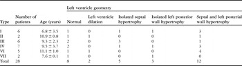
Only 7% of the patients presented ventricular dilation. Septum and posterior wall hypertrophy were diagnosed in 43% and 18% showed signs of isolated septal hypertrophy (Table 1). In all those cases, electrocardiography failed to detect any sign of left ventricle hypertrophy.
Evaluation of left ventricle geometry in each type of mucopolysaccharidosis.

It was possible to evaluate the diastolic function of the left ventricle through the combination of mitral valve and pulmonary venous Doppler profile in 22 patients. Of these, six presented mild dysfunction. However, all the patients had a preserved systolic function (Table 2).
Left ventricle function in each type of mucopolysaccharidosis.

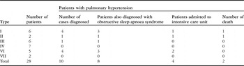
Pulmonary hypertension was detected in 10 patients (36%), of which eight were diagnosed with obstructive sleep apnoea syndrome, by polysomnography. During the study period, four patients were admitted in the Intensive Care Unit and two of them died because of the aggravation of pulmonary hypertension in the presence of a respiratory infection (Table 3).
Pulmonary hypertension in each type of mucopolysaccharidosis.

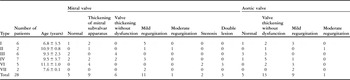
A normal mitral valve was found in 17.8% patients and thickening without dysfunction in 21.4% of patients. Mitral regurgitation occurred in 42.8%, mitral stenosis in 7.2%, and double lesion in 10.8% patients. Thickening of mitral subvalvar apparatus was diagnosed in 32% of the patients. The aortic valve was considered normal in 17.8% and thickened without dysfunction in 46.4% of the patients. Thickening with dysfunction occurred in 35.8% of the patients, all with mild or moderate aortic regurgitation. Table 4 shows the results of the evaluation of the mitral and aortic valves in each type of mucopolysaccharidosis.
Evaluation of mitral and aortic valves in each type of mucopolysaccharidosis.

Of the 17 patients with two or more examinations, 14 (82%) showed echocardiographic worsening, justified by the appearance (4 out of 14) or aggravation (6 out of 14) of valve lesions, appearance (5 out of 14) or progression (6 out of 14) of ventricular hypertrophy, development of left ventricle diastolic dysfunction (1 out of 14), and pulmonary hypertension (4 out of 14).
Comparison between patients who accumulate and those who do not accumulate dermatan sulphate
The number of cases and the age at the last examination were similar, comparing the group that accumulates, 15 patients with a mean age of 8.9 years and a standard deviation of 3 years, and the group that does not accumulate dermatan sulphate, 13 patients with a mean age of 9.4 years and a standard deviation of 3.3 years.
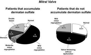
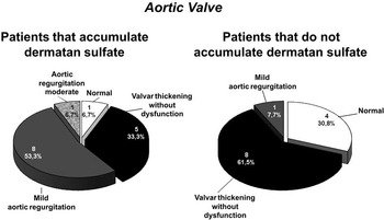
Among patients who accumulate dermatan sulphate, mitral valve dysfunction was found in 93.3% and aortic valve dysfunction in 60%. Among patients who do not accumulate this glycosaminoglycane, only 23% exhibited mitral dysfunction and 7.7% had aortic dysfunction (Figs 1 and 2).
Evaluation of mitral valve in patients that accumulate and that do not accumulate dermatan sulfate.

Evaluation of aortic valve in patients that accumulate and that do not accumulate dermatan sulfate.

There was a strong association between the accumulation of dermatan sulphate and the presence of mitral valve dysfunction (p = 0.0003), aortic valve dysfunction (p = 0.006), mitral and concomitant aortic dysfunction (p = 0.006), and pulmonary hypertension (p = 0.0032).
There was also significant correlation between age and the severity of mitral lesion in both groups: p = 0.001 for those that accumulate and p = 0.012 for those that do not accumulate dermatan sulphate.
Discussion
Practically, all the papers already published concerning cardiovascular involvement in mucopolysaccharidosis included adult patients.Reference Dangel7, Reference Rigante and Segni8, Reference Gross, Julian, Caprioli, Dominguez and Howell16–Reference Nelson, Shields and Mulholand18 The decision to evaluate a paediatric population certainly enabled particular findings in this study.
In agreement with literature, the echocardiographic alterations were more common than the cardiovascular signs and symptoms registered, since thoracic deformities preclude auscultation and exercise limitation can be erroneously attributed to joint lesions or to respiratory failure.Reference Rigante and Segni8
In addition, electrocardiography may not detect ventricular hypertrophy, since the mucopolysaccharide material is likely to be electrically non-conducting.Reference Nelson, Shields and Mulholand18
As has already been shown by other authors, the mitral valve was the most affected structure in spite of the type of mucopolysaccharidosis.Reference Mohan, Hay, Cleary, Wraith and Patel17 A significant correlation between age and the severity of the mitral valve lesion was also verified, varying from discreet thickening to total restriction of the leaflet movement.
Septum and left ventricle posterior wall hypertrophy were more frequently described (43%) than in the group appraised by DangelReference Dangel7 (28%). However, only 7% of the patients presented dilation of the left ventricle against 24% described by Gross et al.Reference Gross, Julian, Caprioli, Dominguez and Howell16
Unlike Mohan et al,Reference Mohan, Hay, Cleary, Wraith and Patel17 who found 13% of the patients with moderate-to-serious systolic dysfunction, the ejection fraction of the left ventricle was normal in this study. Considering that the diastolic dysfunction usually precedes the systolic dysfunction and that myocardial deposits of glycosaminoglycanes potentially affect the ventricular filling,Reference Soliman, Timmermans and Nemes19 it was not surprising to find 25% of patients with mild diastolic dysfunction.
These facts suggest that hypertrophy and diastolic dysfunction could begin at an early stage and that ventricular dilation and systolic dysfunction occur more often in older individuals. Unfortunately, not all patients had Tissue Doppler evaluation during their routine echocardiograms (data not shown). That technique could have identified systolic and diastolic ventricular dysfunctions if universally applied in a larger number of patients.
The factors that are implicated in the genesis of pulmonary hypertension are frequent in patients with mucopolysaccharidosis: left cardiac valve lesions, deposits of glycosaminoglycanes in pulmonary vascular bed, thoracic deformities, frequent pneumonias, and obstructive apnoea.Reference Neufeld and Muenzer20 In fact, 80% of the studied patients with pulmonary hypertension also had apnoea events during polysomnography.
The diagnosis of pulmonary hypertension was, however, frequent (36%) or related to death (two cases) in very few studies.Reference Chan, Li, Yam, Li and Fok21, Reference Walker, Darowski, Morris and Wraith22
Contrary to adult patients, pulmonary hypertension probably has a more important role in morbidity and mortality than left ventricle systolic dysfunction. For this reason, special attention must be given to its early detection and treatment in the paediatric population.
Valve lesions in the mucopolysaccharidosis I group were considered mild when compared to those previously reported by ButmanReference Butman, Karl and Copeland23 and Kraiem et al.Reference Kraiem, Lahidheb and Chehaibi24 Nevertheless, ventricular hypertrophy was detected in 83% and diastolic dysfunction in 50% of patients with type I evaluated in this study.
One of the two patients with type II showed pulmonary hypertension and severe sleep apnoea, as described by Shapiro et al.Reference Shapiro, Strome and Crocker25 This association undoubtedly contributed to his death.
The involvement of the heart in type III was not rare, although the lesions described here were mild, as also reported by Gross et al.Reference Gross, Julian, Caprioli, Dominguez and Howell16
Contradicting Mohan et alReference Mohan, Hay, Cleary, Wraith and Patel17 and PierpontReference Pierpont and Moller26 and in agreement with John et al,Reference John, Hunter and Swanton27 aortic regurgitation was not shown as a characteristic lesion of type IV, and also occurred in eight patients with other types of mucopolysaccharidosis.
The more severe cases of mitral involvement were detected in type VI. Since these were also the oldest patients with a mean age of 11 years, it was not possible to assign the severity of the lesions to that particular type of mucopolysaccharidosis.
No patient with type VII showed aortic root dilation or aortic obstruction, contradicting the conclusions of Schuldt et alReference Schuldt, Hampton and Chu28 and Honjo et al.Reference Honjo, Ishino, Kawada, Ohtsuki and Sano29
Despite the short follow-up period, the progressive character of cardiac lesions and the strong association between the accumulation of dermatan sulphate and the severity of the cardiovascular damage were proven, and these facts were previously suggested by Dangel.Reference Dangel7
Prospective studies should be performed not only to monitor the evolution of the findings described here, but also to determine the impact of the new therapeutics on the natural history of the pathology.