Introduction
Radiotherapy is a treatment technique which uses radiation to kill cancer cells. The aim of radiotherapy is to deliver maximum sufficient dose to the tumour while sparing the normal tissues as much as possible. Breast cancer accounts for almost 12% of the cancer occurrences found internationally.Reference Bray, Ferlay and Soerjomataram1 Radiotherapy is a well-known treatment technique in addition to chemotherapy or hormone therapy for breast cancer patients post-surgery (adjuvant therapy). Adjuvant therapy after mastectomy or lumpectomy has shown to contribute to better tumour control and reduction in the locoregional recurrences.Reference Yang, Chen and Liu2,Reference Clarke, Collins and Darby3 Fisher et al. (2002) found almost a 60% reduction in the local recurrence risk in breast conservative surgeries along with radiotherapy. Also, the survival rates were comparable with mastectomy patients.Reference Fisher, Anderson and Bryant4
Radiotherapy has evolved tremendously in terms of accuracy in treatment delivery and normal tissue dose sparing which in turn reduces cancer mortality and chronic long-term side effects. However, patients dying due to side effects cannot be completely avoided particularly in the case of carcinoma breast, where late cardiac toxicities have been reported even with the improvements in the shielding in advanced radiotherapy techniquesReference Noel and Mazeron5 There is clear evidence of a linear–no threshold relationship between the dose to the heart and the late cardiac toxicities. Therefore, it is important to reduce the heart dose to as low as possible.Reference Darby, Ewertz and McGale6 Pulmonary complications like radiation-induced pneumonitis have also been reported in 1–5% of cases, post-radiotherapy. With conventional tangential radiotherapy, the heart volume overlapping with the radiation field is increased which contributes to the risk of cardiac toxicity, whereas in other advanced delivery techniques like intensity-modulated radiotherapy (IMRT) and volumetric modulated arc therapy (VMAT) may improve the dose distribution between target and non-target tissue, but they may also increase the lower dose volume to the lungs and normal tissues which might increase the chances of secondary malignancies.Reference Rudat, Alaradi and Mohamed7,Reference Hall and Wuu8
Deep inspiration breath-hold (DIBH) technique is widely adopted in left-sided breast cancer patients after breast conservation surgeries and mastectomy. In this method, the breast and chest wall are pushed anteriorly/superiorly and the heart is pushed inferiorly which reduces the volume of heart coming inside the radiation field when compared to normal free breathing (FB) scans. It also increases the lung volume and helps in improving the lung sparing efficiently.Reference Sixel, Aznar and Ung9 DIBH can be done by two methods: one is voluntary DIBH and the other one is moderate DIBH. For voluntary DIBH, patients are instructed about deep inspiration for a period of time and respiratory motion is being monitored by the Real-time Position Management™ (RPM) system (Varian Medical Systems, Palo Alto, CA, USA) using an infrared camera and reflector. During both simulation and treatment, DIBH is continued so that the heart dose can be reduced.Reference Zagar, Kaidar-Person and Tang10
The RPM measures the vertical displacement of abdomen or sternum and provides a relative value with respect to the patient’s breathing baseline.Reference Pepin, Wu and Shirato11 The treatment can be gated so that it can be stopped if the breathing cycle falls outside the preset threshold value. The other method is moderate DIBH, a technique which is used in devices like active breathing control (Elekta, Stockholm, Sweden). These devices use a spirometer which allows for monitoring of air flow throughout the respiratory cycle and stopping airflow at a set threshold volume, causing the patient to hold their breath to maintain this volume.Reference Wong, Sharpe and Jaffray12 In RPM gating, there are two types of gating methods: one is amplitude gating and the other one is phase gating.
In amplitude gating, the gated range is defined by an upper and lower threshold which is known as a breath-hold window (BHW), simulation and treatment is carried out only during this phase. BHW plays a vital role in treatment efficiency because this could lead to treatment uncertainties in terms of target coverage and organ at risk (OAR) sparing if not properly selected. Keeping a shorter window would actually increase the treatment time if the patient was not able to hold the breath for a long time and keeping it longer would give more uncertainty in critical structure doses and target coverage. There are some studies suggesting that a 5 mm BHW would be an optimal window level and this is being widely followed.Reference Rusu, Choi and Thomas13,Reference Hayden, Rains and Tiver14
The treatment can be performed while selecting a 5 mm BHW, only if the inspiration lies anywhere within the window level. Inspiration level may not be uniform during the entire course of the treatment, and it may vary from time to time. It may cause differences in the position, size of the target and critical structures compared to the planned treatment. This can lead to variations in target and critical structure doses and this variation has not been reported anywhere according to our knowledge. In this study, we attempted to evaluate the target and critical structures doses as a result of variations in the respiratory pattern inside a 5 mm BHW for carcinoma of left breast patients.
Methods and Materials
Patient selection
Ten patients of carcinoma left breast post-conservative surgery who were able to hold their breath for minimum 15–20 seconds were selected for this study. All the patients were coached for DIBH procedure.
Immobilisation
All the patients were immobilised in supine position using thermoplastic mask (orfit) with breast board and vacuum cushion bag with both their hands abducted (90° or more) and head turned towards right side. As per our institutional protocol, two orfits were made for each patient: one with normal FB and the other with the DIBH. While preparing the mask for DIBH, the patients were instructed to hold their breath for 15–20 seconds with a short relaxation in between each breath-hold till the end of mask preparation.
CT simulation
Planning CT images were acquired for all the patients using Siemens biograph 16-slice CT scanner (Siemens Medical Systems, Concord, CA, USA). An infrared reflector box was placed 2 cm below xiphoid process perpendicularly facing the infrared camera and this position was marked and used during treatment setup. Before taking the actual scans, the patients were asked to hold their breath under deep inspiration for two or three times to familiarise with the procedure. The Varian RPM system was used to measure the duration and the displacement of breath hold for each patient.
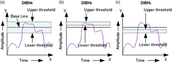
As per our institutional protocol, for all the DIBH patients both FB and the DIBH, CT scans were done. Before doing the DIBH scans, a FB plain CT scan was done for all the patients and after that the patients were asked to breathe normally and the baseline values were set as per the patient breathing pattern (typically around 1–1·5 cm) and window level was set as 5 mm (2·5 mm above and below the baseline amplitude) (DIBHR). For our study, we took another two series of DIBH scans in which instead of 5 mm we kept only 1 mm BHW, one by keeping the lower threshold same (DIBHL) and the other by keeping the upper threshold the same (DIBHH) as DIBHR (as per Figure 1). In this way, we could actually replicate the extreme variations in the breathing pattern inside a 5 mm BHW. All CT series were taken at 3 mm slice thickness and all the images were exported to both the server and the RPM system.
(a) Representation of deep inspiration 5 mm BHW window (DIBHR). (b) 1 mm BHW with upper threshold same as DIBHR (DIBHH). (c) 1 mm BHW with lower threshold same as DIBHR (DIBHL). Shaded region corresponds to beam ON time.

Contouring
In 5 mm BHW DIBH scans, clinical tumour volume which includes the whole breast and supra clavicular nodes was generated by the radiation oncologists as per the Radiotherapy Oncology Group (RTOG) contouring guidelines15 using Eclipse™ treatment planning system version 11·0 (Varian Medical Systems, Palo Alto, CA, USA). Similarly, planning target volumes (PTVs) were generated with 7 mm margins in craniocaudal, radial and anterio-posterior directions to account for the setup variation and patient movement. PTV Evaluation (PTVEval) was created as per the RTOG 1005 guidelines for dose volume histogram (DVH) evaluation.15 All the required critical structures like left lung, heart, left anterior descending artery (LAD), apex of heart, normal breast and spinal cord were also delineated. LAD was further divided as mid LAD and distal LAD, which were also contoured separately.
Planning
For all ten patients, opposing tangential fields were used with field in field technique (forward intensity modulated radiotherapy referred as FIMRT in this study), tangential fields were used to cover the whole breast and anterio-posterior fields were used to cover nodal region with slight tilt to avoid midline structures like spinal cord and oesophagus using treatment planning system. Fields were placed to cover the PTV with adequate margin and to shield the lungs and the heart as much as possible and the number of sub-fields were limited to four in order to reduce the treatment time. Dose calculations were performed using Analytical Anisotropic Algorithm (AAA) with 2·5 mm grid resolution.
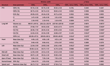
The criteria were set in order to cover the PTVEval volume receiving 95% prescribed dose to be not less than 95%, and volume receiving 98% prescribed dose to be minimum 90% and maximum dose to be less than 110% of the prescribed dose. Critical structures to receive a dose as low as reasonably achievable without spoiling the PTV coverage but not to exceed the tolerance doses of each structure.15,16 A hypo-fractionated dose regimen of 42·6 Gy in 16 fractions was planned for all the patients. Once the plan was finalised, it was replicated for the other two CT series (DIBHL and DIBHH) and the dose was calculated. PTV coverage and critical structure doses were evaluated and tabulated (Table 1).
Summary of treatment planning data of PTV and organ at risk in FIMRT plans

* p Value significant between two scenarios.
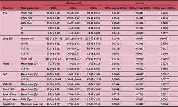
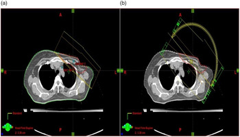
Similarly, VMAT plans were generated using the Eclipse treatment planning system for all the patients. Two partial arcs were used: one arc from 140° to 300° (counter-clockwise) and the other arc from 300° to 140° (clockwise). For optimisation, progressive resolution optimiser-3 was used. Dose calculations were performed using AAA algorithm with grid resolution of 2·5 mm. In these plans, the similar criteria were set as FIMRT. Similarly, the final plan was replicated and the doses were calculated in the other two CT series and the doses were tabulated (Table 2). Figure 2 shows the field arrangements of both the plans.
Summary of treatment planning data of PTV and organ at risk in VMAT plans

* p Value significant between two scenarios.
(a) Field arrangement of FIMRT plan. (b) Field arrangement of VMAT plan.

Plan comparison
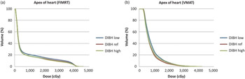
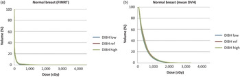
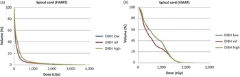
Lung volumes in all three CT datasets were calculated using Eclipse treatment planning system and tabulated. PTVEval covering 90 and 95% of the prescribed dose and D1% (dose to 1% of the PTV) in all three CT series of FIMRT and VMAT plans were also tabulated. Critical structure doses like lung V5% (percentage of the lung volume receiving 5% of the dose) and V10% (percentage of the lung volume receiving 10% of the dose) and V18% (percentage of the lung volume receiving 18% of the dose) for ipsilateral lung, heart mean dose and V13% (percentage of the heart volume receiving 13% of the dose), LAD mean dose and V10% (percentage of LAD receiving 10% of the dose), mid-LAD and distal LAD mean doses, contra-lateral breast D5% (dose to 5% of the breast) and the spinal cord maximum doses were also tabulated for comparison. Figures 3–9 describe the mean DVH of all the structures derived from the planning system.
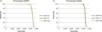
(a) Mean DVH of PTV Evaluation in FIMRT plans. (b) Mean DVH of PTV Evaluation in VMAT plans.

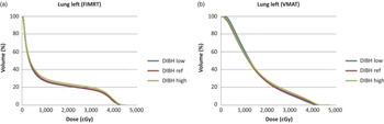
(a) Mean DVH of left lung in FIMRT plans. (b) Mean DVH of left lung in VMAT plans.

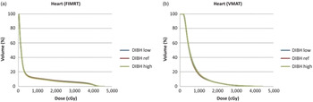
(a) Mean DVH of heart in FIMRT plans. (b) Mean DVH of heart in VMAT plans.

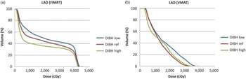
(a) Mean DVH of LAD in FIMRT plans. (b) Mean DVH of LAD in VMAT plans.

(a) Mean DVH of apex of heart in FIMRT plans. (b) Mean DVH of apex of heart in VMAT plans.

(a) Mean DVH of normal breast in FIMRT plans. (b) Mean DVH of normal breast in VMAT plans.

(a) Mean DVH of spinal cord in FIMRT plans. (b) Mean DVH of spinal cord in VMAT plans.

Statistical analysis
We used the paired t test with two tails to find the significance by using Microsoft Office excel software version 2007. Significances of all parameters were found between DIBHL and DIBHR, DIBHR and DIBHH and DIBHL and DIBHH. The null hypothesis was set that all three series are having the same mean with 95% confidence limit. In any event if p value is less than or equal to 0·05, then the differences in the two strategies are statistically significant.
Results
Volume comparison
As per Tables 1 and 2, mean lung volumes for DIBHL, DIBHR, and DIBHH BHW CT series were 960·07 ± 347·01, 1201·22 ± 192·76, and 1357·60 ± 287·63 cc, respectively. There was no significant change in terms of PTV volume covering 90 and 95% of the doses and D1% in all series in both the plans. In case of 50% dose volume inside the lung, significant differences were found in all the series in FIMRT plans and DIBHR versus DIBHH and DIBHL versus DIBHH in VMAT plans. The average change in 50% dose volume inside the lung for DIBHL, DIBHR and DIBHH CT series was 202·89 ± 87·51, 269 ± 86·31 and 309·69 ± 88·39 cc, respectively, for FIMRT plans and 239 ± 87·14, 297·83 ± 53·27 and 346·12 ± 57·06 cc for VMAT plans.
OAR dose comparison
From the DVH analysis, critical structures doses like left lung V5, V10, and V18%, heart V13% and mean doses, LAD mean and V10% doses, normal breast D5% dose and the spinal cord maximum doses were tabulated. Average lung V5% doses of DIBHL, DIBHR and DIBHH doses were 37·98 ± 7·07%, 40·55 ± 6·83% and 42·03 ± 6·73%, respectively, for FIMRT plans and 88·38 ± 3·91%, 83·62 ± 5·52% and 85·96 ± 3·41% for VMAT plans. Similarly, average lung V10% doses were 26·46 ± 7·00%, 28·40 ± 6·42% and 29·95 ± 6·63%, respectively, for FIMRT plans and 62·37 ± 3·11%, 58·47 ± 4·22% and 59·47 ± 1·86% for VMAT plans. Average lung V18% doses were 21·62 ± 6·32%, 23·34 ± 5·90% and 24·88 ± 5·97%, respectively, for FIMRT plans and 30·56 ± 3·39%, 30·54 ± 2·67% and 32·36 ± 2·66% for VMAT plans. p-Values showed significance in V5, V10 and V18% lung doses for DIBHR versus DIBHH (p values = 0·0461, 0·0283 and 0·0213, respectively) and DIBHL versus DIBHH series (p values = 0·0434, 0·0484 and 0·0334, respectively) in FIMRT plans and only in V18% doses of DIBHR versus DIBHH series in VMAT plans (p = 0·0067).
In terms of heart doses, average V13% doses were 9·40 ± 4·35%, 9·98 ± 6·01% and 8·78 ± 3·67% for DIBHL versus DIBHR and DIBHR versus DIBHH and DIBHL versus DIBHH FIMRT plans and 12·39 ± 5·73%, 10·83 ± 8·60% and 11·10 ± 6·70% for VMAT plans. Similarly, average heart mean doses were 4·49 ± 1·63, 4·68 ± 1·69 and 4·53 ± 1·40 Gy in FIMRT and 7·27 ± 0·98, 7·01 ± 1·70 and 7·06 ± 1·23 Gy for VMAT plans. No significances were found in any series in both the plans. LAD mean doses were 21·64 ± 3·59, 19·60 ± 3·60 and 16·81 ± 4·86 Gy for DIBHL, DIBHR, DIBHH for FIMRT plans and 16·53 ± 1·61, 14·46 ± 2·42 and 11·49 ± 1·65 Gy in VMAT plans. All series showed significant change in LAD doses in FIMRT plans (p value= 0·0012, 0·0444 and 0·0048, respectively) and DIBHR versus DIBHH and DIBHL versus DIBHH showed significant changes in VMAT plans (p value = 0·00341, 0·0001, respectively). In case of mid-LAD, no significance in FIMRT plans but DIBHL versus DIBHH showed significant dose reduction in VMAT plans. In case of distal LAD, FIMRT showed difference in DIBHL versus DIBHH and VMAT showed differences in DIBHR versus DIBHH and DIBHL versus DIBHH series. No significances were found in the apex of heart, normal breast and spinal cord doses in any series.
Discussion
There are many studies reporting the heart dose reduction with DIBH compared to FB. Lawler et al. (2017) found that the heart and the LAD sparing are improved with DIBH compared to FB. There were some statistically significant reductions in the left lung V5% and V10% doses alsoReference Lawler and Leech17 Latty et al. (2015) performed a meta-analysis where he reviewed 18 studies and found there have been reports of reductions of almost 26·2–75% in the heart mean dose.Reference Latty, Stuart and Wang18 Similar studies reported a considerable reduction in LAD doses.Reference Pham, Ward and Latty19,Reference Vikström, Hjelstuen and Mjaaland20 Hayden et al. (2012) found that LAD dose could be reduced to 21·9 Gy with DIBH compared to 31·7 Gy with FB and the mean heart dose was found to be around 4 Gy for DIBH compared to 6 Gy with FB.Reference Hayden, Rains and Tiver14 Since there is no definitive safe dose to heart or LAD, many techniques have been reported and implemented to reduce these doses to as low as possible.Reference Pham, Ward and Latty19,Reference Vikström, Hjelstuen and Mjaaland20
The introduction of recent techniques like VMAT along with DIBH further improved PTV coverage and reduced high dose volumes like V20, V30% to heart and mean doses to LAD. But many studies are showing that there was no significant clinical benefit in terms of critical structure doses in VMAT plans compared to tangential field in field or IMRT plans.Reference Badakhshi, Kaul and Nadobny21,Reference Jin, Chen and Deng22 In our study, the heart mean dose that we could achieve was 7 Gy with VMAT, whereas it was well within the institutional tolerance of 5 Gy in FIMRT (4·68 Gy). The lung V5% and V10% doses were very high in VMAT plans compared to FIMRT. This is similar to the result of Badakshi et al. (2013) that VMAT plans were inferior in terms of OAR doses particularly low dose volumes compared to IMRT or three-dimensional conformal radiotherapy technique.Reference Badakhshi, Kaul and Nadobny21 Jin et al. (2013) studied different treatment techniques for left breast and found that VMAT was no superior than FIMRT or tangential IMRT except in high dose volume to critical structures.Reference Jin, Chen and Deng22 Though we recorded some significance in low dose volumes between VMAT and FIMRT, lung V18% and heart mean doses were not clinically much significant which are considered as the main tools in controlling pneumonitis and late cardiac toxicities, respectively.Reference Xu, Guo and Liao23 Though we have produced FIMRT and VMAT plans in our study, our aim was not to compare these since it has been discussed and reported in a number of previous studies.
Our main aim was to study the significance of breathing pattern inside the 5 mm BHW as this has not been reported anywhere as to our knowledge. According to Tables 1 and 2, PTV coverage, conformity and homogeneity did not show any significance in all three window levels which means the variation inside the 5 mm BHW did not affect the PTV coverage. Significant variation in left lung volume was found in DIBHL versus DIBHR and DIBHL versus DIBHH. In left lung, it was found that V5%, V10% and V18% doses were significantly reduced in DIBHL for FIMRT plans while they were significantly high in DIBHH compared to reference level (DIBHR). This might be due to the involvement of more lung volume into the field due to deep inspiration. This trend was not the same in VMAT plans.
Heart mean and V13% doses did not show any statistically significant changes in all window levels in both FIMRT and VMAT plans. The majority of patients with a curved breast had more changes in the lung doses in different levels. A recent study by Loap et al. (2020) showed that the mean heart dose alone could not accurately reflect long-term cardiac toxicity and it is important to check the sub-structure doses if one would want to avoid long-term toxicities.Reference Loap, Fourquet and Kirova24 In our study, LAD doses showed an increment in DIBHL compared to DIBHR and DIBHH in both the plans in a similar trend which might be an important finding of this work since there are many recent studies discussing the importance of LAD dose reduction.Reference Wennstig, Garmo and Isacsson25,Reference Rygiel26 Wennstig et al. (2019) found that coronary intervention increased in patients who have received higher LAD doses, particularly in the mid-LAD portion and it was found that the patients receiving mid-LAD mean doses of 5–20 Gy required more cardiac intervention compared to the patients who received 0–1 Gy. It was also found that more dose to the distal LAD was significant in damaging the vessel but interventions were limited because of its size and location.Reference Wennstig, Garmo and Isacsson25 Taylor et al. (2007) found an increase in myocardial perfusion defects in the region supplied by the LAD 6 months after it was irradiated.Reference Taylor, Nisbet and McGale27
In our study, LAD doses were less in DIBHH window levels compared to the other two in both FIMRT and VMAT plans. It was found that in DIBHH both the plans showed significant reduction in the LAD mean and V10% doses. Similarly when it is contoured as separate segments like mid and distal, there was a significant reduction in mid-LAD and distal LAD mean doses in VMAT plans and only distal LAD mean dose in FIMRT plans. The LAD mean dose values were almost close to published resultsReference Hu, Han and Lei28–Reference Nilsson, Witt Nyström and Isacsson30 in all three phases and it was the least in DIBHH among all. Piroth et al. (2019) derived a DEGRO breast cancer expert panel recommendation which suggests that the mean LAD should be less than 10 Gy which has been closely achieved by VMAT plan in DIBHH.Reference Piroth, Baumann and Budach31 So, if the window level is always in lower threshold while treating, we would be delivering more doses to the LAD in FIMRT and in VMAT plans compared to reference level and we would deliver less doses to the LAD if it was in DIBHH without spoiling the coverage and no clinical significance in other critical structure doses was found. Heart apex, normal breast and spinal cord doses were insignificant in any phase in both the plans.
The limitation of this study is the sample size and an increase in the sample size would give more accurate results. Similarly, the breathing cycle may not vary uniformly for all patients during the entire treatment so it is important to consider the variation during the entire course of the treatment. Thirdly, in our study some patients did not show much variation in the critical structure doses due to the shape of the breast so patient selection criteria should also be a vital parameter.
Modern radiotherapy is progressing towards the hypo-fractionation and dose escalation that involves precise target position, real-time tumour management during both simulation and treatment. DIBH serves this purpose in most of the situations in thoracic tumours where the expected target movement is high. This study addresses the issues with the large volume tumours and this needs to be tested further for small volume lung and liver tumours where stereotactic body radiotherapy can be employed with higher accuracy.
Conclusion
Changes in the 5 mm BHW do not affect the plan quality significantly much but care should be taken in patients who already have some cardiac complications. In such cases, treatment should be done only when the breathing lies in the higher threshold level rather than keeping it in lower threshold to reduce late cardiac complications.
Acknowledgements
None.
Conflict of Interest
The authors have no conflict of interest.
Ethical Standards
This study has been approved by the Institutional Ethical Committee (reference: YSH/2019/0178) and concern of patients was obtained in order to use their data for the study.
Statement of Funding
This study is not funded by any agency and has no financial interest.