Introduction
Unilateral vocal fold paralysis is a common disorder seen in clinical laryngology, with an unknown incidence and prevalence in the general population.Reference Merati, Halum and Smith1 Simpson et al. reported the prevalence of unilateral vocal fold paralysis among patients with voice complaints as 2.57 per cent.Reference Simpson, Cheung and Jackson2
Paralysis of one or both vocal folds may compromise important physiological functions of the larynx, namely breathing, airway protection and phonation. The primary symptoms of unilateral vocal fold paralysis vary, ranging from simple vocal fatigue in mild or well compensated cases, to almost complete aphonia in severe cases, depending on the degree of glottal insufficiency and each patient's unique compensatory phonation strategy.Reference Sulica, Snow and Wackym3 About 30 per cent of patients may be asymptomatic.Reference Havas, Lowinger and Preistley4
Laryngoscopy is important for clinical assessment and for planning the type of intervention required in patients with unilateral vocal fold paralysis. Videostroboscopy provides better image quality, better magnification, and can aid detection of subtle movements of the vocal fold, vocal process and arytenoid. Flexible laryngoscopy provides an image that is more physiological. Hence, both techniques are complementary to each other.
Predicting the outcome of unilateral vocal fold paralysis can aid decision-making regarding the methods used and the timing of surgical intervention. The only investigation currently available that helps in predicting recovery is laryngeal electromyography. However, this investigation is not carried out in routine ENT examinations, and requires expertise for its use and interpretation of findings. A review of the literature focusing on the role of laryngoscopy in predicting recovery in unilateral vocal fold palsy cases that studies are sparse.
Our study aimed to analyse the endoscopic findings of unilateral vocal fold immobility and determine the prognostic value of laryngoscopy in predicting recovery. Patients who did not recover were further analysed to identify those who would regain their phonatory function via the compensatory movement of the normal side. Inter-observer reliability of these findings was also assessed.
Materials and methods
Patients
This was a prospective study conducted at the senior author's voice clinic at the Department of Laryngology, Kerala Institute of Medical Sciences, India, between April 2011 and May 2012.The study protocol was approved by the hospital ethics committee.
Patients who presented with symptoms of voice change, and who, on evaluation, were diagnosed with unilateral vocal fold paralysis, were included in the study after written informed consent was obtained. Patients with unilateral vocal fold immobility due to a progressive lesion (those with a mechanical-cause like laryngeal malignancy or cases with a neurological cause such as multiple sclerosis), or those with arytenoid dislocation, were excluded from the study.
Patients were reviewed every six weeks, for up to six months from the onset of symptoms or until the symptoms recovered if that occurred before six months. The decision regarding surgical intervention was planned after six months from the onset of symptoms.
Endoscopy
Each patient underwent videostroboscopy with a rigid 70° telescope (Karl Storz Pulsar™ I (model number 20140020)) and flexible laryngoscopy (with a Karl Storz ‘chip-on-the-tip’ camera endoscope) during each evaluation, and video recordings were obtained with a charge-coupled device camera.
In all patients, 10 per cent lignocaine spray was used to suppress the gag reflex. Laryngoscopy was conducted during inspiration, whilst patients sustained phonation of the vowel /i /, at a comfortable pitch and volume. The average duration of the recording was 20 seconds. Those recordings which were unclear, as a result of excessive gag reflex or inadequate duration, were excluded from the study.
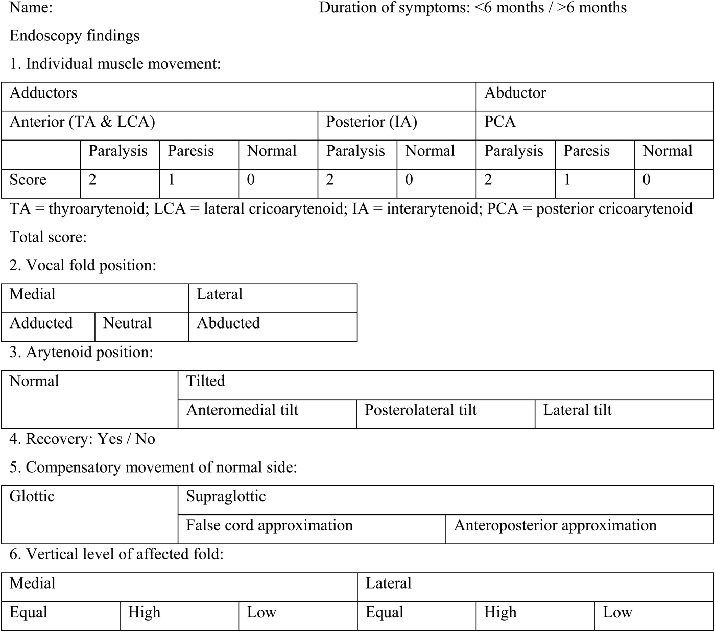
Each video recording was independently evaluated by a senior laryngologist, laryngology trainee and a speech-language pathologist, using a pre-printed proforma (Figure 1). A total of six criteria were assessed, with a formal definition of each criterion based on literature review findings.Reference Blitzer, Jahn and Keidar5–Reference Yumoto, Oyamada, Nakano, Nakayama and Yamashita10 The first three criteria were assessed during each visit and the last three were assessed after six months from the onset of symptoms. The findings were reassessed by all three examiners together and a common consensus was sought.
Proforma used to evaluate endoscopic video recordings.

Individual muscle movement, the first criterion, was assessed for each subject. The muscles were divided into adductors and abductors. Adductors were assessed by asking the patient to phonate. The abductor: Posterior cricoarytenoid, was evaluated on inspiration. The adductors were subdivided into anterior adductors, formed by the thyroarytenoid and lateral cricoarytenoid, and posterior adductors, formed by the interarytenoid. Anterior adductors were assessed in terms of the adduction of the membranous vocal fold (Figure 2). The interarytenoid was assessed in terms of the adduction of the arytenoid ‘hump’ (Figure 3), as described by Fleischer et al.Reference Fleischer, Schade and Hess6 This hump consists of the arytenoid cartilage and its attaching muscles, ligaments and the covering tissue. An associated vocal fold bowing was an added clue that the thyroarytenoid was involved. Reference Blitzer, Jahn and Keidar5 Posterior cricoarytenoid paralysis decreases lateral gliding and abduction (Figure 4). A scoring system was introduced, with a maximum score of 6 for cases of complete paralysis of all muscle groups, and a minimum score of 1 for cases of paresis of a single muscle group.
Anatomical diagram showing anterior adductors, assessed in terms of adduction of membranous vocal fold on phonation (a–e represent the five lines of laryngoscopic orientation7).
Endoscopic views of a (a) normal and (b) paralysed (posterior adductor) interarytenoid muscle, assessed in terms of adduction of the arytenoid ‘hump’ (stars indicate affected sides).
Anatomical diagram showing the posterior cricoarytenoid (abductor) muscle, assessed in terms of movement on inspiration (a–e represent the five lines of laryngoscopic orientation7).
The three positions of the paralysed vocal folds were described according to the five lines of laryngoscopic orientation,Reference Arnold7 as shown in Figure 5. The vocal fold was considered adducted if it was positioned along the median or paramedian line, neutral if along the intermediate line and abducted if laterally placed.
Anatomical diagram showing the three positions of paralysed vocal folds. The vocal fold was considered adducted if it was positioned along the median (c) or paramedian (d) line, neutral if along the intermediate line (b), and abducted if laterally placed (a (partial abduction) or e (full abduction)) (a–e represent the five lines of laryngoscopic orientation7).
The arytenoid positions considered for analysis were normal and tilted positions (Figure 6). If tilted, it can be categorised as: anteromedial displacement, if only the tip of the vocal process is visualised; posterolateral displacement, if the medial surface of the vocal process and body of the arytenoid is observed; and lateral displacement, if the vocal process is visualised.Reference Hong and Jung8
Endoscopic views of arytenoid muscle positions: (a) normal, (b) anteromedial tilt, (c) posterolateral tilt and (d) lateral tilt. (Stars indicate the side of the tilt.)
In this study, recovery was defined as normal or near-normal mobility of the affected vocal fold as compared with the normal side (Figure 7).Reference Sittel, Stennert, Thumfart, Dapunt and Eckel9 The compensatory movement of the normal side was assessed at the level of the glottis and supraglottis where it crosses the midline (Figure 8).Reference Yumoto, Oyamada, Nakano, Nakayama and Yamashita10
Endoscopic views of affected vocal folds (a) before and (b) after recovery.
Endoscopic views showing the compensatory movement of the (a) supraglottis (star) and (b) glottis (star) in the normal vocal fold.
The vertical height of the affected fold could be higher, lower or equal to that of the innervated fold (Figure 9). The most important finding of the video images for determining the height of the paralysed vocal fold was the pattern of contact between the vocal processes of paralysed and normal vocal folds during phonation when both were well visualised.Reference Hong and Jung8
Endoscopic views showing the vertical height of the affected vocal fold, which could be (a) equal to, (b) lower or (c) higher than that of the innervated fold. (Stars indicate affected sides.)
Video rating
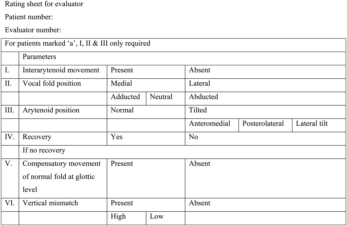
In order to assess inter-observer reliability, each video recording was rated (in terms of the aforementioned six criteria) by three trained laryngologists. These individuals, who were blinded to the clinical details of each patient, were sent a digital video disc containing the video recordings, and a rating sheet (Figure 10) with formal definitions of each criterion.
Evaluator rating sheet, used to assess inter-observer reliability.
For the purpose of statistical analysis, each criterion was analysed in a binary fashion. Each recording was appropriately labelled (using letters and numbers) based on the patient and the follow-up visit. Each examiner could assess the videos as many times as needed for evaluation.
Statistical analysis
The association between two categorical variables was calculated using Fisher's exact test (with Epi Info™ software).11 The significance level was set as p < 0.05. Inter-observer reliability was determined using kappa statistics, as described by FleissReference Fleiss12 (with Reliability Calculator (‘ReCal’) softwareReference Freelon13).
Results
Of the 1841 patients who underwent videostroboscopy for voice complaints, 89 (4.5 per cent) were initially diagnosed with vocal fold paralysis. A total of 66 patients were included in the study, after excluding 15 patients with progressive lesions, 1 patient with cricoarytenoid joint dislocation, and 7 patients for whom the video recording was not of adequate length or visualisation of the larynx was poor because of excessive gag reflex.
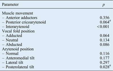
Of the 66 patients, 56 had paralysis and 9 had paresis of the anterior adductors (total of 98.5 per cent); 53 had paralysis and 8 had paresis of the posterior cricoarytenoid (total of 92.42 per cent); and 49 (74.24 per cent) had interarytenoid paralysis. The follow-up results indicated that those patients with an interarytenoid paralysis had less chance of recovery (around 2 per cent) than those with intact interarytenoid function (for whom recovery was 82.35 per cent).
In our study, 30 vocal folds (45.45 per cent) were in a neutral position, 29 (43.93 per cent) were in an abducted position and 7 (10.6 per cent) were in an adducted position.
The position of the arytenoids was normal in 10 of the 66 patients (15.15 per cent) and tilted in 56 of the patients (84.84 per cent). The tilt was anteromedial in 34 cases (60.71 per cent), posterolateral in 14 cases (25 per cent) and lateral in 8 cases (14.29 per cent). Patients with a posterolateral tilt had an associated interarytenoid paralysis and had no recovery.
A bio-statistical analysis, conducted to examine associations between the above three parameters and recovery, revealed that those patients with interarytenoid paralysis (p < 0.001) and posterolateral tilt of the arytenoid (p = 0.028) had less chance of recovery. Vocal fold position had no significant association with recovery (Table I).
Statistical analysis of recovery*
#p < 0.05 (significant)
* Analysed using the Fisher's exact test
Of the 66 patients, 15 (22.73 per cent) recovered and the remaining 51 (77.27 per cent) did not. Of those who did not recover, 13 patients (25.49 per cent) regained phonatory function via compensatory movement of the normal side and the remaining 38 patients (74.51 per cent) required an intervention.
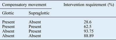
A compensatory movement on the normal side was observed at the level of the vocal fold in 23 out of 48 patients (47.92 per cent) and at the supraglottic level in 35 out of 51 patients (68.63 per cent). Statistical analysis was conducted to assess the association between compensation and intervention. The requirement of intervention was significantly less for those patients who had glottic level compensation (p = 0.002). In isolated glottic compensation cases, only 28.6 per cent required intervention; however, if it was combined with supraglottic compensation, the requirement for intervention increased by 62.5 per cent (Table II). An adducted vocal fold position (66.67 per cent) and normal arytenoid position (80 per cent) increased the chance of glottic level compensation.
Compensatory movement of normal side and intervention requirement
The vertical level of the affected fold was assessed during phonation in the 38 patients who required intervention. For four patients, the level of the vocal fold was not visualised clearly as a result of supraglottic phenomenon. In the remaining 34 patients, the paralysed fold was at the same level as that of the innervated fold in 11 patients, higher in 13 patients and lower in 10 patients. There was no vertical level incompatibility when the vocal fold position was adducted or when the arytenoid was in a normal position.
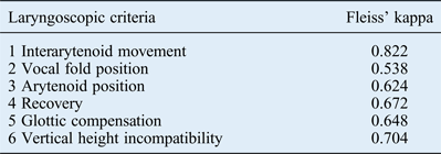
Assessments of inter-observer reliability revealed fair to good agreement for all criteria except interarytenoid movement which had excellent agreement (Table III).
Inter-observer reliability
Discussion
The most significant obstacle in efficient management of patients with vocal fold palsy has been a lack of clear information regarding prognosis and recovery. Rickert et al. highlighted the potential importance of variables such as time of presentation, definition of recovery and duration of follow up, and a simplified concept of paralytic dysphonia.Reference Rickert, Childs, Carey, Murry and Sulica14
Laryngeal electromyography can provide prognostic information about unilateral vocal fold paralysis, especially if it is conducted at least two months after symptom onset.Reference Wang, Chang, Wang and Liu15 It can aid identification of normal innervation, the absence of innervation, reinnervation and even synkinesis, based on characteristic electrical signals. The absence of spontaneous activity, and/or normal or near-normal motor unit potential morphology and recruitment are the most common factors associated with good prognosis. However, this investigation is not carried out in routine ENT examinations, and requires someone with expertise to use and interpret the findings.
A literature search confirmed that there are no standardised, endoscopy-based guidelines on which prognosis can be determined. Hence, this study attempted to explore the value of laryngoscopy in predicting recovery.
The increased percentage of anterior adductor involvement in the study could be explained as due to the associated early presentation of voice complaints. The posterior adductor: the interarytenoid is a midline muscle with rich intramuscular anastomosis with the opposite nerve resulting in its reduced involvement in unilateral vocal fold paralysis. It was found that the scoring system described here was not very helpful in predicting the prognosis or recovery, but was useful for rating the findings.
Throughout the history of laryngology, there has been much debate about the factors responsible for the final position of the vocal fold in unilateral vocal fold paralysis. Based on their observations, Semon and Rosenbach, in 1892, hypothesised that the nerve fibres to abductor muscles are more sensitive to injury than those of adductor muscles so that, in a slowly progressive lesion, the vocal fold assumes a median position first and later a lateral position.Reference Woodson16 However, this conclusion lacked substantiation.Reference Blitzer, Jahn and Keidar5, Reference Woodson16, Reference Crumley17 Later, Wagner and Grossman explained the vocal fold position on the basis of cricothyroid muscle activity. However, with the emergence of laryngeal electromyography, it was found that the final vocal fold position depends on reinnervation and synkinesis (defined as the simultaneous contraction of antagonistic muscles).Reference Blitzer, Jahn and Keidar5, Reference Woodson16, Reference Crumley17 When the adductors and abductors are completely paralysed, vocal fold position ideally should be in neutral position. Nevertheless, the findings of the present study showed that the position varied from one individual to another, independent of the muscle group affected; this can be explained as due to synkinesis.
Hong and Jung classified the position of vocal folds as medial (adducted and neutral) or lateral (abducted).Reference Hong and Jung8 They observed that, out of 39 patients, 25 (66 per cent) had medial paralysis and 13 (33 per cent) had lateral paralysis. In our study, 37 out of 66 patients (56.05 per cent) had medial paralysis and 29 (43.94 per cent) had lateral paralysis. As expected, the position of the vocal fold was not diagnostic of the site of the lesion, nor was it associated with recovery. Nevertheless, the severity of clinical presentation and further management may depend upon vocal fold position. Among those who did not recover in the current study, the chance of glottic level compensation was higher, and the risk of vertical height incompatibility was lower, when the vocal fold was adducted.
Studies focusing on the arytenoid position during phonation seem to be sparse. Hong and Jung described the arytenoid tilt as anteromedial, posterolateral and lateral.Reference Hong and Jung8 The variable arytenoid position could also be explained as due to synkinesis. In our study, the anteromedial tilt was the most common tilt (60.71 per cent), followed by the posterolateral tilt (25 per cent).
The principal determinant of recovery was interarytenoid movement. The exact mechanism underlying this association is unknown. However, the likely explanation would be the unique characteristics in fibre composition and innervation of the interarytenoid muscle that make it resistant to damage, as described by Tellis et al.Reference Tellis, Rosen, Thekdi and Sciote18 It was also noted that all patients with a posterolateral tilt had interarytenoid paralysis. This could be explained as due to the unopposed action of interarytenoid muscle oblique fibres on the normal side. Hence, if interarytenoid paralysis was associated with a posterolateral tilt, the chance of recovery was even worse. Interarytenoid paralysis and posterolateral tilt were predictors of poor recovery in patients with unilateral vocal fold paralysis. This finding is clinically useful for identifying candidates for early definitive intervention. The use of laryngeal electromyography (an objective assessment tool) to confirm the interarytenoid movement would have increased the strength of the study findings; however, this was beyond the scope of the article.
In this study, the outcome measure (in terms of recovery) was normal or near-normal movement of the affected side as compared with the normal side, and not vocal ability. There can be better phonatory function without recovery as a result of the compensatory movement of the normal fold. Sittel et al. have suggested that vocal ability is of great importance in terms of patient outcome.Reference Sittel, Stennert, Thumfart, Dapunt and Eckel9 However, when the accuracy of a prognostic test is investigated, the outcome variable should reflect the system measured. Based on this, we considered vocal fold mobility as the measure of recovery. Laccourreye et al. reported 23 per cent spontaneous recovery.Reference Laccourreye, Papon, Kania, Ménard, Brasnu and Hans19 This is consistent with our study, in which 22.7 per cent of patients recovered.
Yumoto et al. observed over-adduction of the vocal fold on the healthy side (compensatory movements) over the midline during phonation in 40 per cent of subjects.Reference Yumoto, Oyamada, Nakano, Nakayama and Yamashita10 In our study, there was vocal fold over-adduction in 47.92 per cent of cases. It was noted that those patients with vocal fold over-adduction did not require intervention, especially if the position of the affected fold was adducted. Supraglottic compensation was seen in 68.63 per cent of cases, and was common if the vocal fold was in a neutral or abducted position. It was noted that cases of isolated glottic compensation, without supraglottic phenomenon, had less need for intervention. This suggests that therapy should focus on exercises such as the ‘half-swallow boom’, which helps to improve glottic compensation, avoiding supraglottic phenomenon. In the half-swallow boom technique suggested by McFarlane et al. (as cited in Stemple et al.), the patient is asked to take a deep breath and initiate the first part of a swallow.Reference Stemple, Glaze and Gerdeman20 At the peak of a half-swallow, when glottal closure improves, the patient is instructed to phonate by saying ‘boom’. When the patient attains an improved voice quality for ‘boom’, they are educated to say other words and phrases.
The vertical mismatch between the affected vocal fold and the innervated fold depended on the positions of the vocal fold and arytenoid cartilage on the affected side. Anterior sagging of the arytenoid results in inferior displacement of the vocal process and, consequently, a vocal fold positioned lower than the normal one. If the paralysed vocal fold lies at a lower level than the normal vocal fold, the arytenoid adduction could increase that difference. On the other hand, if the vocal process of the paralysed arytenoid is placed higher than the normal vocal fold, the inferior and medial displacement of the vocal process achieved by arytenoid adduction should be beneficial.Reference Hong and Jung8, Reference Rickert, Childs, Carey, Murry and Sulica14 Our findings are comparable with those of Hong and Jung; in their study, the variable vertical level of the affected side was explained based on the biomechanics of arytenoid cartilage movement over the convex facet of cricoid cartilage.Reference Hong and Jung8
When the vertical height was equal, a medialisation laryngoplasty or thyroplasty was planned according to the glottic gap. In cases of vertical height incompatibility, a higher position requires classical arytenoid adduction along with thyroplasty. Various surgical options have been described for cases in which the affected fold is at a lower position. These include a posterior suspension suture with classical arytenoid adduction,Reference Woodson, Sulica and Blitzer21 arytenoidopexy, as described by Zeitel et al.Reference Zeitels, Hochman and Hillman22 and combined type 1 and 4 thyroplasty with arytenoid adduction, as described by Nakamura et al.Reference Nakamura, Isshiki, Kanazawa and Sanuki23
• Few studies have investigated laryngoscopy as a prognostic tool to predict recovery
• Interarytenoid paralysis and posterolateral tilt of the arytenoid were predictors of poor recovery
• Compensatory movement of the normal vocal fold without supraglottic phenomenon aids phonatory function without any intervention
• Intervention, if required, depends on the vertical level of the vocal fold and arytenoid position
• There was high inter-observer reliability for all criteria
Inter-observer reliability was determined using kappa statistics, as described by Fleiss.Reference Fleiss12 Although no uniformly agreed-upon scale exists for Fleiss' kappa, Fleiss described a scale where values more than 0.75 represent excellent agreement, values between 0.40 and 0.75 represent fair to good agreement, and values lower than 0.40 represent poor agreement. The interarytenoid movement, the chief determinant of recovery, had excellent inter-observer agreement in the current study. The other five criteria had fair to good inter-observer agreement.
We have proposed a protocol for the evaluation of unilateral vocal fold paralysis (Figure 11) based on the observations reported above.
Protocol for evaluation and management of unilateral vocal fold paralysis based on laryngoscopy. *Presence of significant life-threatening aspiration on swallowing. †Refers to professionals with increased vocal demands (level 1&2 voice users based on Kauffman's classification)
Acknowledgements
We are grateful to Drs K M Dominic, M Preethy and P Jayanthy for evaluating the video recordings, and to Dr M Gopalan for the illustrations. We would also like to acknowledge Mr S M Nair, for helping us to conduct the statistical analysis, and all the members of the Department of Otorhinolaryngology, Kerala Institute of Medical Sciences Hospital, for their continued help and support throughout this project.