One molecule of urea contains two atoms of N. The synthesis of urea in the mammalian urea cycle requires 1 atom of N to be supplied as ammonia and the other N atom to enter on aspartic acid. It has been suggested that this requirement for an amino acid could mean that hepatic synthesis of urea to detoxify ammonia arising from rumen fermentation consumes amino acids that might otherwise have been retained in tissue or milk protein of growing or lactating animals (Reynolds, Reference Reynolds1992; Lobley et al. Reference Lobley, Connell, Lomax, Brown, Milne, Calder and Farningham1995). In support of the hypothesis, Mutsvangwa et al. (Reference Mutsvangwa, Buchanan-Smith and McBride1999) reported a doubling of 15N-incorporation from [15N]methionine into urea by isolated hepatocytes exposed to increasing concentrations of [14N]ammonia in vitro. However, the majority of the urea synthesized remained unenriched with 15N, no matter which of many amino acids contained the 15N, indicating that the urea-N from aspartate mainly arose, not directly from amino acids, but from ammonia via glutamic dehydrogenase and transamination. Thus, evidence for and against the hypothesis is equivocal.
The possibility that an ammonia load could cause a depression in protein content of milk from dairy cows has been tested by ruminal urea infusion or postruminal ammonia infusions. Administration of 240 g/d urea in two discrete meals per day, as opposed to continuous infusion into the rumen, resulted in a dry matter intake (DMI) depression at lower levels of dietary crude protein (CP) intake (Choung et al. Reference Choung, Chamberlain, Thomas and Bradbury1990). Milk yield declined in a similar manner to the DMI depression but both protein and fat percentages were elevated at the highest doses of urea (Choung et al. Reference Choung, Chamberlain, Thomas and Bradbury1990). Malik et al. (Reference Malik, Nicol and Van Houtert1999) observed a 10% drop in milk protein content and a 24% decrease in protein yield when ewes were infused intravenously with ammonium acetate. Moorby & Theobald (Reference Moorby and Theobald1999) reported no change in milk protein percentage or yield, and a 2% decline in milk casein percentage, during duodenal ammonia infusion of cows. To address the equivocal responses and to evaluate the effect of hyperammonaemia on milk composition, cows were infused intravenously for 10 h with ammonium acetate, and the concentrations and uptakes of milk precursors from circulation by the mammary glands were measured.
Symonds et al. (Reference Symonds, Mather and Collis1981) reported that ammonia toxicity symptoms in dairy cows occurred at a concentration of ammonia in arterial plasma of 800 μm. Choung et al. (Reference Choung, Chamberlain, Thomas and Bradbury1990) observed depressing effects of intraruminal urea infusion on DMI and milk production at approximately 200 μm plasma ammonia. The first objective of the current experiment was to find a jugular infusion rate that would raise plasma ammonia levels to between 200 and 800 μm. Then, we measured the effect of hyperammonaemia during a milking interval on the animal and on milk production and, finally, we examined the effect of a 10-h hyperammonaemia on mammary utilization of milk precursors. To our knowledge, this is the first report of effects of an intravenous ammonia infusion on milk composition in lactating cows.
Materials and Methods
All procedures involving animals were approved by the Animal Care Committee at the University of Guelph. For each experiment, ammonium acetate was the ammonia donor and sodium acetate was the control. Infusates were prepared as 3·33 m solutions, filtered through 0·22-μm cellulose acetate filters and stored in 4-l autoclaved bottles prior to use. Actual weights of solutions infused were recorded at hourly intervals during infusion.
Experiment 1: hyperammonaemia
Four multiparous Holstein cows of body weight (BW) 669±20 kg and in the second trimester of pregnancy were housed individually in tie stalls, milked twice a day at 8.00 and 16.00, fed twice daily for ad-libitum intake with a total mixed ration (21·6% CP, 31·5% neutral detergent fibre) based on first-cut lucerne chaff, and given free access to water bowls. Cows were each fitted with indwelling jugular catheters. One catheter was for infusion and the other for blood sampling. Ammonium acetate was infused for 1 h at 567 mmol/h, which was calculated to be 50% of the maximum hepatic detoxification rate of 1·84 mmol/min per kg liver (Symonds et al. Reference Symonds, Mather and Collis1981). Venous blood was sampled into heparinized tubes at 10-min intervals over the 60 min of infusion and for the following 30 min. Samples were centrifuged and the plasma was removed by pipette and frozen for later analysis.
Experiment 2: feed intake and milk production
The four cows used in Experiment 1 were infused into one jugular catheter with ammonium acetate or sodium acetate at the same rates as in Experiment 1 for four 1-h periods, each separated by 1 h without infusion. The ammonium and sodium acetates were administered on consecutive days in a randomized sequence for each cow. The intermittent infusion protocol ensured that the total amount of ammonia entering the circulatory system was high without exceeding toxic levels.
Cows were milked twice daily. Feed was weighed into an empty manger in the morning and the infusion protocol began. At the end of 8 h, infusions were terminated, cows were milked, and feed remaining in the manger was weighed to calculate 8-h intakes.
Experiment 3: mammary arteriovenous differences
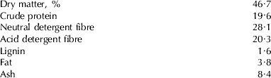
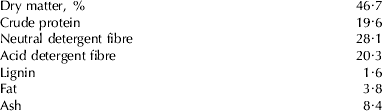
Four cows in their second lactations were fitted with catheters in a jugular vein, a subcutaneous mammary abdominal vein, and an intercostal artery acording to Mackle et al. (Reference Mackle, Dwyer, Ingvartsen, Chouinard, Ross and Bauman2000). All animals were fed with a total mixed ration based on corn and lucerne silages (Table 1) for ad libitum intake. Feed intakes were measured daily and feed was sampled every second day. Infusions into the jugular catheter commenced at the completion of the morning milking at 8.00 and continued until cows were milked at 18.00. Ammonium acetate was infused at 360 mmol/h, which was estimated to raise the plasma ammonia concentration to 200 μm for the entire 10-h interval between morning and afternoon milkings. Sodium acetate was infused, according to the same protocol, the day before or after ammonium acetate infusion at an equimolar rate. Milk was sampled from the afternoon milking to represent the effect of treatment.
Chemical analysis (% of dry matter unless stated otherwise) of the total mixed ration fed in Experiment 3
Every hour after the start of the infusion, 10-ml blood samples were collected into heparinized tubes from arterial and mammary vein catheters simultaneously. Samples were centrifuged and the plasma was removed and frozen for later analysis.
Analytical procedures
Milk samples were analysed by infrared spectroscopy for protein, fat and lactose. Plasma samples were analysed spectrophotometrically for glucose (kit 510-A; Sigma Chemical Co., Oakville, Ontario), triglycerides (Sigma kit 336), non-esterified fatty acids (NEFA C kit; Wako Chemicals GmbH, Neuss, Germany), acetate (Boehringer Mannheim kit; R-Biopharm GmbH, Darmstadt, Germany), β-hydroxybutyrate (Cant et al. Reference Cant, DePeters and Baldwin1993), urea (Sigma kit 640-B) and ammonia (Sigma kit 171-C).
Concentrations of amino acids in plasma were analysed by the method of MacKenzie (Reference MacKenzie1987). Briefly, 0·5 ml sample with 0·3 ml 2·5 mm-norleucine was deproteinized with 1·2 ml 12% (w/v) TCA and centrifuged at 2000 g for 20 min. The supernatant was run through a cation-exchange column, vacuum-dried and converted to N-heptafluorobutyryl n-propyl ester derivatives according to Culea & Hachey (Reference Culea and Hachey1995). Derivatives were separated on a model 6890 GC linked to a 5973N quadrupole MS (Agilent Technologies, Mississauga, Canada) and quantified using norleucine as the internal standard.
Mammary uptakes of plasma metabolites during the final hour of infusion were calculated as the product of arteriovenous concentration difference and plasma flow rate. Mammary plasma flow (MPF) was estimated from the quotient of arteriovenous concentration difference of Phe+Tyr and uptake of Phe+Tyr from plasma, assuming that 96·5% of Phe+Tyr output in milk protein is equal to Phe+Tyr uptake from plasma (Cant et al. Reference Cant, DePeters and Baldwin1993). Mammary extraction of a plasma metabolite, calculated as the quotient of its arteriovenous difference and arterial concentration, is a consequence of both transmembrane transport and plasma flow. As an alternative to calculating extraction, then, the product of the first-order rate constant for capillary uptake (k) and the total volume of perfused capillaries (Volcap⋅Ncap) was calculated as k⋅Volcap⋅Ncap=−ln(1− extraction)×MPF, according to Cant & McBride (Reference Cant and McBride1995).
Statistical analyses
For Experiment 2, variance in each observation (Yijk) was analysed by using the following model, with the general linear models procedure of SAS Institute Inc. (2000) :

where
μ … =mean,
cowi=ith cow effect (1=1 to 4)
perj=jth period effect (j=1 to 2),
trtk=kth treatment effect (k=1 to 2), and
εijklm=error.
For Experiment 3, treatment effects were detected by an analysis of covariance (Neter et al. Reference Neter, Wasserman and Kutner1985). For all observations except plasma ammonia and urea, the categorical treatment effect in the above model was replaced by the continuous effect of plasma ammonia concentration. In place of the cow effect, observations of DMI and milk production and composition the day before infusions commenced served as covariates for those variables during infusions. Likewise, observations of plasma metabolite concentrations at 8·00, immediately prior to the onset of infusions, served as covariates for metabolite concentrations during the final hour of infusion. Variances in MPF and amino acid concentrations were analysed with a categorical cow effect. Significance was declared at P⩽0·05 and tendencies were declared at 0·05<P⩽0·15.
Results
Experiment 1
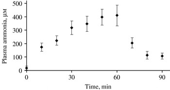
The single hour of infusion of ammonium acetate was conducted to find a jugular infusion rate that would elevate plasma ammonia to a concentration between 200 and 800 μm. The increase in concentration of ammonia was rapid, reaching a mean of 411 μm within 1 h of the start of infusion at 567 mmol/h (Fig. 1). Animals appeared normal throughout. Ammonia concentrations dropped rapidly within 30 min of cessation of the infusion to around 107 μm. Concentrations of ammonia above baseline from 0 to 90 min, [A]t, were used in Excel® Solver to estimate the plateau ammonia concentration above baseline, [A]max, and rate constant for first-order clearance from plasma, k, by fitting the equations: [A]max (1− e−kt) when t ⩽60, and [A]60e−k(t−60)) when t >60. The rate constant, k, was 0·054/min for a half-life of 12·9 min. [A]max was estimated to be 377 μm. The volume of ammonia distribution was calculated from the quotient of the infusion rate of 567 mmol/h and k[A]max to be 465 l, or 69·5% of BW. This percentage agrees closely with the total body water content of lactating dairy cows (Brown et al. Reference Brown, Taylor, DePeters and Baldwin1989) and reflects rapid distribution of ammonia throughout. Clearance was calculated as 60 k×volume to be 1504 l/h.
Mean (±se) jugular plasma ammonia concentrations measured during and after 1 h of infusion into the contralateral jugular vein of ammonium acetate at 567 mmol/h (n=4; Experiment 1).

Experiment 2
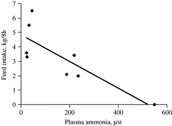
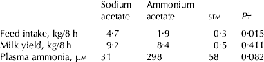
Because a single hour of ammonia infusion at 567 mmol/h appeared safe for the cows, the procedure was repeated four times during a milking interval with a 1-h washout between each infusion hour. There was a significant decline in feed intake during the interval studied but no significant decline in milk production compared with infusion of sodium acetate in the same fashion (Table 2). Plasma ammonia concentration in one cow rose to twice that of the other cows on the ammonia infusion, and that cow also consumed no feed during the 8-h experiment. The linear regression between plasma ammonia concentration and feed intake was significant (P=0·014) and exhibited a coefficient of determination of 0·66 (Fig. 2).
Relationship between feed intake and plasma ammonia concentrations in four cows (Experiment 2) infused with 567 mmol/h ammonium acetate and sodium acetate on consecutive days in four 1-h intervals, each followed by a 1-h washout; linear regression (solid line) is y=−0·0092x+4·8 (P=0·014, r 2=0·66).

Average feed intake, plasma ammonia concentration, and yield of milk from four cows (Experiment 2) after four 1-h periods of infusion into the jugular vein of 567 mmol/h ammonium acetate or sodium acetate, each followed by a 1-h washout
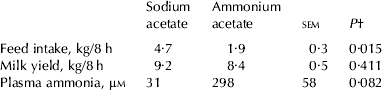
† Probability of no effect of ammonium compared with sodium acetate
Experiment 3
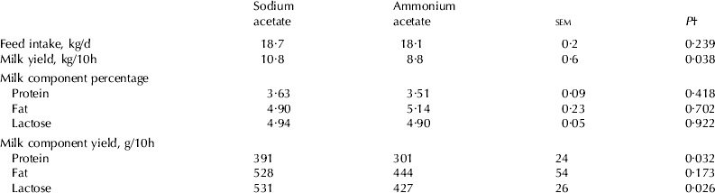
Effects of hyperammonaemia on uptake and utilization of milk precursors by the mammary glands were studied with a continuous infusion of ammonium acetate throughout the entire interval between am- and pm-milking. The infusion rate was 60% of that used in Experiments 1 and 2 in an attempt to minimize the feed intake depression that could confound the ability to test the hypothesis that hepatic amino acid utilization affects milk synthesis. The hyperammonaemia caused a 20% decrease in milk, protein and lactose production with no effect on percentage composition of milk or the yield of milk fat (Table 3).
Average feed intake and yield and composition of milk from four cows (Experiment 3) after 10 h of infusion into the jugular vein of 360 mmol/h ammonium acetate or sodium acetate
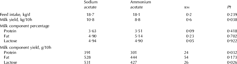
† Probability of no continuous effect of arterial plasma ammonia concentration
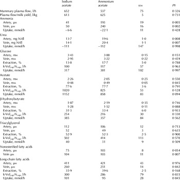
Arterial and venous concentrations of ammonia and urea were both elevated by the ammonia infusion. The increase in plasma ammonia concentration, predicted from the infusion rate of 360 mmol/h and the clearance rate of 1504 l/h estimated in Experiment 1, was 239 μm and the observed increase was 152 μm. The difference suggests that clearance was faster in Experiment 3 than in Experiment 1. Arterial concentrations of glucose and NEFA tended to increase (Table 4). There were no differences in arterial concentrations of acetate, β-hydroxybutyrate or triacylglycerol.
Average plasma flow and balance of plasma metabolites across the udder of four cows (Experiment 3) during the last hour of a 10-h infusion into the jugular vein of 360 mmol/h ammonium acetate or sodium acetate

† For urea and ammonia, probability of no categorical effect of infusion of ammonium compared with sodium acetate; and for all other metabolites, probability of no continuous effect of arterial plasma ammonia concentration
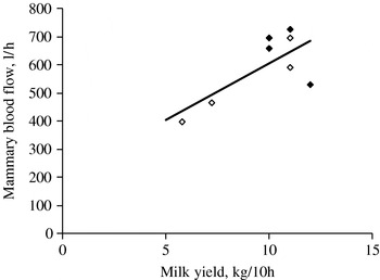
The 18% reduction in average MPF was not statistically significant (Table 4), although it was significantly (P=0·046) related to the level of milk production over the 10-h infusion (Fig. 3). Extractions of metabolites were not affected by the hyperammonaemia but the k⋅Volcap⋅Ncap product for acetate removal by the mammary glands tended to be 19% lower with ammonia infusion. Where the change was of the same magnitude as for MPF, the drop in k⋅Volcap⋅Ncap probably represents changes in capillary perfusion volume Volcap⋅Ncap and not in the efficiency of uptake, k. Mammary uptakes of acetate tended to be reduced by hyperammonaemia. Uptakes of other metabolites were not affected. On a net basis, ammonia and urea were released by the mammary glands although the rates were not significantly different from zero.
Relationship between mammary blood flow and milk yield in four cows (Experiment 3) infused with 360 mmol/h ammonium acetate (open diamonds) and sodium acetate (closed diamonds) for 10-h periods on consecutive days; linear regression (solid line) is y=40·3x+202 (P=0·046, r 2=0·51).

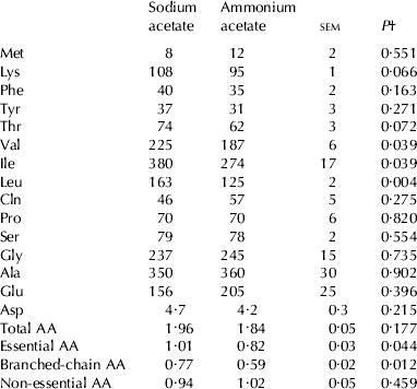
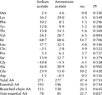
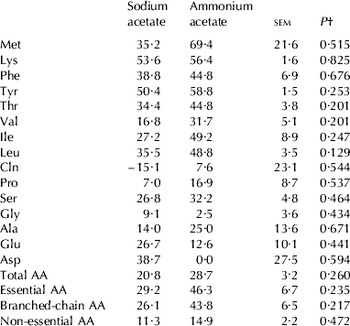
Arterial concentrations of all three branched-chain amino acids (BCAA) were reduced during hyperammonaemia (Table 5). There was also a tendency for Lys and Thr concentrations to decrease, which resulted in a decrease in the summed concentration of all essential amino acids. Uptakes of amino acids were not affected (Table 6). Percentage extraction of Leu tended to increase (Table 7).
Average concentrations of amino acids (AA, μm) in arterial plasma of four cows (Experiment 3) during the last hour of a 10-h infusion into the jugular vein of 360 mmol/h ammonium acetate or sodium acetate
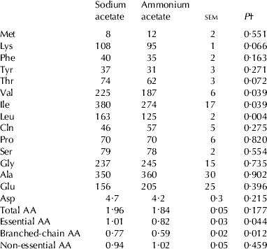
† Probability of no continuous effect of arterial plasma ammonia concentration
Average mammary uptakes (mmol/h) of amino acids (AA) from plasma in four cows (Experiment 3) during the last hour of a 10-h infusion into the jugular vein of 360 mmol/h ammonium acetate or sodium acetate

† Probability of no continuous effect of arterial plasma ammonia concentration
Average extraction percentages of arterial amino acids (AA) by the mammary glands of four cows (Experiment 3) during the last hour of a 10-h infusion into the jugular vein of 360 mmol/h ammonium acetate or sodium acetate

† Probability of no continuous effect of arterial plasma ammonia concentration
Discussion
Ammonia toxicity arises when portal ammonia is not removed by the liver and is allowed to accumulate in the systemic circulation, primarily affecting function of the central nervous system through shifts in amino acid metabolism (Bachmann et al. Reference Bachmann, Braissant, Villard, Boulat and Henry2004). Although ruminant animals, as a consequence of pregastric microbial fermentation, absorb ammonia at rates that approach or even exceed the capacity of the liver to remove it (Symonds et al. Reference Symonds, Mather and Collis1981), the effects of hyperammonaemia on milk synthesis in lactating ruminants have not been well characterized. When lactating ewes were infused abomasally with 46 mmol/h ammonium bicarbonate, milk production declined by 15%, protein yield by 24%, and milk fat percentage increased by 14% (Malik et al. Reference Malik, Nicol and Van Houtert1999). Plasma ammonia concentrations were not measured but if ammonia were cleared at 0·054/min from a volume of distribution of 69·5% of BW as in Experiment 1, then arterial concentration of ammonia would have been 46÷(60·0·054·0·695·BW)=0·307 mm, or close to 10-times normal. Moorby & Theobald (Reference Moorby and Theobald1999) infused 200 mmol/h ammonium acetate into the duodenum of lactating cows and observed only a 2% decrease in milk casein percentage, a 4% increase in milk fat percentage, and no change in yields of milk or its components. Again, plasma ammonia concentrations were not measured. To use the data from Experiment 1 to estimate the level of hyperammonaemia reached in the work of Moorby & Theobald (Reference Moorby and Theobald1999) we must take into account the different site of ammonia infusion. The jugular infusion given by Malik et al. (Reference Malik, Nicol and Van Houtert1999) and by ourselves bypasses the normal route of ammonia entry from the digestive tract into circulation, which is via the portal vein from which the liver can extract a large proportion. First-pass extraction of portal ammonia by the porcine liver was estimated as 73% by fitting dilution curves of [13N]ammonia following pulse doses (Keiding et al. Reference Keiding, Munk, Roelsgaard, Bender and Bass2001). Keiding et al. (Reference Keiding, Munk, Roelsgaard, Bender and Bass2001) also estimated from the arteriovenous concentration difference measurements of Lobley et al. (Reference Lobley, Connell, Lomax, Brown, Milne, Calder and Farningham1995) and Symonds et al. (Reference Symonds, Mather and Collis1981) that first-pass extraction in sheep and lactating dairy cows was approximately 75%. Assuming a clearance of 1504 l/h as in Experiment 1 and 75% extraction of portal ammonia on first pass through the liver, 200 mmol/h duodenal ammonia (Moorby & Theobald, Reference Moorby and Theobald1999) would increase arterial concentrations by 33 μm, or approximately 2-fold. In contrast, arterial ammonia concentrations were increased 5-fold in our Experiment 3 and an estimated 10-fold in the experiment of Malik et al. (Reference Malik, Nicol and Van Houtert1999). It appears from the limited results that the higher the peripheral ammonaemia, the more likely milk yield and, possibly, protein percentage will be depressed.
Metabolic response
The 5-fold hyperammonaemia for 10 h caused 20% decreases in yields of milk, protein and lactose. Feed intake during the infusion interval was observed to decrease in Experiment 2. In Experiment 3, feed intake was monitored over a 24-h period that included the 10-h infusion interval and the 14-h recovery period. Intakes over 24 h were not depressed but this does not preclude the possibility that elevated intakes after the infusion compensated for an intake depression during infusion. Ammonia infusion into lactating ewes and cows did not affect intake (Malik et al. Reference Malik, Nicol and Van Houtert1999, Moorby & Theobald, Reference Moorby and Theobald1999) but urea supplementation has depressed intake (Choung et al. Reference Choung, Chamberlain, Thomas and Bradbury1990). The intake depression we observed in Experiment 2 may be regarded as a protective response which lessens the rate of ammonia entry from the rumen. It might also have been responsible for the depression in milk yield, given that milk production decreases approximately 20% within 6–12 h of starting to fast (McGuire et al. Reference McGuire, Bauman, Dwyer and Cohick1995; Toerien & Cant, Reference Toerien and Cant2007).
Whether intake depression or some other contributing factor from ammonium acetate was responsible for the decline in milk production can be evaluated by determining whether the metabolic response of the cows corresponded to an intake depression effect or a hyperammonaemia effect. The response of lactating ruminants to a severe feed restriction or fast is not rapid owing to the continued outflow of nutrients from the reticulorumen. Typically, plasma concentrations of glucose decrease little, and acetate and gluconeogenic amino acid concentrations decrease until such time as the milk yield falls precipitously, after 18–24 h (Lomax & Baird, Reference Lomax and Baird1983; McGuire et al. Reference McGuire, Bauman, Dwyer and Cohick1995; Ndibualonji et al. Reference Ndibualonji, Dehareng and Godeau1997; Toerien & Cant, Reference Toerien and Cant2007). Plasma NEFA concentrations rise throughout a fast but elevated ketone body concentrations are not apparent until 24–30 h (McGuire et al. Reference McGuire, Bauman, Dwyer and Cohick1995; Toerien & Cant, Reference Toerien and Cant2007). Concentrations of essential amino acids in plasma increase as they are used less for protein synthesis and are mobilized from muscle (Ndibualonji et al. Reference Ndibualonji, Dehareng and Godeau1997). MPF and uptakes of milk precursors decrease in conjunction with the declining rate of milk synthesis (Linzell, Reference Linzell1967; Chaiyabutr et al. Reference Chaiyabutr, Faulkner and Peaker1980). Lactose percentage in milk decreases while protein and fat percentages increase (Linzell, Reference Linzell1967; Drackley et al. Reference Drackley, Veenhuizen, Richard and Young1991; Friggens et al. Reference Friggens, Emmans, Robertson, Chamberlain, Whittemore and Oldham1995). Similarities between the response to hyperammonemia observed here and the response to a restricted intake include the increase in plasma NEFA concentrations with no change in β-hydroxybutyrate, and the matching of MPF and acetate uptake with the declining milk yield. Differences include an increase in plasma glucose concentration, a decrease in concentrations of several essential amino acids, and a lack of change in milk protein, fat and lactose percentages and mammary uptakes of amino acids and energy metabolites other than acetate. These differences suggest that 10 h of ammonia infusion was not long enough for an intake depression to have affected the cows' metabolism.
Similarities between the hyperammonaemia response we observed and hyperammonaemia responses in dairy cows and other species include the increase in plasma glucose concentration (Emmanuel & Edjtehadi, Reference Emmanuel and Edjtehadi1981; Symonds et al. Reference Symonds, Mather and Collis1981; Barej et al. Reference Barej, Harmeyer, Drost and Libau1982; Choung et al. Reference Choung, Chamberlain, Thomas and Bradbury1990; Holeček et al. Reference Holeček, Šprongl and Tichý2000) that has been attributed to decreased rates of glucose utilization in the body (Emmanuel & Edjtedahi, Reference Emmanuel and Edjtehadi1981) and/or adrenaline-induced glycogenolysis (Slawski et al. Reference Slawski, Barej and Wiechetek1984; Wiechetek et al. Reference Wiechetek, Podgurniak, Zabielski and Podgurniak1989), an increase in plasma NEFA concentrations that persists in adrenalectomized animals (Slawski et al. Reference Slawski, Barej and Wiechetek1984; Wiechetek et al. Reference Wiechetek, Podgurniak, Zabielski and Podgurniak1989), decreased plasma concentrations of BCAA that have been attributed to increased Gln synthesis and decreased protein degradation in muscle (Leweling et al. Reference Leweling, Breitkreutz, Behne, Staedt, Striebel and Holm1996; Holeček et al. Reference Holeček, Šprongl and Tichý2000), and decreases in plasma concentrations of Lys and Thr (Holeček et al. Reference Holeček, Šprongl and Tichý2000) that have not yet been fully explained.
It has been suggested that detoxification of free ammonia to urea and Gln consumes essential amino acids that might otherwise have been used for growth or lactation (Reynolds, Reference Reynolds1992; Lobley et al. Reference Lobley, Connell, Lomax, Brown, Milne, Calder and Farningham1995). The role of the liver has been called into question by arteriovenous difference data that showed that the additional urea output during mesenteric infusion of 1100 μmol/min ammonium bicarbonate into sheep was readily accounted for by the hepatic removal of ammonia from circulation, without any need for additional amino acid catabolism (Milano & Lobley, Reference Milano and Lobley2001). Isotopomer experiments have shown that both N atoms on the urea molecule can derive from ammonia (Mutsvangwa et al. Reference Mutsvangwa, Buchanan-Smith and McBride1999). This small influence of the liver may explain why milk synthesis was not affected by duodenal ammonia infusion (Moorby & Theobald, Reference Moorby and Theobald1999) that did not exceed the hepatic capacity for removal, while synthesis of milk components was depressed by jugular ammonia infusions that elevated peripheral concentrations of ammonia 5–10-fold (Experiments 2 and 3 and Malik et al. Reference Malik, Nicol and Van Houtert1999).
Gln concentration in circulation typically increases during hyperammonaemia, which is thought to be due to Gln synthesis in muscle, where Ala and the BCAA, Leu, Ile and Val, provide much of the C skeleton and some N (Leweling et al. Reference Leweling, Breitkreutz, Behne, Staedt, Striebel and Holm1996; Bachmann et al. Reference Bachmann, Braissant, Villard, Boulat and Henry2004). Accordingly, Ala and the BCAA concentrations decrease during hyperammonaemia (Leweling et al. Reference Leweling, Breitkreutz, Behne, Staedt, Striebel and Holm1996; Holeček et al. Reference Holeček, Šprongl and Tichý2000). In addition to muscle, portal-drained viscera (Milano & Lobley, Reference Milano and Lobley2001) and the brain (Ott et al. Reference Ott, Clemmesen and Larsen2005) also consume BCAA during hyperammonaemia, presumably to provide C for Gln synthesis. In Experiment 3, we found that BCAA, along with Lys and Thr, decreased in arterial concentration in hyperammonaemic dairy cows. However, uptake of these amino acids from circulation by the mammary glands was not affected. These five amino acids are all part of the group II essential amino acids that are taken up by the mammary glands in excess of their output into milk (Mepham, Reference Mepham1982) so that deficiencies are relatively rare. Removal of approximately half of the Leu, Ile and Val from the absorptive amino acid supply had no effect on yields of milk and protein from early lactation cows (Weekes et al. Reference Weekes, Luimes and Cant2006). Thus, though we find evidence that ammonia detoxification in non-hepatic tissues alters the arterial supply of essential amino acids for milk synthesis, there is no evidence that the decreases in amino acid concentrations were responsible for the decline in milk component production during hyperammonaemia.
Milk synthetic response
The effect of ammonium acetate infusion on the profile of circulating metabolites in Experiment 3 shares more in common with responses to hyperammonaemia in non-lactating, non-ruminant species than with responses to fasting in lactating ruminants. We conclude that the decrease in milk yield was not caused by a depression in feed intake but, alternatively, the depressions in intake and milk yield are independent and may constitute two prongs of a coordinated response to hyperammonaemia.
MPF was estimated from Phe+Tyr arteriovenous differences by a method that assumes that these two amino acids are not taken up from red blood cells or peptides, and they are transferred without loss into milk proteins. These issues have been discussed by others using Phe+Tyr to estimate MPF (Cant et al. Reference Cant, DePeters and Baldwin1993; Mackle et al. Reference Mackle, Dwyer, Ingvartsen, Chouinard, Ross and Bauman2000; Thivierge et al. Reference Thivierge, Bernier, Dubreuil and Lapierre2002) and the possibility of deviation from the assumptions, which could shift the accuracy by 10–20%, is considered acceptable to detect treatment differences. For example, the contribution of peptides to mammary Phe+Tyr supply would have to change by several fold to be responsible for a 20% difference in MPF. However, because of the error inherent in calculating a difference between two measures of Phe+Tyr concentration in plasma, the MPF estimate is not very precise. According to the se of 75 l/h in Experiment 3 (Table 4), the difference in MPF between treatments would have to be 178 l/h to be detected as significant at P<0·05 with a power of 0·80. This means that if MPF decreased to the same degree as milk yield, i.e,. 20% or 130 l/h, it would not be detectable. Regression analysis shows that MPF was significantly related to milk yield (Fig. 3) which, given the decline in milk yield, may suggest that MPF was actually decreased by hyperammonaemia.
A similar argument involving se could be applied to the metabolite uptakes from plasma except that none of the uptake means, barring acetate, were arithmetically decreased by ammonium acetate infusion. Thus, errors do not appear to be masking a true effect of treatment. Amino acid uptakes were not decreased during hyperammonaemia, yet arterial concentrations of many essential amino acids were depressed, which manifested itself, for Leu, as an increased extraction percentage (Table 7). The only uptake affected was that of acetate. Aside from acetate, then, the decrease in milk yield and MPF was not accompanied by or consequent to a decrease in mammary utilization of milk precursors.
It is possible that hormones mediated the 20% decline in milk yield. Adrenaline release from the adrenal glands is stimulated by ammonia (Slawski et al. Reference Slawski, Barej and Wiechetek1984) and exogenous adrenaline reduces milk yield (Gorewit & Aromando, Reference Gorewit and Aromando1985; Leenanuruksa & McDowell, Reference Leenanuruksa and McDowell1985). Exogenous insulin can stimulate milk synthesis (Mackle et al. Reference Mackle, Dwyer, Ingvartsen, Chouinard, Ross and Bauman2000) but plasma insulin concentration is generally increased during hyperammonaemia (Strombeck et al. Reference Strombeck, Rogers and Stern1981; Slawski et al. Reference Slawski, Barej and Wiechetek1984) so insulin does not make a likely candidate to explain the decreased milk yield. To our knowledge, effects of hyperammonaemia on the galactopoietic hormones growth hormone and IGF-1 have not been studied.
Conclusion
Malik et al. (Reference Malik, Nicol and Van Houtert1999) infused 46 mmol/h ammonium bicarbonate into the jugular vein of lactating ewes, to an arterial concentration of ammonia we estimate to have been 10-times normal. The observed 24% decrease in milk protein yield supports the hypothesis that ammonia detoxification alters availability of amino acids for productive purposes. Moorby & Theobald (Reference Moorby and Theobald1999) created a much smaller hyperammonaemia in lactating dairy cows by duodenal infusion of 200 mmol/h ammonium acetate to an estimated arterial concentration twice normal. There was no effect on milk production, a 4% increase in milk fat percentage and only a 2% decline in casein percentage. They concluded that ammonia detoxification had a negligible impact on amino acid supplies for productive purposes. We infused 360 mmol/h ammonium acetate into the jugular vein of dairy cows to an arterial concentration 5-times normal at 193 μm, and observed a 20% decrease in milk, protein and lactose yields. Circulating concentrations of several essential amino acids decreased but mammary uptakes of amino acids and energy metabolites, excluding acetate, were not affected by the hyperammonaemia. Mammary acetate uptake decreased in proportion to the drop in milk yield and mammary plasma flow. A depression in feed intake may have caused the decrease in milk production. Further research should focus on the possible role of adrenaline in mediating the depression in milk synthesis during hyperammonaemia.
The authors thank Sarah Armstrong, Donna Benschop, Kathleen (Brown) Shore, Jodi Calberry, Peter deVries, Susy Kilby, Sue (Edmonds) Sinclair, Bev Livingston, Paul Luimes, Angela Martin, Lori Sheehan, Melanie Sippel, Chanelle Toerien, Tammi Weekes and Erin Winter for assistance with animal care, sampling and laboratory analysis. Financial support for this work was provided by NSERC Canada and the Ontario Ministry of Agriculture and Food. Norm Purdie was in receipt of a Dairy Research Development Corporation postgraduate award.