1. Introduction
The Devonian period is a crucial interval of time for understanding the diversification of the echinoid stem group. Stem group echinoids reach their peak diversity in Carboniferous times (Kier, Reference Kier1965; Smith, Reference Smith1984); however, this heightened Carboniferous diversity has its roots in the Devonian period. The Palaechinidae, Archaeocidaridae, Proterocidaridae and Lepidesthidae, which constitute the most diverse families of echinoids in the Carboniferous period, and indeed the entire Late Palaeozoic, are all first known from Devonian strata (Whidborne, Reference Whidborne1898; Thomas, Reference Thomas1921; Belanski, Reference Belanski1928; Dehm, Reference Dehm1961; Baird et al. Reference Baird, Gryta, McKenzie, Over, Pulawski and Sullivan2009). The proterocidarids, which are charismatic stem group echinoids with flattened tests and expanded adoral surfaces and pore pairs, are amongst the most abundant Devonian echinoids in the fossil record. This is thanks, in part, to the existence of a Lagerstätte preserving large numbers of the genus Hyattechinus in the Famennian of Pennsylvania (Jackson, Reference Jackson1912; Baird et al. Reference Baird, Gryta, McKenzie, Over, Pulawski and Sullivan2009). Hyattechinus is little known from outside this Lagerstätte in Devonian strata, although it is more commonly recorded from the Mississippian of North America and Western Europe (Jackson, Reference Jackson1929; Hawkins, Reference Hawkins1935; Kier, Reference Kier1965; Thompson & Denayer, Reference Thompson and Denayer2017).
Jackson (Reference Jackson1929), in his excellent monograph of the Mississippian echinoids of Belgium, mentioned a single specimen of an undescribed species from the Devonian of Devon, UK, housed in the Natural History Museum, London (then the British Museum Natural History). In addition to referring this species to Hyattechinus, he stated, ‘It is hoped to describe this [specimen] in a later publication’ (Jackson, Reference Jackson1929, p. 30). Sadly, Jackson never managed to describe the specimen, which has remained in the collections of the NHM for the 89 years since his monograph on the Belgian Palaeozoic echinoids was published. We herein describe the specimen, which comprises the only known individual of the new species Hyattechinus anglicus n. sp. and is the first Devonian occurrence of Hyattechinus from outside of North America. This expands the palaeobiogeographic range of Hyattechinus in the Devonian period, and indicates that the genus had a fairly wide distribution along the southern margin of Laurussia during Famennian times.
2. Geological setting and stratigraphy
The specimen of Hyattechinus anglicus was collected by J. G. Hamling and donated to the Natural History Museum in October 1916. The collections details accompanying the specimen state ‘Pilton Beds, Top Orchard, Pilton, Devon’, and the specimen is preserved as a mould in a grey-green mudstone. The ‘Pilton Beds’, which have also been called the ‘Pilton Shale’, are now formally designated the Pilton Mudstone Formation (Leveridge & Hartley, Reference Leveridge, Hartley, Brenchley and Rawson2006). The Pilton Mudstone Formation forms the upper part of the Exmoor Group and straddles the Devonian–Carboniferous boundary (Waters et al. Reference Waters, Waters, Barclay and Davies2009). The Pilton Mudstone Formation comprises grey to grey-green mudstones and siltstones, thin to thickly bedded sandstones and beds and lenses of limestone. The base of the Pilton Mudstone Formation is defined as the first appearance of mudstone beds within the more dominant sandstones (which prevail in the underlying Famennian Baggy Sandstones Formation). Higher up in the Pilton Mudstone Formation, the mudstones become dominant, with the sandstones, siltstones and limestones becoming progressively less frequent (Waters et al. Reference Waters, Waters, Barclay and Davies2009).
The Pilton Mudstone Formation was subdivided into three units (A–C) by Goldring (Reference Goldring1957, Reference Goldring1970) with a fourth unit (D) added by Prentice (Reference Prentice1960). The precise Devonian–Carboniferous boundary is identified as the boundary between units A and B where the coarse sandstones disappear and mudstones dominate (Whittaker & Leveridge, Reference Whittaker and Leveridge2011 and references therein). Understanding the distribution of the various units of the Pilton Mudstone Formation is complicated by its limited outcrop and the fact that it is highly folded and faulted. However, after extensive investigation, Unit A of the Pilton Mudstone Formation and indeed all Pilton Mudstone Formation outcrops known north of the River Taw have been identified as belonging to the uppermost Famennian Wocklumeria Ammonoid Zone based on various different palaeontological lines of evidence (see summary of Lane, Maples & Waters, Reference Lane, Maples and Waters2001 and references therein).
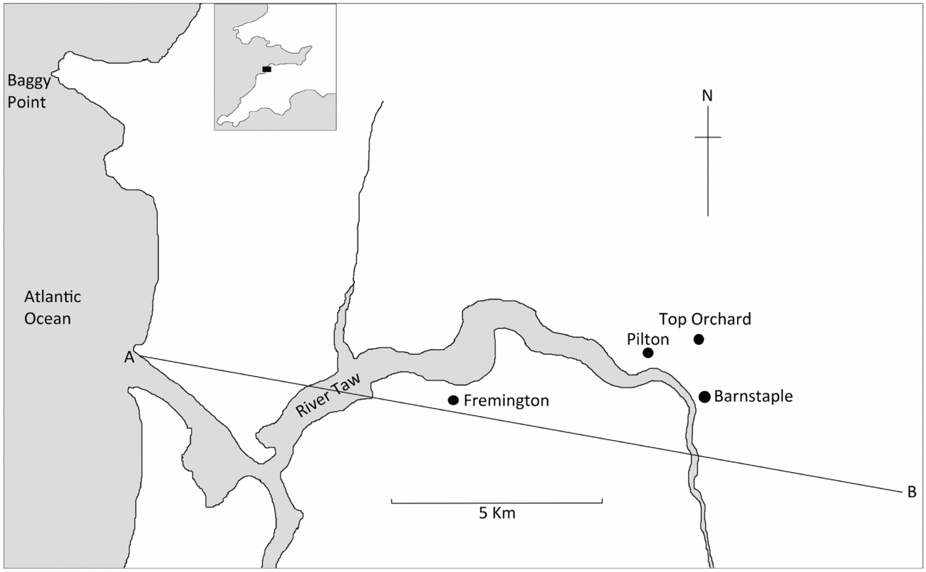
Unit A of the Pilton Mudstone Formation was further subdivided into three subunits (1–3) (Goldring, Reference Goldring1955). Based on observations of the Pilton Mudstone Formation from both Croyde Bay and at Poleshill Quarry, Barnstaple, Lane, Maples & Waters (Reference Lane, Maples and Waters2001) judged that all pelmatozoan fossils collected historically from north of the River Taw around Barnstaple (by J. G. Hamling and other collectors around the early 1900s) were from the fossil-rich subunit A3 (with A1 and A2 being devoid of articulated echinoderm fossils). ‘Top Orchard’ as found on the label with the specimen of H. anglicus is probably an abbreviated name for Top Orchard Quarry, a quarry situated c. 1 km east of the village of Pilton (now part of Barnstaple), to the north of the River Taw (Fig. 1). The lithology preserving H. anglicus, a siliciclastic mudstone with a distinct grey-green appearance, closely matches that of other echinoderms collected from Top Orchard Quarry and thus, for these reasons, and those stated above, we conclude that the specimen of H. anglicus was collected from subunit A3 of the Pilton Mudstone Formation and is from the Wocklumeria Zone of uppermost Devonian age, rather than from the slightly younger Tournaisian (Mississippian) mudstones.
Simplified map of the area from where the specimen of Hyattechinus anglicus n. sp. was collected. Line A–B marks the southernmost limit of the North Devon Basin and approximately the Pilton Mudstone Formation. Dots mark the location of relevant settlements and outcrops.

The Pilton Mudstone Formation forms part of the rock succession of the upper North Devon Basin. The North Devon Basin is the most northerly outcrop area of the British Rhenohercynian Zone. The Rhenohercynian Zone formed on the Avalonian Terrane and extended c. 1000 km from eastern parts of North America (Newfoundland) in the west, through SW England and across northern Europe into Germany to the east (Leveridge & Hartley, Reference Leveridge, Hartley, Brenchley and Rawson2006). The geological history of the British Rhenohercynian Zone is complex, as it was intimately associated with the closure of both the Iapetus and later the Rheic oceans and the subsequent continental collisions. As such, its structural interpretation is controversial (see summary of Leveridge & Shail, Reference Leveridge and Shail2011). However, from Devonian to Mississippian times, the British Rhenohercynian Zone, which comprises all of the SW English Devonian (Holder & Leveridge, Reference Holder and Leveridge1986), was formed from a series of fault-controlled basins on a southern-facing passive continental marginal on the southern flank of the Avalonian Terrane, during its docking with Laurentia (and so, in part, forming Laurussia) (Golonka & Gawęda, Reference Golonka, Gawęda and Sharkov2012). Continuous subduction of the Rheic Ocean to the south caused tensional stresses in the part of Avalonia that is SW England and formed a series of basins that filled with sediment and now form the outcrops observed in Devon and Cornwall. These basins were active at different times, with the North Devon Basin active from Middle Devonian to Mississippian times. The British Rhenohercynian basins only moved to their current contiguous position with respect to South Wales (and the rest of Britain) during middle–late Carboniferous times, following considerable transverse movement along the Bristol Channel–Bray Fault (Shail & Leveridge, Reference Shail and Leveridge2009). The North Devon Basin experienced mostly shallow marine and terrestrial conditions and comprises a series of marine and terrestrially derived sedimentary rocks (Leveridge & Hartley, Reference Leveridge, Hartley, Brenchley and Rawson2006).
The Pilton Mudstone Formation was therefore deposited during a gradual transgression from terrestrial deltaic and shallow marine environments to more open marine conditions, as evidenced by the increased dominance of mudstones and the appearance of a normal marine fauna including trilobites, crinoids, goniatites and brachiopods (Whittaker & Leveridge, Reference Whittaker and Leveridge2011). Therefore, subunit A3 of the Pilton Mudstone Formation was deposited in a transgressive shallow marginal shelf sea setting, on the southern slope of the Avalonian Terrane about 30°S (southern part of the Laurussia megacontinent) within the Rheic Ocean (Golonka & Gawęda, Reference Golonka, Gawęda and Sharkov2012). We therefore assume that the echinoid was living in neritic conditions when it was buried.
3. Systematic palaeontology
Class ECHINOIDEA Leske, Reference Leske1778
Stem group ECHINOIDEA
Family Proterocidaridae Smith, Reference Smith1984
Type genus. Proterocidaris de Koninck, Reference de Koninck1882
Other genera. Pronechinus Kier, Reference Kier1965, Hyattechinus Jackson, Reference Jackson1912, Jacksonechinus Lambert, Reference Lambert1936, Fournierechinus Jackson, Reference Jackson1929, Perischodomus M'Coy Reference M'Coy1849, Pholidocidaris Meek & Worthen, Reference Meek and Worthen1869, Perischocidaris Neumayr, Reference Neumayr1881, Protoechinus Austin, Reference Austin1860.
Genus Hyattechinus Jackson, Reference Jackson1912
Type species. Hyattechinus beecheri Jackson, Reference Jackson1912
Other species. Hyattechinus pentagonus Jackson, Reference Jackson1912, Hyattechinus elegans Jackson, Reference Jackson1929, Hyattechinus laudoni Kier, Reference Kier1965, Hyattechinus toreumaticus Hawkins, Reference Hawkins1935, Hyattechinus dixoni Hawkins, Reference Hawkins1935, Hyattechinus rarispinus (Hall, Reference Hall1868), and now Hyattechinus anglicus sp. nov.
Diagnosis. We herein provide an amended diagnosis for the genus Hyattechinus. Proterocidarid with test flattened and pentagonal or rounded in plan view. Two columns of ambulacral plates on adapical surface and two columns of enlarged ambulacral plates on adoral surface. Interambulacral plates at ambitus on adapical surface arranged into 7 to 14 columns. On oral surface, number of plate columns decreases adorally. A single basicoronal interambulacral plate in contact with peristome.
Occurrence. Hyattechinus is known from the Famennian of Pennsylvania, USA, and now Devon, UK, and from the Tournaisian of Pembrokeshire, Wales, Tournai, Belgium, and Montana, USA.
Hyattechinus anglicus sp. nov.
Figures 2–4
Diagnosis. Hyattechinus with at least seven columns of plates at the adoral ambitus. Interambulacral plates with distinct cruciform and two-barred cross shape. All interambulacral plates bearing a single, relatively large tubercle and numerous smaller secondary tubercles. On the adoral surface, two most adambulacral columns of interambulacral plates with imperforate, non-crenulate primary tubercles. Adapical ambulacral plates with numerous small secondary tubercles, and a single imperforate non-crenulate larger tubercle between the perradial suture and the pore pair. On the oral ambulacral plates, at least three tubercles between the perradial suture and the peripodial ring.
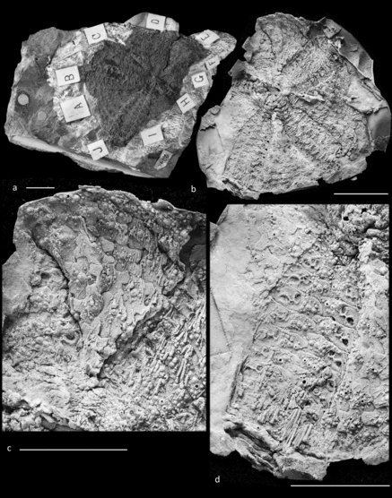
Holotype. The holotype is housed in the Natural History Museum, London (NHMUK) and is specimen number NHMUK PI E 12262. This specimen consists of the part and counterpart of a mould of the adoral surface of the test, and portions of the adapical surface. Latex moulds of the specimen have revealed four and a half of the ambulacra of the oral surface, and three interambulacra. The peristome and tips of the oral surface of the teeth are also visible. A latex mould of the counterpart shows portions of an ambulacra and associated interambulacra from the adapical surface of the test, and part of the internal surface of the adoral surface of the test, including hemipyramids and teeth.
Description. Test pentalobate in outline, depressed in profile and thus wider than high (Figs 2a, b, 3a, b). The test is c. 60.3 mm in diameter. Test plating imbricate, and interambulacral plates imbricating over ambulacral plates. At the ambitus, one ambulacral area is 16.3 mm wide and the adjacent interambulacral area is 21.7 mm wide, thus interambulacral areas are wider than the ambulacral areas at the ambitus. About midway between the ambitus and the peristome, however, ambulacral areas are wider than interambulacral areas.
Photographs of external view of oral surface of specimen NHMUK PI E 12262 of Hyattechinus anglicus n. sp. from the Top Orchard Quarry, Pilton, Devon, UK, Unit A3 of the Pilton Mudstone Formation, Exmoor Group, Wocklumeria Zone, Famennian, Upper Devonian. (a) External mould of oral surface. Scale bar is 10 mm. (b) Latex mould of oral surface showing peristome, interambulacral areas and ambulacral areas. Note how ambulacral areas are expanded and wider than interambulacral areas, and note the presence of serrations on the tip of the tooth and the absence of non-ambulacral plates on the peristome. Scale bar is 10 mm. (c) Close up of oral interambulacral area and portion of ambulacral area. Median interambulacral plates have numerous processes and bear primary and secondary tubercles (compare Fig. 4a). Ambulacral plates also bear numerous secondary tubercles and spines. Scale bar is 10 mm. (d) Close up of latex mould of oral ambulacral areas. Note numerous tubercles and differentiation of size between larger and smaller tubercles. Plates appear to have been covered with numerous small spines. Additionally, note enlarged pore pairs and peripodial rings. Scale bar is 5 mm.

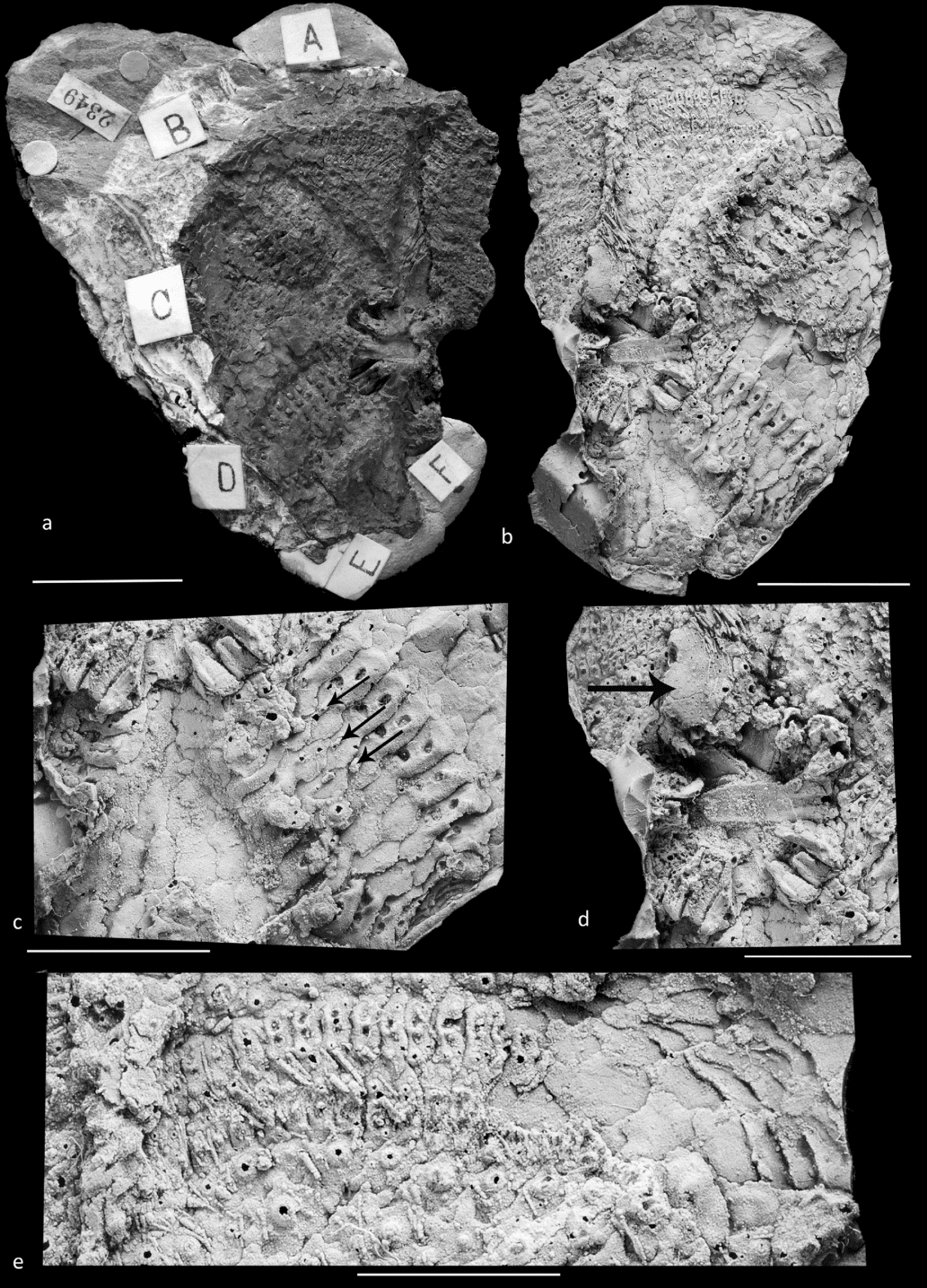
The apical disc is fragmentary; however, there does appear to be one large plate which is probably the madreporite (Fig. 3d). This plate is covered with tiny tubercles and is at least 6.4 mm long at its widest point. Smaller associated plates may be ocular or periproctal plates. The Aristotle's lantern is preserved, and is composed of, at the very least, teeth, hemipyramids, epiphyses and rotulae (Fig. 3d). The lantern is 20.2 mm in diameter. No compasses are visible, but this does not mean that they are absent. The foramen magnum is deep and wide, c. 5.2 mm wide. The individual teeth are c. 2.3 mm wide and at least 13.5 mm high. The tips of the teeth are serrate (Fig. 2b), and thus may be compound lamellar sensu Reich & Smith (Reference Reich and Smith2009). Rotulae are fragmentary, but c. 4.4 mm long and 1.4 mm wide. This taxon has no perignathic girdle. The peristome is 16.8 mm in diameter. Peristomial ambulacral plates are arranged into two columns in each area (Fig. 2b). A measured plate is 3.3 mm in diameter and 0.5 mm high. The ambulacral pores are 1.6 mm away from the perradial suture. Plates are elongate, and towards the interradial edge of the plates they bend orally before coming into contact with the adjacent ray of ambulacral plates.
Photographs of internal view of oral surface and portion of adapical surface of test of specimen NHMUK PI E 12262 of Hyattechinus anglicus n. sp. from the Top Orchard Quarry, Pilton, Devon, UK, Unit A3 of the Pilton Mudstone Formation, Exmoor Group, Wocklumeria Zone, Famennian, Upper Devonian. (a) Internal mould of oral surface and portions of external mould of adapical surface. Scale bar is 10 mm. (b) Latex mould of internal surface of oral interambulacra and ambulacra (bottom right and left), Aristotle's lantern and teeth (centre) and external surface of adapical interambulacra and ambulacra (upper right). Note the protrusions enclosing the radial water vessel on oral ambulacral plates proximal to the peristome. Scale bar is 10 mm. (c) Close up of latex mould of internal surface of oral surface. Note the protrusions (indicated by arrows), which enclose the radial water vessel, and the imbrication of the interambulacral plates over ambulacral plates. Scale bar is 5 mm. (d) Close up of latex mould of internal view of Aristotle's lantern and portions of the external surface of the apical system including the probable madreporite (indicated with an arrow). Scale bar is 5 mm. (e) Close up of latex mould of external surface of adapical coronal plating. Note that not all interambulacral plates bear primary tubercles. Furthermore, ambulacral pore pairs are much smaller than those on oral surface (compare Fig. 2d) and lack enlarged peripodia. Scale bar is 5 mm.

The ambulacra are straight, and in each ambulacral area plates are arranged into two columns of plates throughout their entire length (Figs 2d, 4b–d). Each ambulacral plate is pierced by two pore pairs. Ambulacral plates are imbricate. The nature of the imbrication is such that, from the exterior of the test, more adapical plates imbricate on top of more adoral plates. Plates also imbricate on top of more adoral plates in the opposite ambulacral column along the perradial suture. Where the width of the ambulacral area was measured on the adapical surface it was 9.6 mm wide and on the oral surface it was 14.9 mm wide, indicating an expansion of c. 1.5 times from the adapical to adoral surface.
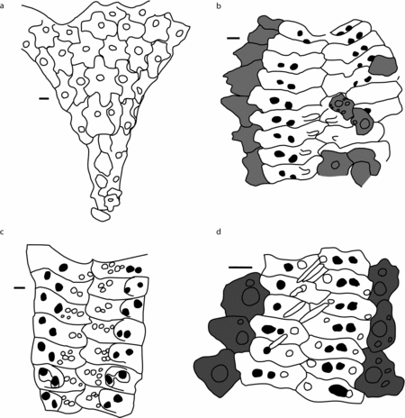
Camera lucida drawings of specimen NHMUK PI E 12262 of Hyattechinus anglicus n. sp. (a) Oral interambulacral area of H. anglicus. Note the two-barred cross-shaped plate in the centre of the interambulacral area. More median plates imbricate over more adambulacral. The more oral area has been crushed and plates appear to have fractured and moved. Secondary tubercles not included in drawing. The outline marks the border of the interambulacrum. (b) Interior view of oral ambulacra and interambulacra. Note the processes enclosing the radial water vessel on the more adoral plates. Tuberculate interambulacral plates are shifted from their original position, and may be from the adapical surface. (c) Exterior view of oral ambulacral plates. Note the peripodial rings which enclose the pore pairs. Ambulacral plates bear numerous tubercles. (d) Adapical ambulacral area and adambulacral interambulacral plates. Tubercles situated between pore pairs and perradial suture bear spines, which have shifted slightly. Adambulacral plates bear primary tubercles. In (b, d) ambulacral plates are shown in white, interambulacral plates in grey. All scale bars are 1 mm and drawings are oriented with more adoral plates at the bottom of the image. Open plate lines indicate ambiguity regarding morphology.

Ambulacral plates on the adapical surface are about three times as wide as they are high (Figs 2, 4d). A raised interporal partition is present between the ambulacral pores on each plate. As opposed to being sunken into the plate, the periphery of the pores seems to be raised off the plate surface such that the pore pair is in relief against the rest of the plate. Numerous small tubercles are present on the plates (Fig. 4d). Where it is visible, most plates show one small tubercle between the perradial suture and the pore pairs, and another slightly adoral to the pore pairs between the pore pairs and the adradial suture. These tubercles are imperforate and non-crenulate. In addition to these two tubercles, smaller tubercles may be present on the plate surface.
On the oral surface, plates are about five times wider than high. Pore pairs are in large, circular peripodia (Figs 2d, 4c), which on a plate 11 mm wide is 2.9 mm in width. The peripodial ring is faint and appears to be better developed on the adapical, adoral and perradial sides of the plate than on the adradial side. Pore pairs are oriented obliquely on plates, with the perradial pore pairs located more adorally within the peripodial ring (Fig. 4c). As on the adapical plates, the edge of the pores is elevated in relief with respect to the rest of the peripodial ring floor. This elevated pore rim also connects to a raised interporal partition between the two pores with the peripodium. Three or more larger tubercles are present on plates between the peripodial ring and the perradial suture, usually in a line parallel to the adoral plate edge. These tubercles are imperforate and non-crenulate, and bear spines. A variable number of smaller imperforate non-crenulate tubercles are also present on most plates. The interior surface of oral ambulacral plates is smooth, except for the most oral plates, which bear a small peg-like protrusion between the pore pairs and the perradial suture (Figs 3c, 4b). On the adradial edge of ambulacral plates a large flange protrudes adorally and adradially underneath the adjacent interambulacral plates. This flange is particularly visible on plates about midway between the ambitus and the peristome.
Interambulacral areas on the oral surface are triangle shaped and narrow adorally (Figs 2c, 4a). At the border with the peristome, there appears to be a single basicoronal plate, though the details of the plating towards the peristomial border are obscured and plates appear to have shifted from their original positions. The next row of plates adapically from the peristome which confidently represents the original plating structure has four plates abreast. The next adapical row has five columns of plates, then six, then seven (Figs 2c, 4a). The number of columns of plates at the ambitus is unknown, though is likely at least seven. On the adoral surface, plates imbricate adambulacrally. In the row with five columns of plates, the median-most plate imbricates over plates adambulacrally in both directions (Fig. 2c). The two median plates adapical and adambulacral to this plate in turn imbricate over the plates located adapically to them; however, the plates adambulacral to either of these two median plates imbricate under the plates in the next most adapical row (Fig. 2c). This pattern seems to exist throughout the oral interambulacral.
The median interambulacral plates in the five-columned row, and six-columned row on the adoral surface, are 20-sided, with a shape that is similar to a two-barred cross (Figs 2c, 4a). These plates have six processes, with two projecting in the adambulacral direction on either side of the plate and one process each projecting adorally and adapically. A single plate in the most median column of the interambulacral area imbricates over plates in all directions, except for possibly adorally, where the plates have shifted from their original position. Median plates in the seven-columned row are cruciform in shape. The two adambulacral projections present in the more adoral plates appear to have fused, resulting in large, cross-shaped plates. The more adambulacral plates in this row do, however, still display a two-barred cross morphology, as least on their adambulacral edges. The precise shape of adambulacral plates is difficult to ascertain, given the nature of preservation of the specimen, but they appear to have a more rounded adambulacral edge, without any prominent processes. All interambulacral plates bear a single larger imperforate non-crenulate tubercle in the centre of the plates (Fig. 4a). They are also covered in numerous small imperforate, non-crenulate secondary tubercles. Most of these tubercles are concentrated around the primary tubercle, but some are also present on the plate processes.
Adapical interambulacra are not well preserved, except for one partial area. The shape of plates is difficult to ascertain, but there do appear to be some processes as is the case with the oral interambulacral plates. The two most adambulacral rows have large primary tubercles. They are non-crenulate and imperforate. Other plates have a single larger tubercle in the centre, though these are at least half the size of the primary tubercles found on the adambulacral plates. Some scattered primary tubercles are also present on non-adambulacral plates, but the test is too fragmentary to distinguish any pattern regarding their organization. Smaller non-crenulate, imperforate secondary tubercles are also present on interambulacral plates.
Small, short primary and secondary spines cover interambulacral and ambulacral areas of the test, and the peristome and apical disc. Primary and secondary spines are only differentiated based upon their size. The longest primary spine on the test, which is missing both its proximal and distal ends, is 3.6 mm long. Spines are striate and taper distally. Spines lack a milled ring, and the spine base consists solely of a small swelling or bump, which is the widest part of the spine. Spines appear to have been solid in cross-section and radially symmetrical. Pedicellariae unknown.
Remarks. H. anglicus is amongst the oldest known species of Hyattechinus, along with H. beecheri, H. pentagonus and H. rarispinus, which are also known from the Famennian (Jackson, Reference Jackson1912). H. angilcus is differentiated from these taxa in having much more cruciform interambulacral plates, and in having more tuberculate test plates. The very cruciform interambulacral plates differentiate it from all other species of Hyattechinus; however, they do appear to be similar to the hexagonal adoral interambulacral plates of H. elegans Jackson, Reference Jackson1929 (Thompson & Denayer, Reference Thompson and Denayer2017, fig. 2B), H. pentagonus Jackson, Reference Jackson1912 (Jackson, Reference Jackson1912, fig. plate 59, fig. 5) and H. toreumaticus Hawkins, Reference Hawkins1935 (Hawkins, Reference Hawkins1935, plate 14, fig. 2). As opposed to other species of Hyattechinus, the tubercles of H. anglicus also do not appear to be sunken. The primary tubercles of H. elegans and H. laudoni are clearly sunken, and are within a distinct areole, while those of H. anglicus protrude from the surface of the plate surrounded by a cluster of secondary tubercles. H. anglicus also has large numbers of secondary tubercles relative to other species of Hyattechinus, such as H. laudoni, which only has two or three secondary tubercles (Kier, Reference Kier1965, plate 60, fig. 3).
H. anglicus, like other species of Hyattechinus, bears small perradial protrusions on the interior surface of the most adoral ambulacral plates. This protrusion may have been associated with oral enclosure of the radial water vessel, or may be a vestigial structure related to this function in the ancestors of Hyattechinus. So far these protrusions have been identified in H. laudoni, H. elegans, H. rarispinus and H. beecheri (Jackson, Reference Jackson1912, Reference Jackson1929; Kier, Reference Kier1965). In H. anglicus they are only present on the first four adoral plates, while in H. laudoni they appear to be present almost all the way to the ambitus (Kier, Reference Kier1965, plate 59, fig. 2). Furthermore, like other species of Hyattechinus where the tips of the teeth are visible, and like many other proterocidarids, H. angilcus has serrate teeth, which may be compound lamellar.
Etymology. Hand-written notes from R. T. Jackson stored with the specimen at the NHM indicate he had intended to name it Hyattechinus anglicus. We here honour Jackson's intentions and thus name this species anglicus owing to its recovery from English strata.
Occurrence. The type and only known specimen comes from the Famennian Pilton Mudstone Formation, Top Orchard Quarry, Pilton, Devon, UK.
4. Discussion
4.a. Echinoids from the type Devonian
The Upper Devonian of the UK yields a diverse echinoderm fauna. While crinoids are abundant (Lane, Maples & Waters, Reference Lane, Maples and Waters2001; Donovan & Fearnhead, Reference Donovan and Fearnhead2014), echinoids are exceptionally rare. The rarity of stem group echinoids relative to other echinoderms in the type Devonian is likely the product of their propensity to rapidly disarticulate (Thompson & Ausich, Reference Thompson and Ausich2016), and their general lack of abundance in Palaeozoic ecosystems relative to other echinoderm groups. With the description of Hyattechinus anglicus, the total diversity of echinoids from the Pilton Mudstone Formation of the type Devonian now stands at three species. Previously, Whidborne described two species: the lepidesthid Lepidesthes devonicans Whidborne, Reference Whidborne1896 and the essentially indeterminate Pholidocidaris acuaria Whidborne, Reference Whidborne1898. Furthermore, an isolated spine attributed to an echinoid is also present in the older Givetian strata of the Lummaton Shell Beds (Donovan, Fearnhead & de Winter, Reference Donovan, Fearnhead and de Winter2017). Despite the rarity of echinoids, the fauna of the Pilton Mudstone Formation thus preserves at least three different genera from two different families, which is a fairly diverse echinoid assemblage when compared to other Devonian faunas.
4.b. Palaeobiogeographic distribution of Famennian Hyattechinus
The classic Hyattechinus Lagerstätte, from near Meadville, Pennsylvania (Jackson, Reference Jackson1912), is known from the Famennian ‘Drake Well Formation’. The Hyattechinus-bearing strata have long been considered to be Mississippian in age (Jackson, Reference Jackson1912; Kier, Reference Kier1965); however, recent stratigraphic and biostratigraphic work indicates that the ‘Drake Well Formation’ yields conodonts indicative of the Palmatolepsis gracilis expansa and Siphonodella praesulcata zones (Baird et al. Reference Baird, Gryta, McKenzie, Over, Pulawski and Sullivan2009), the two youngest conodont biozones of the Famennian, and thus the Devonian (Becker, Gradstein & Hammer, Reference Becker, Gradstein, Hammer, Gradstein, Ogg, Schmitz and Ogg2012). This Lagerstätte contains three species of Hyattechinus: H. pentagonus, H. rarispinus and H. beecheri. The ‘Drake Well Formation’ was deposited in the Appalachian Basin on the southern margin of Laurussia and bounded to the east by the uplands of the Appalachian Peninsula (Kent, Reference Kent, Woodrow and Sevon1985; Woodrow, Reference Woodrow, Woodrow and Sevon1985; Golonka & Gawęda, Reference Golonka, Gawęda and Sharkov2012). H. anglicus is known from the North Devon Basin, which was part of the Rhenohercynian Basin (Holder & Leveridge, Reference Holder and Leveridge1986), marginal to the Rheic Ocean, southeast of the margin of Laurussia (Golonka & Gawęda, Reference Golonka, Gawęda and Sharkov2012), and was also deposited during latest Devonian time. While the Hyattechinus Lagerstätte from the Appalachian Basin was also deposited on the southern margin of Laurussia, it was further (some 3000 km) to the southwest and at a higher palaeolatitude (50°S) (Golonka & Gawęda, Reference Golonka, Gawęda and Sharkov2012). Thus, it appears that Hyattechinus occurred in at least two localities along the southern margin of Laurussia during late Famennian time, and may have had a fairly wide distribution along the margin of the Rheic Ocean.
Hyattechinus is not the only echinoderm to have been present in the Famennian of both the Appalachian Basin and North Devon Basin, as similarities have also been noted between their crinoid faunas (Lane, Maples & Waters, Reference Lane, Maples and Waters2001). Waters & Webster (Reference Waters, Webster and Königshof2009) divided Famennian pelmatozoan faunas into the Rheic (Europe, North America and North Africa) and Palaeotethys (NW and SW China, Australia and Iran) realms. They attributed differences in the taxonomic composition of these faunas in part to differences in tolerance for clastic sediments, as the Rheic realm contained a diverse fauna of cladid crinoids, which are more tolerant to siliciclastic environments than other crinoid clades (Kammer & Ausich, Reference Kammer and Ausich1987). Hyattechinus, so far, appears to be a constituent of the Rheic realm, with only Famennian occurrences along the southern margin of Laurussia. Furthermore, both H. anglicus from the North Devon Basin and the Hyattechinus from the Appalachian Basin are known from siliciclastic sediments, and the abundance of Hyattechinus in the ‘Drake Well Formation’ Lagerstätte suggests that this genus was also tolerant of siliciclastic environments. Currently no echinoids are described from the Famennian of Palaeotethys, and future fieldwork will be necessary to determine if Hyattechinus is unique to the Rheic realm. Furthermore, in contrast to Devonian Hyattechinus, Mississippian species are known predominantly from carbonate environments (Jackson, Reference Jackson1929; Hawkins, Reference Hawkins1935). Additional Famennian Hyattechinus, if they are found, will allow for testing of the hypothesis that Devonian Hyattechinus preferred siliciclastic environments.
Acknowledgements
J.R.T. was funded by the US National Science Foundation Grant IOS1240626 to D. J. Bottjer and E. H. Davidson. J.R.T. also thanks D. J. Bottjer for reading over an earlier version of the manuscript. We thank A. Kroh and an anonymous reviewer for their comments, which helped to improve the clarity of the manuscript.