Introduction
Benign positional vertigo is due to movement of otolithic debris, either free in a semicircular canal duct (canalithiasis) or attached to the cupula (cupulolithiasis).Reference Parnes, Agrawal and Atlas1 Consider the most common type of benign positional vertigo, posterior semicircular canal canalithiasis: as the head is tilted backwards (clinically in the Dix–Hallpike test), the free otoconia in the posterior canal duct move away from the ampulla, creating ampullofugal endolymph flow which displaces its cupula, producing increased activity of the posterior canal primary vestibular afferents and an excitatory nystagmus with vertical-torsional quick phases beating upwards and towards the affected ear. Lateral semicircular canal canalithiasis produces positional vertigo when the patient is supine, with horizontal nystagmus beating towards the affected (lowermost) ear, whereas lateral canal cupulolithiasis produces horizontal nystagmus beating towards the uppermost ear. Intriguingly, there are no convincing reports of posterior canal cupulolithiasis.
Since the early 1960s, it has been known that it is possible to inactivate (but not de-afferent) a semicircular canal by surgically obstructing its duct.Reference Money and Scott2 Once the canalithiasis theory of benign positional vertigoReference Epley3 took hold, it became logical to consider attempting to abolish posterior canal benign positional vertigo by occluding the posterior canal duct. The main risk of such an operation was to hearing, and the first report of successful posterior canal occlusion for posterior canal benign positional vertigo was in a deaf ear in 1990,Reference Parnes and McClure4 and a year later in a hearing ear.Reference Parnes and McClure5
We performed our first posterior canal occlusion in a hearing ear in November 1991.Reference Pohl, Baloh and Halmagyi6 Since then, we have performed two to three posterior canal occlusions each year. Here, we report the benefits of this procedure for controlling benign positional vertigo, and its risks to hearing and balance.
Methods
We reviewed data from 53 patients (55 ears) who had a posterior canal occlusion between 1991 and 2011 as treatment for intractable posterior canal benign positional vertigo. During this 20-year period, we saw and treated 5364 patients with posterior canal benign positional vertigo in our balance disorders clinic. All posterior canal occlusion patients met the following criteria: (1) persistent or relapsing, distressing or disabling, unilateral posterior canal benign positional vertigo; (2) poor or no response to repositioning manoeuvresReference Fife, Iverson, Lempert, Furman, Baloh and Tusa7, Reference Helminski, Zee, Janssen and Hain8 performed by at least one of the authors over at least a six month period (by poor response we mean at least monthly relapses); and (3) hearing in the unaffected ear equal to, or better than, that in the affected ear.
For these 53 patients, we reviewed the following: (1) clinical features and duration of symptoms before posterior canal occlusion; (2) clinical outcome after posterior canal occlusion (i.e. reported hearing loss or spontaneous vertigo, recurrence of ipsilateral benign positional vertigo, development of contralateral benign positional vertigo, and/or development of imbalance); (3) audiometry results before and after posterior canal occlusion; and (4) caloric test results before and after posterior canal occlusion.
Audiometry
We reviewed the air-conduction pure tone threshold audiograms (0.25 to 8 kHz) for each ear, taken before and after posterior canal occlusion. Bone-conduction pure tone thresholds were also measured in those patients whose air-conduction thresholds were above normal for age, and in those with middle-ear disease. All patients had an audiogram before posterior canal occlusion; those who reported a hearing change one week after posterior canal occlusion had progress audiograms. For those 24 patients whose first audiogram after posterior canal occlusion showed a >20 dB increase in hearing threshold at any frequency, we tried to obtain subsequent audiograms; we were able to do this for 19 patients.
Vestibular testing
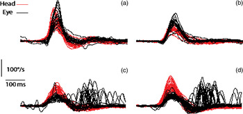
All patients had clinical horizontal and vertical head impulse testing before and after posterior canal occlusion. After posterior canal occlusion, 12 had measurements of vertical semicircular canal vestibulo-ocular reflex gain, as assessed with scleral search coilsReference Aw, Halmagyi, Pohl, Curthoys, Yavor and Todd9, Reference Cremer, Halmagyi, Aw, Curthoys, McGarvie and Todd10 and more recently (for six patients) with video-oculography (ICS; GN Otometrics, Taastrup, Denmark)Reference MacDougall, Weber, McGarvie, Halmagyi and Curthoys11 (Figure 1). All except three frail, elderly patients had standard clinical caloric testingReference Halmagyi, Yavor and McGarvie12 (usually bithermal – some patients could not tolerate four irrigations) before posterior canal occlusion. After posterior canal occlusion, all patients who reported vertigo or had a clinically positive lateral canal head impulse testReference Halmagyi and Curthoys13 at one week, indicating inadvertent damage to lateral canal function, had progress caloric testing. Some patients with hearing loss but no vertigo after posterior canal occlusion also had progress caloric testing.
Video head impulse test results for the vertical semicircular canals of a patient with bilateral posterior semicircular canal occlusion, for (a) left anterior, (b) right anterior, (c) left posterior and (d) right posterior semicircular canals. A reduced vestibulo-ocular reflex gain (approximately 0.4) is seen from both posterior semicircular canals, with multiple overt ‘catch up’ saccades, but a normal vestibulo-ocular reflex gain is seen from the anterior semicircular canals, with no ‘catch up’ saccades. Head = head velocity; eye = eye velocity

Surgical procedure
All 53 patients were operated upon by the one surgeon (DVP). The surgical technique began with a cortical mastoidectomy adequate to expose the lateral and posterior semicircular canals. The posterior semicircular canal was skeletonised carefully over its presenting curvature. A 3 mm fine diamond burr was then used to ‘blueline’ the posterior circular canal at its convex curvature. Progressively smaller diamond paste burrs were used to create a dome at the proposed fenestra site. The dome should be thicker centrally and thinned as far as the endosteum peripherally. An obtuse stapedectomy hook was then used to gently lift the dome and expose the endosteum. Frequently, there was minor weeping of perilymph at this time. The posterior semicircular canal could then be plugged. The material used was a combination of bone dust and Tisseel (Immuno, Vienna, Austria). The resulting material had a plasticine-like quality and could be gently inserted into the opening. Additional small amounts were inserted until such time as firm resistance was encountered, indicating that the endolymph lumen had been compressed. The bony dome removed at fenestration was then replaced and a small layer of temporalis fascia glued into position. Care was taken to remove blood and soft tissue from the air-containing mastoid cavity, as adhesions to the fenestra site can give rise to pressure-induced vertigo.
At the start of the procedure, 40 mg of frusemide was given, and frusemide treatment was continued throughout the first post-operative week in order to minimise post-operative hydrops caused by compression of the endolymphatic space.
Results
There were 35 women and 18 men, with a mean age at posterior canal occlusion of 59 years (range, 31 to 88 years). The mean duration of symptoms was six years (range, one to 27 years); however, some patients had suffered benign positional vertigo for many years before they came to us. The mean duration of follow up was 4.3 years (range, 0.5 to 13 years).
Seven patients had unilateral ear disease on the side of the benign positional vertigo which might have caused or contributed to their benign positional vertigo:Reference Karlberg, Hall, Quickert, Hinson and Halmagyi14, Reference Lee, Ban, Lee and Kim15 two had Ménière's disease, one had previous vestibular neuronitis, two had mastoid middle-ear surgery and one had previous herpes zoster infection of the vestibulo-cochlear nerve. One patient had an incidental ipsilateral vestibular schwannoma, and one patient had cerebellar atrophy (but no cerebellar signs). Eight patients had post-traumatic benign positional vertigo. Two patients needed bilateral (sequential) posterior canal occlusion after developing intractable posterior canal benign positional vertigo in the opposite ear; thus, we performed 55 posterior canal occlusions.
Control of positional vertigo
The posterior canal benign positional vertigo was abolished in all 55 operated ears of all 53 patients. Of the 53 patients, eight subsequently developed posterior canal benign positional vertigo in the opposite ear and two developed lateral canal benign positional vertigo in the posterior canal occlusion ear; each responded to the appropriate particle repositioning manoeuvre.Reference Parnes and McClure5 One patient had ongoing non-positional vertigo with headache which was treated as migrainous vertigo.Reference Millen, Schnurr and Schnurr16
Two patients developed post-surgical complications: one a posterior canal fistula needing repair,Reference Cremer, Migliaccio, Pohl, Curthoys, Davies and Yavor17 and the other a wound infection.
Hearing
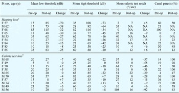
We were able to review audiograms from both before and after 49 of the 55 posterior canal occlusion procedures. We were not able to retrieve post-operative audiograms for six of the posterior canal occlusions, but none of these six patients had complained of hearing loss in the operated ear. In 24 patients, the first audiogram after posterior canal occlusion showed more than 20 dB sensorineural hearing loss at one or more frequencies. Further audiograms showed that this hearing loss tended to improve. Nine patients suffered a final sensorineural hearing loss of more than 20 dB at low frequencies (mean threshold at 500, 1000 and 2000 Hz) or more than 25 dB at high frequencies (mean threshold at 4000, 6000 and 8000 Hz), or at both (Table I). A further five patients suffered a 20–24 dB loss at high frequencies which was not clinically noticeable.
Final hearing and caloric test loss

*On audiometry, >20 dB hearing loss at low frequency and/or >25 dB hearing loss at high frequency. †Deterioration in caloric test results, for 30° and 40° irrigation. Y = years; pre-op = pre-operative; post-op = post-operative; F = female; M = male; – = worse after surgery; += better after surgery; NA = results not available
Balance
Most patients reported mild, transient imbalance in the week after surgery. Fifteen patients developed prolonged imbalance after posterior canal occlusion, but only five of these showed deterioration of vestibular function on caloric testing.
Vestibular function
Fifty patients had caloric testing before surgery and 37 had caloric testing after surgery. Two patients underwent caloric testing after but not before posterior canal occlusion.
None of the 13 patients who had caloric testing before but not after posterior canal occlusion had any vestibular symptoms after surgery, and none had a positive clinical horizontal head impulse test.Reference Halmagyi and Curthoys13
Of the 50 patients who had caloric tests before posterior canal occlusion, 14 patients had a canal paresis of more than 25 per cent on the affected side: two had known Ménière's disease, one had had vestibular neuritis and another herpes zoster of the vestibulo-cochlear nerve. Of the 37 posterior canal occlusion patients who had caloric testing after as well as before surgery (Table I), 10 showed deterioration in caloric sensitivity in the operated ear: the final canal paresis ranged from 43 to 100 per cent. Absolute loss of caloric sensitivity (i.e. the sum of 30 and 44oC responses) from the posterior canal occlusion ear ranged from 9 to 92°/second. Interestingly, four of these patients complained of no spontaneous vertigo or symptoms of an acute vestibular syndrome, even though they had suffered acute vestibular damage; two complained of acute vertigo and nausea, and in four patients we were unable to determine whether acute spontaneous vertigo was present. Only five of the 10 patients who lost caloric sensitivity had any permanent imbalance, which was mild and subjective in each case.
Discussion
Our study confirms that posterior canal occlusion is an effective treatment for posterior canal benign positional vertigo:Reference Leveque, Labrousse, Seidermann and Chays18–Reference Dingle, Hawthorne and Kumar27 the posterior canal benign positional vertigo was abolished from all 55 operated ears of our 53 patients.
What of the risks to hearing and balance?
The immediate sensorineural hearing loss that occurred in some patients after posterior canal occlusion improved over time and sometimes fluctuated, perhaps responding to diuresis, suggesting that it was related to endolymphatic hydrops. Of the 53 patients, 24 initially had more than 20 dB hearing loss, but only nine patients were left with more than 20 dB loss at low frequencies or more than 25 dB loss at high frequencies. In the end, 19 per cent suffered a permanent, greater than 20 dB, low frequency hearing loss or a greater than 25 dB, high frequency hearing loss: four patients at both high and low frequency, two patients only at low frequencies and three patients only at high frequencies.
Those who lost hearing tended not to lose their caloric responses, and vice versa. Of the 10 patients who lost some vestibular function as confirmed by caloric testing, three had some pre-existing loss; seven had normal caloric function before posterior canal occlusion and a total or subtotal loss afterwards. Interestingly, four of the 10 patients who lost caloric function post-operatively did not suffer the symptoms of acute vestibular loss (spontaneous vertigo and nausea). Fifteen patients were left with a mild but permanent subjective imbalance after posterior canal occlusion, but only five of these had abnormal caloric responses. The imbalance seems to be similar to that experienced by patients who have a permanent total unilateral vestibular loss, and might reflect inadequate brainstem compensation for the reduced peripheral vestibular input during head movements.Reference Halmagyi, Weber and Curthoys28
In summary, approximately one posterior canal occlusion patient in every six was left with a noticeable, permanent hearing loss, and approximately one patient in every 10 was left with some degree of balance loss.
A potential limitation of this study was that we were not able to find follow-up audiograms for six patients or follow-up caloric test results for 13 patients (of the 50 who underwent pre-operative testing). However, none of these patients complained of post-operative deafness or vertigo, and on clinical examination at the first post-operative visit none had developed any loss of hearing or of vestibular (i.e. lateral semicircular canal) function in the operated ear.
Could posterior canal occlusion predispose to the development of benign positional vertigo from the lateral or anterior semicircular canal of the operated ear, or would this happen as a matter of chance in patients with otoconiolithiasis? It is hard to know. Lateral canal benign positional vertigo eventually developed in two of our patients in the posterior canal occlusion ear and, happily, responded to particle repositioning manoeuvres. It should also be recognised that benign positional vertigo can be bilateral: eight of our posterior canal occlusion patients developed posterior canal benign positional vertigo in the opposite ear – six responded to particle repositioning manoeuvres, whereas two needed a second posterior canal occlusion procedure.
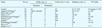
There have been nine previous reports of posterior canal occlusion as treatment for benign positional vertigo,Reference Agrawal and Parnes19–Reference Dingle, Hawthorne and Kumar27 with study cohorts ranging from four to 44 ears and a total of 159 ears (Table II). The cure rate for benign positional vertigo from the operated posterior canal is agreed to be close to 100 per cent. Most patients experience transient hearing loss. The reported incidence of permanent sensorineural hearing loss ranges from nil in one series of five patientsReference Seo, Hashimoto, Saka and Sakagami26 to 31 per cent in another of 32 patients.Reference Kisilevsky, Bailie, Dutt and Rutka20 Imbalance after posterior canal occlusion is commonly reported, but there is little reporting of actual caloric test results.
Hearing and balance loss after posterior semicircular canal occlusion: reported results

*Mean or range, or both. †On audiometry, >20 dB at low frequencies or >25 dB at high frequencies, or both. Pts = patients; SNHL = sensorineural hearing loss; FU = follow up; mth = months; T = temporary, P = permanent; ND = not documented; n = number of patients
Benign positional vertigo is believed to be due to endolymph flow induced by otoconial debris in a semicircular canal duct moving under the influence of gravity. Occluding the posterior canal stops endolymph flow, and consequently stops posterior canal benign positional vertigo permanently.Reference Parnes, Agrawal and Atlas1 Thus, the basic concept of posterior canal occlusion for benign positional vertigo is simple enough, and the procedure works remarkably well in practice: it is virtually guaranteed to abolish posterior canal benign positional vertigo. Furthermore, the procedure is not simply an elaborate labyrinthectomy: in our series, five in six patients lost no hearing and nine in 10 lost no balance.
However, there are still some unanswered questions about why and how posterior canal occlusion is so effective at permanently abolishing benign positional vertigo. For example, why does not the posterior canal benign positional vertigo recur if there are otoconia trapped between the site of occlusion and the cupula? Even if the endolymph could no longer move within the duct, why could not the otoconia attach to the cupula to produce posterior canal cupulolithiasis and benign positional vertigo with nystagmus beating vertically down and torsionally away from the affected ear (a pattern almost never seen)? In addition, vestibulo-ocular reflex data indicate that although semicircular canal occlusion permanently impairs semicircular canal function in the cat,Reference Money and Scott2 guinea pigReference Gilchrist, Curthoys, Burgess, Cartwright, Jinnouchi and MacDougall29 and human,Reference Aw, Halmagyi, Pohl, Curthoys, Yavor and Todd9 it does not do so in at least two species of monkey (rhesus and cynomolgus),Reference Yakushin, Kolesnikova, Cohen, Ogorodnikov, Suzuki and Della Santina30 at least in response to high-acceleration stimulation of the lateral semicircular canals. However, it is not certain whether the permanent impairment of the posterior canal vestibulo-ocular reflex in humans after posterior canal occlusion indicates total or partial loss of posterior canal sensitivity, and whether sensitivity might increase in time. Some sensitivity could return, as during posterior canal occlusion surgery the hydrostatic pressure transmitted to the ampulla might detach the cupula from the ampulla, contributing to loss of sensitivity.Reference Rabbitt, Boyle and Highstein31 The occlusion is of course permanent, but the cupula might reattach and restore some canal sensitivity.
• In benign positional vertigo unresponsive to repositioning techniques, posterior canal occlusion has almost 100 per cent success
• The presented, large series had a 100 per cent cure rate
• Post-operatively, approximately 83 and 90 per cent of patients developed no hearing or vestibular loss, respectively
These questions about how posterior canal occlusion cures benign positional vertigo might be answered by a longitudinal study of the vestibulo-ocular reflex before, soon after and a long time after posterior canal occlusion, utilising modern video head impulse testing (Figure 1).Reference MacDougall, Weber, McGarvie, Halmagyi and Curthoys11
Conclusion
Intractable benign positional vertigo unresponsive to repositioning techniques is disabling. There are approximately 150 reported cases of posterior canal occlusion performed to treat intractable posterior canal benign positional vertigo, with an almost 100 per cent cure rate.
The current study reports the largest such series to date, comprising 55 posterior canal occlusion procedures performed in 53 patients as treatment for intractable posterior canal benign positional vertigo. Our results confirm that posterior canal occlusion is a highly effective treatment for this condition. As regards hearing and balance outcomes, in our series approximately five in every six patients had no hearing loss and approximately nine in every 10 had no vestibular loss, post-operatively.