Introduction
Superior semicircular canal dehiscence syndrome is characterised by the presence of sound- and/or pressure-induced vertigo.Reference Minor, Solomon, Zinreich and Zee1–Reference Minor3 A few patients may also report the presence of chronic imbalance, autophony or oscillopsia.Reference Minor, Solomon, Zinreich and Zee1, Reference Minor2 The treatment of patients with incapacitating symptoms involves repair of the dehiscence, either through plugging of the superior semicircular canal or resurfacing utilising the middle cranial fossa approach.Reference Minor2–Reference Kertesz, Shelton, Wiggins and Galstonbury5 Our case is unique in that we performed transmastoid plugging of the superior semicircular canal dehiscence under local anaesthesia. This approach has not previously been described in the literature.
Case report
A 37-year-old man presented to our clinic with the chief complaints of autophony, oscillopsia and chronic imbalance of nine months' duration. The autophony was described as an echoing sensation while speaking. He suffered from imbalance and dizziness, particularly on inserting his finger in his right ear and on exposure to loud sounds. This was associated with oscillopsia. He had had multiple episodes of falling down when exposed to loud sounds. He was particularly troubled by a chronic sense of imbalance. He also complained of decreased hearing in the right ear for seven to eight months. There was no history of any prior otological disease.
Serological tests for syphilis were negative. On examination, there was no evidence of a patulous eustachian tube.
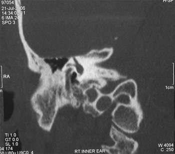
Audiometric tests revealed a moderate high frequency sensorineural hearing loss (SNHL) in the right ear and normal hearing in the left ear (Figure 1). High resolution computed tomography scanning of the temporal bone (Figure 2) revealed a dehiscence of the right superior semicircular canal, without any other significant pathology in the middle and inner ear. On magnetic resonance imaging, no pathology was detected in the brain or middle ear.
Pre-operative audiogram.

Coronal high resolution computed tomography scan showing dehiscence of superior semicircular canal in the right inner ear.

Electronystagmography revealed absence of spontaneous nystagmus, and the presence of vertical torsional nystagmus (slow phase component directed upward and clockwise) on exposure to loud sound and pressure stimuli to the right ear, with a subjective feeling of oscillopsia and dizziness. There was no significant asymmetry on caloric testing. Hence, a diagnosis of superior semicircular canal dehiscence syndrome was made.
Operative technique
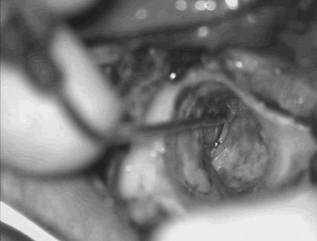
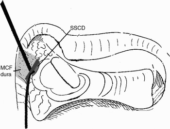
The patient was operated upon under local anaesthesia and sedation. A cortical mastoidectomy was performed via the postauricular approach and the tegmen was skeletonised. The lateral semicircular canal was identified and the subarcuate cell tracts were exenterated in order to skeletonise the superior semicircular canal. The middle cranial fossa dura over the area of the defect was carefully elevated from the area of the arcuate eminence with a blunt suction probe, care being taken not to accidentally suck the membranous labyrinth. The patient complained of a strong vertiginous sensation when a blunt probe was moved over the anterior end of the groove (towards the ampulla of the superior semicircular canal). The bone over the dome of the superior semicircular canal was drilled out (Figures 3 and 4). Bone paté was gently packed into the fistula and down both the limbs so as to block the lumen completely. It was then reinforced with bone wax, tissue seal (subcutaneous fibro-fatty tissue) and a slice of cartilage. The middle fossa dura was gently released to drape over the tissue seal. The wound was closed in layers.
Intra-operative photograph showing a blunt probe over the dehiscent superior semicircular canal and a suction tip retracting the middle fossa dura.

Diagram showing the exposure through the transmastoid approach. SSCD = superior semicircular canal dehiscence; MCF = middle cranial fossa

Result
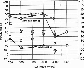
Post-operatively, the patient had immediate resolution of his symptoms of sound- and pressure-induced vertigo and oscillopsia, but complained of unsteadiness which lasted for a few days. He experienced transient SNHL which improved over time (Figure 5). He received a short course of intravenous steroids and antibiotics.
Post-operative (post-op) audiogram traces at 1 week, showing transient hearing loss across all frequencies, and at 2 months, showing significant improvement.

At 12 months' follow up, the patient was completely well and free of all his symptoms of oscillopsia, autophony and imbalance.
Discussion
Superior semicircular canal dehiscence syndrome is a condition characterised by the presence of sound- and/or pressure-induced vertigo or oscillopsia due to the dehiscence of bone overlying the superior semicircular canal.Reference Minor, Solomon, Zinreich and Zee1–Reference Minor3 Usually, the patient will have vestibular or auditory symptoms or both, or, very rarely, auditory symptoms alone.Reference Minor2–Reference Mikulec, Poe and McKenna4 Patients with superior semicircular canal dehiscence syndrome suffer vertigo and oscillopsia (i.e. apparent motion of objects that are known to be stationary in the vertical plane) induced by loud noises (Tullio's phenomenon) and/or by stimuli causing pressure changes in the middle ear or cranial cavity. The dehiscence in the temporal bone overlying the superior canal results in the presence of a ‘third mobile window’ into the inner ear, although this may not always be symptomatic.Reference Minor, Solomon, Zinreich and Zee1 This defect causes the canal to be responsive to sound and/or pressure changes in the membranous labyrinth, and may be responsible for ‘inner ear conductive hearing loss’.Reference Minor, Carey, Cremer, Lustig, Streubel and Ruckenstein6 The evoked eye movement seen is generally upward torsional nystagmus in the plane of the affected canal, i.e. the superior canal.Reference Minor2
Our patient suffered from debilitating symptoms, prompting us to offer him surgery to alleviate them. Superior semicircular canal dehiscence repair can be performed in a manner similar to posterior semicircular canal plugging, i.e. by fenestration and plugging via a transmastoid approach, with the rationale that simple obstruction rather than obliteration may suffice to alleviate the symptoms of canal dehiscence.Reference Mikulec and Poe7 We chose to plug the superior semicircular canal dehiscence with bone paté as this is more efficacious than resurfacing, as reported by other authors.Reference Minor2, Reference Mikulec, Poe and McKenna4 We planned to use the transmastoid approach for surgery so as to avoid opening the cranial cavity and subsequent retraction of the temporal lobe of the brain. The plugging of the superior semicircular canal was highly effective in this case; the patient reported significant post-operative improvement, especially regarding sound- and pressure-induced dysequilibrium. The subjective feeling of imbalance was present after surgery but resolved spontaneously. The patient showed a transient post-operative deterioration in hearing across all frequencies, but this subsequently improved significantly (Figure 5).
• Plugging of superior semicircular canal dehiscence via the transmastoid approach offers a reliable and effective solution for patients with superior semicircular canal dehiscence syndrome
• A transmastoid approach can be adequate and avoids the morbidity of craniotomy; it can be undertaken under local anaesthesia
• Due to the risk of possible sensorineural hearing loss, it is advisable that this procedure be undertaken with caution and with a guarded prognosis
Sensorineural hearing loss is a known complication of plugging any semicircular canal. The incidence of SNHL after plugging the posterior semicircular canal to treat benign positional vertigo is less than 5 per cent, and it is expected that superior semicircular canal dehiscence plugging should also be similarly safe.Reference Agarwal and Parnes8
The symptom complex of superior semicircular canal dehiscence syndrome needs to be differentiated from those of eustachian tube dysfunction, perilymph fistula and Ménière's disease, in order to avoid unnecessary middle-ear surgery in some cases.
Conclusion
Plugging of the superior semicircular canal via the transmastoid approach offers a reliable and effective solution for patients with superior semicircular canal dehiscence syndrome. A transmastoid approach can be adequate and avoids the morbidity of craniotomy. Performance of the procedure under local anaesthesia enables the fistula to be identified accurately and with certainty, as the patient will complain of vertigo when the fistula site is handled. However, due to the risk of possible SNHL, it is advisable that the procedure should be performed with caution, with a guarded prognosis, and only when the symptoms are debilitating.