INTRODUCTION
Ascaris is a large parasitic nematode of the small intestine of major human health importance in many countries. About 1·4 billion people are infected with the parasite globally, of which ~530 million are in China and 100–200 million are clinically affected children (WHO, 1996; Peng et al. 1998a; O'Lorcain and Holland, 2000; Stephenson, 2002). Ascaris infection of the pig is also of economic significance due to production losses linked to reduced feed conversion efficiency and the condemnation of ‘milk-spot’ livers at slaughter (Stewart and Hale, 1988).
There has been continued controversy as to whether Ascaris lumbricoides Linnaeus, 1758 from humans represents a different species to Ascaris suum Goeze, 1782 from pigs. A range of studies has attempted to address this question, such as early experimental cross-host species transmission (Takata, 1951; Galvin, 1968), and morphological (Sprent, 1952; Ansel and Thibault, 1973; Maung, 1973; Kurimoto, 1974), karyotypic (Mutafova, 1983; He et al. 1986), immunological and/or biochemical investigations (e.g. Kennedy et al. 1987; Nadler, 1987; Christie et al. 1990; Abebe et al. 2002). Given the absence of any reliable morphological characters which distinguish human Ascaris from pig Ascaris, it was unclear whether ‘cross-host infections’ reported previously relate to Ascaris lumbricoides in pigs or Ascaris suum in humans, particularly in regions endemic for ascariasis in both hosts, although molecular investigations in the USA and Denmark show evidence that cross-infection of Ascaris from one host to another can occur (Anderson, 1995, 2001; Nejsum et al. 2005). The controversy has also raised questions regarding the epidemiology of the two operational taxonomic units of Ascaris, given that effective control programmes against Ascaris are based on knowledge and understanding of transmission patterns. For instance, it had been unclear as to whether Ascaris-infected pigs represent a significant reservoir for infection to humans in endemic and non-endemic countries and, thus, whether pig ascariasis should be controlled at the same time as any control programmes targeting the human community where pigs co-exist (and vice versa) (Peng and Zhou, 2001).
DNA techniques provide useful complementary tools to study the genetics and epidemiology of Ascaris (Anderson et al. 1993, 1995; Anderson and Jaenike, 1997). For example, Anderson et al. (1995) identified 42 distinct mitochondrial haplotypes in 265 Ascaris worms taken from humans and pigs in two villages of Guatemala, showing that parasites of the same haplotype were found more frequently than predicted by chance for single individuals. Extending previous work (Peng et al. 1998b; Zhu et al. 1998, 1999), Peng et al. (2003a) undertook a comprehensive molecular-epidemiological study in China using a mutation scanning-sequencing approach. These workers studied nucleotide variation in the first internal transcribed spacer (ITS-1) of nuclear ribosomal DNA within and among large numbers of Ascaris individuals from humans (n=486) and pigs (n=329) from 6 endemic regions in China, and examined the frequency of different genotypes of Ascaris in relation to host species and geographical origin. There were significant differences in the frequency of genotypes between human- and pig-derived Ascaris, suggesting that, in endemic countries, cross-infection between the two host species occurs at a low level. The preponderance of genotype G1 in humans and of genotype G3 in pigs led to the proposal that each of the two genotypes has an affinity to a particular host species. The findings from a subsequent study (Peng et al. 2005) employing markers in the cytochrome c oxidase subunit 1 (cox1) and NADH dehydrogenase subunit 1 (nad1) genes of mitochondrial DNA (mtDNA) supported this proposal in that particular mitochondrial haplotypes had a particular affiliation to the pig or human host. Recent studies suggest that host, environmental and/or parasite factors may have led to this apparent affiliation (Wakelin and Bradley, 2002; Peng et al. 2003a). In the present study, by excluding environmental effects or variation relating to the exposure of hosts to eggs of different genotypes of Ascaris, and minimizing genetic variation in the host species, experimental infections in pigs and mice were carried out using eggs of two different genotypes of Ascaris to test the proposal that there is a particular host affiliation.
MATERIALS AND METHODS
Collection of adult Ascaris, preparation of eggs and viability of larvae
Female adults of Ascaris were collected from humans after treatment with pyrantel pamoate (Hangzhou Minsheng Pharmaceutical Group Co., Ltd, 10 mg/kg, 1 dosage) from Changyi township, Xinjian County, Jiangxi Province, China, and from pigs from a local slaughter house (Peng et al. 2003b). Eggs from the distal part of the uterus from individual adult females of Ascaris were isolated and incubated as reported previously (Jungersen et al. 1996) until 95% of the eggs developed into the infective stage, and were then stored at 4–10 °C for subsequent use. The genotypes of Ascaris individuals and eggs were determined using molecular approaches (described below).
Experimental animals and maintenance
Rabbits (New Zealand White; male; 1·5 kg body weight) were purchased from the Centre of Laboratory Animals of Jiangxi Medical College, and specific pathogen-free (SPF) C57BL/6 mice (females and males; 20–25 g body weight) were obtained from this Centre or the Shanghai Slac® Centre of Laboratory Animals. Both species were provided ad libitum with sterilized feed and water during the entire experimental period. Helminth-free pigs (Yorkshire×Danish Landrace; males; 20 kg) were purchased from the Jiangxi Agricultural University or Hong Yue Stock Farm, Nanchang and maintained for an ‘observation’ period of 40 days. To prevent accidental infection with Ascaris during this period, pigs were treated twice with ivermectin (0·2 mg/kg) and once with albendazole (600 mg per pig; supplied by Jiangxi Provincial Station of Prevention and Control for Diseases of Domestic Animals) prior to experimental infections. Three faecal samples from pigs (taken 5–6 days apart) were examined for helminth eggs, and no eggs were detected. Throughout the entire experimental period, pigs were kept in purposely-constructed and quarantined facilities (in which pigs had not been housed previously).
Experimental design and infections
The infectivity of each batch of Ascaris eggs (each from a single adult female of Ascaris) of a particular genotype was verified by infecting rabbits and/or mice (gastric inoculation of 1000 eggs per animal). If larvae were detected using the Baermann funnel technique (Slotved et al. 1997) in the lungs from inoculated animals 7 days after inoculation, the batch of eggs tested was considered infective.
In Experiment 1 (Table 1), mice (n=50 total) were individually inoculated orally with 1000 eggs representing Ascaris genotypes G1 and G3 respectively. All mice were euthanased 7 days after inoculation (cf. Lewis et al. 2006) to recover larvae from the lungs and livers using the Baermann funnel technique (Slotved et al. 1997). Also, randomly selected livers from infected mice were crushed and examined for larvae, and spleens weighed and examined for pathological changes (congestion and petechia) indicating infection/inflammation.

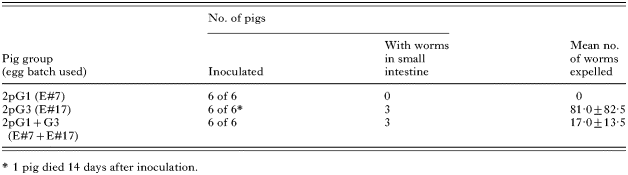
In Experiment 2 (Table 2), pigs (n=18 total) in groups 2pG1, 2pG3 and 2pG1+G3 were individually inoculated orally with 2000 eggs of genotype G1, G3 and both genotypes G1 and G3 (mixed in equal proportions), respectively (see Tables 1 and 2). On days 60, 69 and 76, faecal samples (n=10–12) were collected randomly from the pen of each group for microscopic examination for Ascaris eggs using a conventional flotation method (Roepstorff and Nansen, 1998). On day 76, all pigs were euthanased, and all Ascaris worms in the small intestine collected and enumerated.

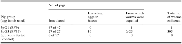
In Experiment 3 (Table 3), pigs (n=86 total) in groups 3pG1 and 3pG3 were individually inoculated orally with 2000 eggs of genotypes G1 and G3, respectively. Twelve pigs served as uninfected controls for the detection of any accidental infection of Ascaris. On day 60 after inoculation, all pigs were treated with pyrantel pamoate (1·8 g per pig) and a faecal sample (3–5 g) was collected rectally from each pig for microscopic examination of Ascaris eggs (cf. Roepstorff and Nansen, 1998). Worms expelled from pigs in their faeces (within 48 h of treatment) were collected and enumerated.

Molecular analyses
Genomic DNA was isolated from the musculature from individual adult specimens of Ascaris or from pooled eggs (from the uteri of female individuals or from faeces). The preparation of DNA samples from Ascaris from different host individuals was spatially and temporally separated. In brief, individual adult worms were washed extensively in physiological saline, pH 7·3, and stored in Carnoy's solution (60% ethanol, 30% chloroform and 10% acetic acid; v/v) at 4 °C until required for DNA isolation. A small section (5 mm) of each worm was excised from the mid-body region, dried on filter paper and then soaked in water to re-hydrate the nematode tissues. DNA was then released from each nematode section or eggs by a standard method of sodium dodecyl-sulphate/proteinase K (Boehringer-Mannheim) treatment (Gasser et al. 1993), purified over a spin column (Wizard™ DNA Clean-Up, Promega) and eluted in 30 μl of reverse-osmosis purified, deionized water. DNA from human blood or pig musculature (healthy individuals) was also prepared using the same procedures as for parasites.
A ~300 bp ITS-1 rDNA region (pITS-1) known to contain at least 6 variable nucleotide positions in Ascaris sp. (see Zhu et al. 1999) was amplified by PCR (Saiki et al. 1988) from individual worms or pooled eggs. PCR reactions (25 μl) were carried out in 10 mM Tris-HCl, pH 8·4; 50 mM KCl; 3 mM MgCl2; 250 μM of each dNTP; 25 pmol of each of the 2 primers NC5 (forward: 5′-GTAGGTGAACCTGCGGAAGGATCATT-3′) and XZ6R (reverse: 5-ATAGCTCGGTGAAGCGTAGA-3) (cf. Zhu et al. 1999), with 1 unit of Taq polymerase (Promega) in a thermocycler (480, Perkin Elmer Cetus). PCR cycling conditions were 94 °C for 5 min (initial time delay), then 94 °C for 30 sec (denaturation), 55 °C for 30 sec (annealing) and 72 °C for 30 sec (extension) for 30 cycles, followed by a final extension at 72 °C for 5 min. One μl (5–10 ng) of genomic DNA was added to each PCR reaction. After thermocycling, individual amplicons (10 μl) were mixed with an equal volume of loading buffer (10 mM NaOH, 95% formamide, 0·05% of both bromophenol blue and xylene cyanole) and the intensity of selected samples verified on ethidium bromide-stained 2% (w/v) agarose gels using TBE (65 mM Tris-HCl, 27 mM boric acid, 1 mM EDTA, pH 9; Bio-Rad, Richmond, CA, USA) as the buffer and ΦX174-HaeIII (Promega, WI, USA) as a size marker.
SSCP analysis was performed as described previously (Gasser et al. 2004). In brief, individual pITS-1 amplicons were mixed with an equal volume of loading buffer (10 mM NaOH, 95% formamide, 0·05% of both bromophenol blue and xylene cyanole). After denaturation at 94 °C for 15 min and snap cooling on a freeze block (−20 °C), individual samples (12 μl) were loaded into the wells of pre-cast GMA™ S-50 gels (96×261 mm; product no. 3548, Elchrom Scientific) and subjected to electrophoresis for 17 h at 74 V and 7·4 °C (constant) in a horizontal SEA2000™ apparatus (Elchrom Scientific) connected to a MultiTemp III (Pharmacia) cooling system. After electrophoresis, gels were stained for 20 min with ethidium bromide, destained in water for the same time and then photographed upon ultraviolet transillumination.
For selected samples from different SSCP gels, the entire ITS-1 region was amplified by PCR using primers NC5 and NC13R (reverse; 5′-GCTGCGTTCTTCATCGAT-3′) (Zhu et al. 1999), column-purified (Wizard™ PCR Prep, Promega) and subjected to PCR-RFLP or/and automated sequencing. For PCR-RFLP, column-purified ITS-1 amplicons (3 μl) were digested directly with 10 units (1 μl) of the restriction endonuclease Hae III (Promega) in 20 μl for 2 h at 37 °C (cf. Zhu et al. 1999). Digests were resolved on 2% (w/v) agarose gels, stained with ethidium bromide and photographed (with reference to the ΦX174-Hae III marker). Automated cycle-sequencing (ABI BigDye chemistry v.1; in a PE Applied Biosystems 377 sequencer) was carried out in both orientations using primers NC5, NC13R and 2 internal ones, WP3F (forward: 5′-TCGCAGTGGCTTGAACCGGG-3′ and XZ5R (reverse: 5′-CGCCGACTGCTATTACATCA-3′). Nucleotide sequences were compared with sequences published previously (Zhu et al. (1999); Accession numbers AJ000895 and AJ000896, Peng et al. (2003a); Accession numbers AJ554036–AJ554043).
Statistical analysis
Where considered appropriate, a standard statistical analysis was conducted. The raw data were transformed using a common logarithm and the means compared using one-way analysis of variance (ANOVA, using the program SPSS v12.0.1).
RESULTS
Genotypic identification of Ascaris eggs
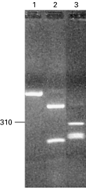
Batches of eggs from Ascaris of human and pig origins were genetically defined as genotypes G1 and G3, respectively, based on DNA sequencing and/or PCR-RFLP of the pITS-1. The pITS-1 sequences of egg batches E#7 and E#17 were consistent with those reported previously for genotypes G1 (Accession numbers AJ000895 and AJ554036) and G3 (Accession numbers AJ000896 and AJ554043), respectively (cf. Zhu et al. 1998; Peng et al. 2003a). Based on the PCR-RFLP profiles for these 2 batches of eggs, the genotypes of 2 other batches of eggs (including E#9 and E#13; see Fig. 1) were determined.

Fig. 1. RFLP analysis of the ITS-1 amplified by PCR from genomic DNA from eggs isolated from individual Ascaris females. The eggs were used to infect mice and pigs. An undigested amplicon (lane 1), and amplicons digested with the endonuclease Hae III (lanes 2 and 3). The profiles displayed for egg batches E#7 and E#9 represented genotype G1 (lane 2) and for batches E#13 and E#17 represented genotype G3 (lane 3). The DNA sequences of amplicons representing the 2 profiles were consistent with Accession numbers AJ000895 and AJ554036 (genotype G1) and Accession numbers AJ000896 and AJ554043 (genotype G3) (Zhu et al. 1998; Peng et al. 2003a), respectively. The size indicated (left) is in bp.
Ascaris infections in mice
In the first experiment, 7 days after intragastric inoculation with Ascaris eggs of genotype G1 or G3, the lungs from all mice and the livers from randomly selected mice (n=10) were examined for Ascaris larvae. Mice in Group 1mG1 harboured more larvae in both lungs (44·4±26·3) and livers (3·7±4·7) compared with those in Group 1mG3 (Table 1). There was a significant difference between the groups in the number of larvae recovered from the lungs (F=39·22, P<0·000) and from the livers (F=5·914, P=0·007) (see Table 1). Lungs with petechial bleeding and enlarged, congested spleens were more often detected in Group 1mG1 than in Group 1mG3. Also, the mean spleen weight of randomly selected mice (n=6–8) was significantly different between the 2 groups (F=16·972, P<0·001) (Table 1).
Ascaris infections in pigs
In the second experiment (Table 2), 60, 69 and 76 days after the oral inoculation of pigs, Ascaris eggs were detected in all faecal samples from pigs in Groups 2pG3 and 2pG1+G3, but no eggs were detected for Group 2pG1. All pigs were euthanased after 76 days, and the worms in the small intestines enumerated. None of the 6 pigs in Group 2pG1 harboured Ascaris, 3 of the 5 pigs in Group 2pG3 (1 pig died 14 days after inoculation) harboured 243 worms, and 3 of the 6 pigs in Group 2pG1+G3 harboured 51 worms (Table 2). The genotypes of the Ascaris eggs obtained from the faeces from pigs with a patent infection and Ascaris worms recovered from the pigs after autopsy were established using molecular methods.
The pITS-1 amplified from genomic DNA isolated from Ascaris eggs from the faeces from Group 2pG3 and Group 2pG1+G3 were sequenced. The eggs from both groups of pigs were demonstrated by sequencing to represent genotype G3. The pITS-1 amplicons produced from DNA isolated from 50 worms from Group 2pG1+G3 and the same number from Group 2pG3 were subjected to SSCP analysis. No variation in profile was detectable among all 100 amplicons on SSCP gels. PCR-RFLP analysis of a subset of these amplicons (n=80) also revealed profiles that were all consistent with genotype G3, irrespective of group (cf. Fig. 1). For multiple individual worms (n=17; sample nos 61, 63, 66, 69, 71, 76, 80, 81, 84, 85, 87, 89, 93, 95, 101, 102 and 106) representing Group 2pG3 (and from different SSCP gels), the pITS-1 was amplified and sequenced. The same was done for selected adult Ascaris (n=12; sample nos 12, 17, 19, 22, 23, 28, 32, 33, 38, 42, 46 and 55) collected from pigs from Group 2pG1+G3. The sequencing supported the SSCP and PCR-RFLP results in that there was no variation in the pITS-1 at the 6 variable positions (nos 20, 21, 128, 129, 173 and 246; cf. Peng et al. 2003a) for all 29 amplicons sequenced and, hence, all worms analysed were of the genotype G3.
In the third experiment (Table 3), 60 days after inoculation, faecal samples from individual pigs were collected for microscopic examination. Eggs were neither detected in any of the samples from the 12 uninfected control pigs (Group 3pU) nor in those from infected pigs in Group 3pG1, but were detected in most (59%) of the 27 pigs in Group 3pG3. Within 48 h of pryrantel pamoate treatment, 1 immature Ascaris worm was expelled from 1 pig in Group 3pG1. In contrast, a total of 305 adult Ascaris was expelled from pigs in Group 3pG3 (Table 3).
DISCUSSION
The present study utilized genetically defined eggs from Ascaris from human or pig to investigate their ability to infect helminth-free rabbits or mice and pigs and to establish as adult worms in pigs. Experiments using susceptible mice (C57BL strain; see Lewis et al. 2006) and pigs revealed significant differences in the number of Ascaris larvae or adults recovered from these hosts for the different genotypes. For instance, while no worms were detected in Group 2pG1 and only 1 immature worm was collected from 1 out of 47 pigs in Group of 3pG1, a large number of worms was detected in Groups 2pG3 and 3pG3. Therefore, Ascaris eggs of genotype G1 of human origin did not establish readily in experimental pigs, whereas those of genotype G3 from pigs did. These findings are in accordance with those from a recent molecular-epidemiological investigation in China, showing that the prevalence of genotype G1 in pigs is very low (2·4%, n=329) and inferring a limited cross-infection of Ascaris between human and pig (Peng et al. 2003a). Although a limited number of different egg batches from single Ascaris females (n=4) of the 2 genotypes were used in the present infection experiments, the results support the hypothesis that Ascaris of human origin does not readily infect pigs. This statement is reinforced by other findings from China that eggs of genotypes other than G1 (being consistent with genotype G2 from human Ascaris; see Peng et al. 2003a) do not establish as adults in pigs (n=28) following inoculation with the same egg dose as employed herein (using appropriate control groups) (Peng et al. unpublished findings). It is also supported by previous molecular-epidemiological studies by us and other workers (Anderson, 2001; Peng et al. 2003a, 2005). Nonetheless, in spite of the evidence, a further study, using more egg batches from single female worms representing the different Ascaris genotypes (cf. Peng et al. 2003a) from the 2 host species is required to reinforce current evidence for a host affiliation of different genetic variants of Ascaris. Given the extent of variation in mitochondrial DNA present within and among Ascaris populations (cf. Peng et al. 2005), it would also be worthwhile exploring the ability of Ascaris of different haplotypes (of human and pig origins) to infect pigs.
The present results suggest that the host origin of Ascaris eggs affects the establishment, survival and/or maturation of the parasite in a specific, but in an unknown way. While parasite factors could include variation in the physiology and/or biochemistry among different genotypes of Ascaris, host factors such as genes associated with susceptibility (Wakelin and Bradley, 2002; Williams-Blangero et al. 2002) and/or the immune responses in the host (Cooper, 2002; Jungersen, 2002) could relate to the ability of particular Ascaris genotypes to establish preferentially in pigs and others in humans. The distinctiveness in recovery rates of Ascaris adults or larvae with different genotypes from pigs or mice, the variation in spleen weight of mice infected with eggs of genotypes G1 (from human) and G3 (from pig) all suggest that future investigations of the relationship of the host origin/genotype/haplotype of Ascaris, host susceptibility/resistance and immune responses are needed.
Support from the Wellcome Trust, the National Natural Science Foundation of China (W.P.), Elchrom Scientific AG (R.B.G.) and the University of Melbourne is gratefully acknowledged. Thanks also to R. Lewis and C. Holland for guidance on the mouse model, and to C. Holland and I. Beveridge for constructive comments on the manuscript.