Around 100 years ago, Carlos Chagas published a seminal study in the history of parasitology (Ref. Reference Chagas1). He described for the first time the parasite Trypanosoma cruzi, the aetiological agent of American trypanosomiasis, also known as Chagas disease. During a campaign to fight malaria in the Minas Gerais state of Brazil, Carlos Chagas identified a haematophagous bug associated with diseased individuals living in poor dwellings. In the intestine of the bug, he found flagellated parasites, which he named T. cruzi after his mentor Oswald Cruz, who led a pioneering biomedical research institution in Rio de Janeiro. Together, they established cultures and experimental infections with T. cruzi and fulfilled the Koch's postulates (Ref. Reference Evans2) for Chagas disease. Carlos Chagas observed that the haematophagous bugs were present only in inhabited houses, suggesting that humans were their main blood source. He also found T. cruzi parasites in the blood of sick people, and soon correlated the parasitaemia (level of parasites in the blood) with some symptoms of the disease, such as fever, anaemia, lymphadenopathy and splenomegaly, and he described the cardiac form of the disease (Ref. Reference Chagas1). Finally, primates and other laboratory animals, such as dogs, rabbits and guinea pigs, were infected and all developed parasitaemia, demonstrating the T. cruzi pathogenicity (Ref. Reference Chagas1).
In a few years Carlos Chagas had described: (1) the aetiological agent, T. cruzi, and its complex life cycle; (2) the invertebrate host responsible for parasite transmission; (3) the vertebrate host reservoirs; (4) the symptoms and signs of human disease; and (5) the likely social impact of the disease (Refs Reference Chagas1, Reference Coura and Dias3). A century after the discovery of Chagas disease, significant progress has been made in understanding different aspects of the epidemiology, immunology and cell biology events that are important in the host–parasite interaction, in both vertebrate and invertebrate hosts (Refs Reference Coura and Dias3, Reference Brener and Gazzinelli4, Reference Tarleton5). Furthermore, the parasite genome has been sequenced (Ref. Reference El-Sayed6). Importantly, control of natural transmission of Chagas disease has been achieved in some countries by targeting the bug with insecticide (Ref. Reference Coura and Dias3). However, the pathogenesis of Chagas disease remains largely unknown, and there are still no effective vaccines or drugs to prevent or treat chronic infection with T. cruzi (Refs Reference Khaw and Panosian7, Reference Urbina8).
T. cruzi life cycle
It is estimated that T. cruzi emerged as a species over 150 million years ago, originally infecting primitive mammals. The first contact with humans probably occurred much more recently, 30 000–15 000 years ago, when humans first reached the American Continent (Refs Reference Guhl9, Reference Pena and Santos10). T. cruzi is a single species that comprises several lineages that have distinct morphology, infectivity, virulence and pathogenicity (Ref. Reference de Freitas11). The parasite displays different developmental stages during its life cycle. In the stomach of haematophagus bugs of the Reduviidae family responsible for the natural transmission of T. cruzi, the epimastigotes proliferate by cell division. At the distal part of the intestine, metacyclogenesis transforms the parastite into the metacyclic trypomastigote, the infective form for the vertebrate host. During blood sucking, the insect defaecates on the skin and the metacyclic trypomastigotes enter the mammalian hosts when they scratch their skin or rub their eye. The parasites then invade host cells, transforming into amastigotes. Amastigotes replicate and differentiate into trypomastigotes, disrupting host cells and infecting other cells. Although it can replicate in various cell types, T. cruzi has a particular tropism for cardiac, skeletal and smooth muscle cells (Refs Reference Chagas1, Reference Brener12). Finally, the haematophagus bugs are infected by ingesting trypomastigotes in the blood from infected hosts, thus completing the T. cruzi life cycle (Ref. Reference Brener12).
Epidemiology and transmission of Chagas disease
In nature, over 100 mammalian species from different orders have been reported to be infected with T. cruzi, which is transmitted by hemiptera from the family Reduviidae such as Triatoma, Rhodnius and Panstrongylus. Two different cycles for T. cruzi transmission exist: the sylvatic cycle related to wild hemiptera and wild vertebrate hosts; and the domestic cycle, which is dependent on home-dwelling hemiptera and primarily involves humans and household animals. The connection between the two transmission cycles is made by infected rats, mice, bats, opossum and hemiptera that migrate to the human environment as a result of the destruction of forests (Ref. Reference Coura and Dias3). The infected Reduviidae haematophagus vectors that potentially transmit T. cruzi to humans are found in most countries of the American Continent, within the boundaries of southern states of the USA to the southern part of Argentina (Ref. Reference Coura and Dias3). Today, T. cruzi is responsible for the infection of ~10 million individuals, and more than 80 million people are at risk of infection across the Latin American continent (Ref. Reference Coura and Dias3). Natural transmission of Chagas disease has been controlled in many countries by insecticide targeting of haematophagus bug populations, as well as improved socioeconomic status and quality of dwelling in Latin America (Refs Reference Coura and Dias3, Reference Moncayo and Silveira13). However, individuals living in various countries of Central and South America are still exposed to the infected vectors that are responsible for the dissemination of T. cruzi infection. In addition, different ways of transmission, such as oral infection and transmission after blood transfusion or organ transplant, have become more frequent (Ref. Reference Moncayo and Silveira13). Importantly, it has been estimated that 700 000 infected people are living outside of Latin America; thus, blood transfusion and organ transplants are becoming a new threat for T. cruzi infection in nonendemic countries (Ref. 14).
The pathogenesis of Chagas disease
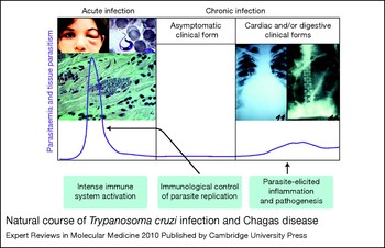
As illustrated in Figure 1, after an incubation period of 2–3 weeks, T. cruzi infection is manifested by the presence of a large number of parasites in the bloodstream and various tissues. The acute infection is accompanied by an excessive activation of the immune system that includes cytokinaemia (high plasma levels of cytokines), intense activation of B and T cells, lymphoadenopathy, splenomegaly, and intense inflammatory processes associated with parasite nests within tissues. The acute phase is followed by the development of an effective acquired immunity, leading to the control of parasitaemia and parasitism (parasite levels in tissues). The lifelong chronic phase is maintained with scarce parasitaemia and parasitism.
Natural course of Trypanosoma cruzi infection and Chagas disease. The figure illustrates the course of T. cruzi infection and development of Chagas disease in humans. The acute phase is normally symptomatic and associated with high parasitaemia, tissue parasitism and strong activation of the immune system (with cytokinaemia, polyclonal activation of B and T cells, lymphoadenopathy and splenomegaly, and an intense inflammatory reaction associated with tissue parasitism). The photo images illustrate: a child with Romaña sign, the site of T. cruzi entry in the eye; a trypomastigote form in the bloodstream; and an amastigote nest in the heart tissue. During the chronic phase, the parasitaemia and tissue parasitism are very low and most patients are asymptomatic; however, 20–30% of chronic chagasic patients may evolve the symptomatic forms – that is, cardiac and digestive clinical forms – which are associated with intense local inflammation and scarce parasitism at the affected tissues. The photo images illustrate an enlarged heart and a megacolon in chronic chagasic patients (for clinical information see Ref. Reference Rassi, Rassi and Marin-Neto15). The purple line indicates change in parasitaemia and tissue parasitism during the course of T. cruzi infection. The illustrations of chagasic patients were obtained, with permission, from the World Health Organization website (http://apps.who.int/tdr/publications/tdr-image-library; accessed July 2010).

At the beginning of the chronic phase, infection with T. cruzi is asymptomatic in most patients. However, as chronic Chagas disease advances, the clinical course becomes variable, ranging from an absence of symptoms to severe disease with cardiovascular and/or gastrointestinal involvement (Ref. Reference Rassi, Rassi and Marin-Neto15). The heart pathology is often associated with an intense myocarditis, and frequently leads to cardiomegaly, congestive heart failure and arrhythmias (Refs Reference Gutierrez16, Reference Bilate and Cunha-Neto17). In the digestive forms of the disease, focal lymphocytic infiltrates and neuronal degeneration with denervation are characteristics of megaoesophagus and megacolon pathology (Ref. Reference Coura and Borges-Pereira18).
Fundamental questions regarding the pathogenesis of Chagas disease remain unexplained. For instance, why do different patients develop the cardiac, digestive, cardiodigestive or asymptomatic clinical forms of the disease? Another intriguing aspect is the intense inflammatory process associated with relatively few parasites (Ref. Reference Bilate and Cunha-Neto17). This finding in autopsies of symptomatic patients led to the concept that an autoimmune component was involved in the genesis of chagasic myocarditis (Refs Reference Leon and Engman19, Reference Cunha-Neto20). However, studies using more-sensitive techniques, such as the polymerase chain reaction (PCR) and immunocytochemistry, demonstrated a positive correlation between the number of parasites and the severity of cardiac (Refs Reference Gutierrez16, Reference Jones21, Reference Higuchi22) as well as digestive (Ref. Reference Vago23) disease, challenging the autoimmune hypothesis for Chagas disease. The significant geographical variation in the prevalence and severity of the different forms of the disease (Ref. Reference de Freitas11) indicates the importance of T. cruzi genetic variation, since it does not present a clear correlation with any pattern of ethnic or human genetic differences. By contrast, findings from different laboratories indicate a crucial influence of host genetic background on resistance and pathogenesis of experimental infections with T. cruzi. Indeed, Chagas disease is now seen as the product of the interaction of two highly variable genomes: those of the parasite and the human host (Ref. Reference Macedo, Oliveira and Pena24). In this review, we summarise studies indicating the critical role of both innate and acquired immunity in host resistance to T. cruzi infection and pathogenesis of Chagas disease.
Innate immunity and T. cruzi infection
Despite controversy surrounding the mechanism of pathogenesis of Chagas disease (Refs Reference Tarleton25, Reference Andrade26, Reference Golgher and Gazzinelli27), recent studies indicate that T. cruzi parasites are physically associated with sites of inflammation in the heart (Refs Reference Jones21, Reference Higuchi22, Reference Vago23) and oesophagus (Ref. Reference Vago23). While acquired immunity following T. cruzi infection has been thoroughly studied, the innate immune response has not been subjected to similar scrutiny. Nevertheless, before acquired immunity takes place, the innate immune system seems to have a fundamental role in at least two important aspects of Chagas disease: the control of parasite replication and spread in host tissues; and the inflammatory reaction in infected host tissues, thought to be a major cause of tissue damage and organ dysfunction.
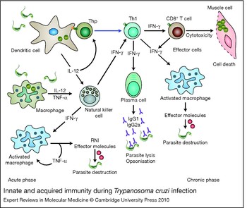
The role of cytokines such as interleukin (IL)-12, tumour necrosis factor (TNF)-α and interferon (IFN)-γ (encoded by IL12, TNF and IFNG genes, respectively), as well as of reactive nitrogen intermediates (RNIs), on host resistance during the early stages of infection and also in the genesis of myocarditis is established (Refs Reference Brener and Gazzinelli4, Reference Golgher and Gazzinelli27). Furthermore, cells with roles in innate immunity, such as natural killer (NK) cells, dendritic cells and macrophages, are key elements in the initial control of T. cruzi replication (Fig. 2). Over the past decade, we have focused on: (1) defining protozoan-derived pathogen-associated molecular patterns (PAMPs), which are targets for innate immune receptors; (2) identifying relevant innate immune receptors in the host–parasite interaction and disease outcome; and (3) developing prophylactic/therapeutic interventions employing protozoan-derived PAMPs.
Innate and acquired immunity during Trypanosoma cruzi infection. In the initial stage of T. cruzi invasion, innate immunity plays a crucial role in host resistance to infection: acting as the first barrier, the cells from the innate immune system (macrophages, NK cells and dendritic cells) produce cytokines (IL-12, TNF-α and IFN-γ) and effector molecules [reactive nitrogen intermediates (RNIs) and IFN-inducible GTPases] that control parasite replication. At the same time, innate immune cells, particularly dendritic cells, make the bridge between the innate and acquired immunity, producing cytokines (IL-12) necessary for differentiation and clonal expansion of T helper 1 (Th1) CD4+ as well as CD8+ T cells and plasma B cells. IFN-γ produced by CD4+ Th1 or CD8+ T cells activates effector mechanisms in macrophages to destroy both amastigotes and phagocytosed trypomastigotes, whereas cytotoxic activity displayed by CD8+ T cells destroys cells containing intracellular amastigotes. Antibodies produced by B cells lyse the extracellular trypomastigote form or facilitate the phagocytosis of parasites opsonised with IgG. Abbreviations: IFN, interferon; IL, interleukin; NK, natural killer; Thp, Th precursor cell; TNF, tumour necrosis factor.

PAMPs and TLR activation by T. cruzi parasites
The early elimination of invasive microorganisms is the primary function of innate immunity. Macrophages and dendritic cells can recognise traces of microbial components and orchestrate an early immune defence. These same cells also have an important role in shaping long-term acquired immunity. Of the various families of innate immune receptors, which include helicases, inflammasomes and Nod-like receptors (NLRs), the Toll-like receptors (TLRs) have been the focus of most studies in T. cruzi infection. TLRs are a primary means by which the innate immune system recognises and rapidly responds to the presence of microbes (Ref. Reference Takeuchi and Akira28). TLRs are crucial for many aspects of this process, including the recruitment of phagocytes to infected tissue and killing of invasive pathogen (Ref. Reference Takeuchi and Akira28). However, when activated in excess, TLRs may mediate pathology, as in the case of septic shock induced during infection with Gram-negative bacteria (Refs Reference Dobrovolskaia and Vogel29, Reference Lien and Ingalls30). To date, TLRs have been implicated in defence against all known categories of microorganism that cause human diseases, including protozoan parasites (Ref. Reference Gazzinelli, Ropert and Campos31). Indeed, as discussed below, TLRs are central mediators of innate immune activation during infection with T. cruzi.
The TLR family is composed of 11 and 13 members in humans and mice, respectively (reviewed in Ref. Reference Takeuchi and Akira28). The main microbial molecules that activate such receptors have been identified as components from the cell wall or membrane of bacteria: TLR2 and TLR1 are activated by triacylated lipopeptide; TLR2 and TLR6 by diacylated lipopeptide; TLR4 by lipopolysacharide (LPS); and TLR5 by flagellin. In addition, nucleic acids from viruses and bacteria have also been identified as major TLR agonists; for example, TLR3 is activated by viral double-stranded DNA, TLR7 and TLR8 by viral single-stranded RNA, and TLR9 by unmethylated CpG motifs.
TLRs share a cytoplasmic domain of high homology to the IL-1 receptor (IL-1R), known as the TLR/IL-1R (TIR) domain (Refs Reference Gay and Keith32, Reference Rock33). TLRs cannot signal without an intact TIR domain (Ref. Reference O'Neill, Fitzgerald and Bowie34). Furthermore, the TIR domains of TLRs interact with cytoplasmic adaptor molecules that also contain TIR domains. The best-studied example of a TIR-domain-containing adaptor protein is the myeloid differentiation primary-response gene 88 (MYD88), which transduces signals for all of the known TLRs, except TLR3 (Ref. Reference Takeuchi and Akira35), as well as the IL-1, IL-18 and IL-33 receptors. Hence, mice deficient in functional MYD88 are basically devoid of TLR functions (Ref. Reference Takeuchi and Akira35). At least three additional TIR-domain-containing adaptors exist: MYD88-adaptor-like protein (Mal; also known as TIRAP), TIR-domain-containing adaptor protein inducing IFN-β (TRIF, also known as TICAM1) and TRIF-related adaptor molecule (TRAM1) (Refs Reference Fitzgerald36, Reference Fitzgerald37). Mal/TIRAP is required for TLR2 and TLR4 functions, whereas TRIF is employed by TLR3 and TLR4 (Refs Reference Yamamoto38, Reference Horng39). TRAM was shown to be required only for TLR4 function (Ref. Reference Fitzgerald37).
T. cruzi GPI-mucins and GIPLs
Several studies have identified glycosylphosphatidylinositol (GPI) anchors – glycolipid additions to the C-termini of proteins that ensure membrane localisation – from Plasmodium falciparum, Leishmania major, Toxoplasma gondii, Trypanosoma brucei and T. cruzi as molecules that activate lymphoid or myeloid cells (Ref. Reference Almeida and Gazzinelli40). Analysis of GPI anchors isolated from trypomastigote-derived mucin-like glycoproteins (GPI-mucins) revealed the presence of unsaturated fatty acid chains, as opposed to conventional saturated fatty acid chains, and chemical removal of unsaturated fatty acid from GPI anchors abrogated their ability to activate macrophages and elicit the production of pro-inflammatory cytokines (Refs Reference Almeida41, Reference Campos42). Similarly, periodate oxidation, which opens saccharide rings, also inactivated GPI-mucin function, indicating the requirement of carbohydrates for the pro-inflammatory activity of the T. cruzi-derived GPI-mucins (Ref. Reference Almeida41).
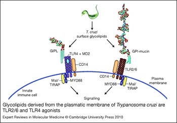
Importantly, TLR2 was identified as the main receptor mediating macrophage activation by GPI-mucins (Refs Reference Campos42, Reference Ropert43). Our experiments also showed that the innate immune receptor activated by GPI-mucins is a heterodimer composed of TLR2 and TLR6 and has CD14 as a co-receptor (Ref. Reference Gazzinelli and Denkers44) (Fig. 3). However, the response of macrophages from Tlr2 −/− mice is only partially impaired when exposed to live trypomastigotes. By contrast, no cytokine synthesis (Ref. Reference Campos45) is observed in macrophages from Myd88 −/− mice (which lack functions for most TLRs except TLR3) exposed to live trypomastigotes. These results suggest the involvement of other TLRs, and possibly other parasite molecules besides GPI-mucins, in macrophage activation by T. cruzi.
Glycolipids derived from the plasmatic membrane of Trypanosoma cruzi are TLR2/6 and TLR4 agonists. The glycosylphosphatidylinositol anchors derived from mucin-like glycoproteins (GPI-mucins) and glycoinositolphospholipids (GIPLs) – the dominant glycoconjugates present on the T. cruzi surface – are Toll-like receptor (TLR) agonists able to elicit the production of pro-inflammatory cytokines by innate immune cells. GPI-mucins are TLR2/6 agonists, which signal to host cells via the MYD88 or Mal/TIRAP pathway, whereas GIPLs are TLR4 agonists and trigger the MYD88, Mal/TIRAP, TRIF/TICAM and TRAM pathways (latter two not shown). CD14 is a co-receptor that concentrates and transfers GPI mucins and GIPLs to TLR2/6 and TLR4, respectively. MD2 binds to the extracellular domain of TLR4, and is essential for recognition and activation. Adaptor abbreviations: MYD88, myeloid differentiation primary-response gene 88; Mal/TIRAP, MYD88-adaptor-like protein; TRAM, TRIF-related adaptor molecule; TRIF/TICAM, TIR-domain-containing adaptor protein inducing IFN-β.

Besides the abundantly expressed GPI-mucins, free GPI anchors – also referred to as glycoinositolphospholipids (GIPLs) – are found on the plasma membrane of all T. cruzi stages. As with any other GPI anchor, the GIPLs share a conserved hydrophilic motif; however, unlike GPI, the T. cruzi GIPL lipid moiety contains mainly ceramide (Ref. Reference Almeida and Gazzinelli40). A complex formed by CD14, TLR4 and MD2 (LY96) is the crucial receptor for macrophage activation and pro-inflammatory activity of GIPL-ceramide (Ref. Reference Oliveira46). In support of this finding, macrophages from C3H/HeJ mice that have a point mutation in the TIR domain of TLR4 do not respond to GIPLs containing ceramide and are hyporesponsive to free ceramide (Ref. Reference Thieblemont and Wright47).
In conclusion, highly purified (i.e. LPS-free) GPI-mucins and GIPL-ceramide induce the synthesis of pro-inflammatory cytokines by macrophages via distinct receptors, namely the TLR2–TLR6 heterodimer and TLR4, respectively.
T. cruzi CpG motifs
Parasite genomic DNA has also been shown to play a role in pro-inflammatory responses during infection with protozoan parasites. Similar to bacterial CpG DNA motifs, DNA from T. cruzi, T. brucei and Babesia bovis can stimulate macrophages and dendritic cells. Protozoan genomic DNA contains sufficient levels of CpG motifs to cause moderate activation of host cells, and treatment of parasite DNA with DNase or methylase destroys the DNA pro-inflammatory activity on macrophages and dendritic cells (Ref. Reference Shoda48).
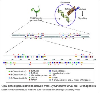
In silico analysis of T. cruzi genome sequences (Ref. Reference El-Sayed6) revealed a high number of CpG motifs (Fig. 4) that were similar but not identical to those previously defined in bacterial DNA (Ref. Reference Krieg49). The CpG motifs were not evenly distributed in the T. cruzi genome, but preferentially associated with regions of the genome that contain T. cruzi-specific genes, including those encoding surface proteins such as mucin-like glycoprotein (TcMUC) genes (Ref. Reference Bartholomeu50). The immunostimulatory CpG motifs were also associated with multiple copies of the gene encoding the retroelement VIPER (Ref. Reference Bartholomeu50), inserted in the trypanosomatid founder lineage after the divergence of Leishmania and Trypanosoma (Ref. Reference Lorenzi, Robledo and Levin51) and sharing a common ancestor with RNA viruses (Ref. Reference Xiong and Eickbush52).
CpG-rich oligonucleotides derived from Trypanosoma cruzi are TLR9 agonists. Parasite DNA from parasites destroyed in the endolysosome compartment is released and activates Toll-like receptor 9 (TLR9). An in silico analysis of the T. cruzi destroyed genome identified CpG-rich regions, which contain A-, B- and C-classes of CpG motifs. [Three main classes of the immunostimulatory CpG DNA motifs that activate human TLR9 have been described. A-class CpG oligonucleotides (ODNs) have been defined as a weaker inducer of pro-inflammatory cytokines, but a strong inducer of type I interferons. B-class CpG ODNs are the most potent inducers of pro-inflammatory cytokines, and B cell responses. C-class CpG ODNs combine the immunostimulatory properties of both the A-class and the B-class.] For detailed nucleotide sequences, see Ref. Reference Akira, Takeda and Kaisho66. The representation of genomic regions in the figure is reproduced, with permission, from Ref. Reference Akira, Takeda and Kaisho66 (© 2008. The American Association of Immunologists, Inc.).

The pro-inflammatory activity of T. cruzi DNA has been shown to be mediated by TLR9 (Ref. Reference Bafica53), a TLR known to be activated by unmethylated DNA containing CpG motifs (Ref. Reference Hemmi54). Importantly, infection with T. cruzi was shown to trigger high levels of nuclear factor κB (NF-κB) via TLR9. TLR9 translocates to lysosomes during invasion/uptake of T. cruzi by dendritic cells, and a potent immunostimulatory activity is observed when highly unmethylated T. cruzi genomic DNA is delivered to the endolysosomal compartment of cells that express TLR9. Together these results indicate that unmethylated CpG motifs from the T. cruzi genome are key parasite targets, which become available to TLR9 when parasites are destroyed in the lysosome-fused vacuoles (Ref. Reference Bartholomeu50). We also have recent in vivo and in vitro evidence that parasite RNA may also be recognised by host TLR7, which should happen as for TLR9, at the level of endolysosomal network (B. Caetano and R. Gazzinelli, unpublished).
TLR-independent pathways
Despite the importance of TLRs indicated by the studies mentioned above, T. cruzi can also activate innate immune responses independently of TLRs. T. cruzi is able to induce IFN-β by a TLR-independent pathway that requires both TANK-binding kinase (TBK) 1 and IFN regulatory factor (IRF) 3 (Refs Reference Chessler55, Reference Chessler56). Also, NFATC1 (nuclear factor of activated T cells, cytoplasmic) mediates TLR-independent IFN production (Refs Reference Chessler55, Reference Kayama57). This alternative signalling pathway can also induce other molecules such as IFN-inducible GTPase LRG47 (IRGM) that play an important role in control of T. cruzi infection (Refs Reference Chessler55, Reference Santiago58). Another study showed that the elevation of intracellular Ca2+ in cells infected with T. cruzi is responsible for calmodulin–calcineurin activation, which triggers a pathway mediated by NFATC1, leading to IFN-γ production in a TLR-independent manner (Ref. Reference Kayama57). A recent study also showed that NLRs are also activated and play a significant role in host resistance to experimental infection with T. cruzi (Ref. Reference Silva59).
The role of TLRs and their adaptors during in vivo infection with T. cruzi
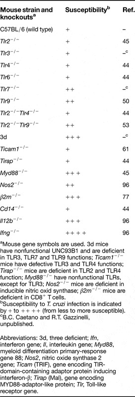
Gene-knockout mouse models lacking function of specific TLRs or their signal-transducing adaptor molecules (especially the Myd88 −/− mice) have been used to shed light on the role of TLR pathways in in vivo infections with protozoan parasites. Importantly, both IL-1 and IL-18 receptors use the MYD88 adaptor to signal, and thus could be responsible for the immunological defect in Myd88 −/− mice. Nevertheless, neither IL-1 nor IL-18 has been reported to be a critical determinant in host resistance to T. cruzi (Ref. Reference Graefe60). Hence, an impaired response of Myd88 −/− mice to T. cruzi is compelling evidence that TLRs are engaged by this pathogen. Indeed, different studies reveal that infection with trypomastigotes in Myd88 −/− mice results in impaired production of pro-inflammatory cytokines (IL-12 and IFN-γ), as well as enhanced parasitaemia and accelerated mortality (Refs Reference Gazzinelli and Denkers44, Reference Campos45, Reference Bafica53). However, Myd88 −/− mice are less susceptible to infection than are Ifng −/− mice. Furthermore, although they respond to a lesser extent than wild-type mice, Myd88 −/− mice infected with T. cruzi produced significant amounts of IL-12 and IFN-γ. Thus, we propose that IL-12 and IFN-γ are induced by both MYD88-dependent and -independent mechanisms during T. cruzi infection (Refs Reference Campos45, Reference Bafica53).
Furthermore, Ticam −/− mice (lacking TRIF) are not more susceptible to T. cruzi infection, whereas the double-knockout MyD88 −/−Ticam −/− mice showed increased parasitaemia and accelerated mortality compared with the already susceptible Myd88 −/− mice (Ref. Reference Koga61). These studies suggest that during infection with T. cruzi, TLRs that recognise T. cruzi components might employ MYD88 and TRIF as adaptor molecules. Since TLR4 recognises T. cruzi-derived GIPLs, it is a clear candidate for a TLR that employs TRIF and is involved in host resistance to T. cruzi. However, we found that C57BL/6 mice that lack specific receptor(s) involved in recognition of GPI anchors or GIPLs – that is, Tlr2 −/−, Tlr4 −/−, Tlr6 −/−, Cd14 −/−and double-knockout Tlr2 −/−Tlr4 −/− mice – did not show an impaired pro-inflammatory response or enhanced susceptibility to acute infection with T. cruzi (Ref. Reference Gazzinelli and Denkers44) (Table 1). This might be explained by the redundant role of TLRs that recognise GPI anchors (i.e. TLR2–TLR6 and TLR4) and nucleic acids (i.e. TLR7 and TLR9) from T. cruzi parasites. Nevertheless, the TLRs that recognise GPI anchors might have an important role in eliciting TNF-α production and cardiac inflammation during chronic infection with T. cruzi. Furthermore, association of a Mal/TIRAP polymorphism with outcome of Chagas disease suggests a possible role of TLR2 and TLR4 in the pathogenesis of Chagas disease (Ref. Reference Ramasawmy62).
Host susceptibility to Trypanosoma cruzi infection

aMouse gene symbols are used. 3d mice have nonfunctional UNC93B1 and are deficient in TLR3, TLR7 and TLR9 functions; Ticam1 −/− mice have defective TLR3 and TLR4 functions; Tirap −/− mice are deficient in TLR2 and TLR4 function; Myd88 −/− have nonfunctional TLRs, except for TLR3; Nos2 −/− mice are deficient in inducible nitric oxid synthase; β2m −/− mice are deficient in CD8+ T cells.
bSusceptibility to T. cruzi infection is indicated by + to ++ ++ (from less to more susceptible).
cB.C. Caetano and R.T. Gazzinelli, unpublished.
Abbreviations: 3d, three deficient; Ifn, interferon gene; Il, interleukin gene; Myd88, myeloid differentiation primary-response gene 88; Nos2, nitric oxide synthase 2 gene; Ticam (TRIF), gene encoding TIR-domain-containing adaptor protein inducing interferon-β; Tirap (Mal), gene encoding MYD88-adaptor-like protein; Tlr, Toll-like receptor gene.
3d mice lack functional UNC93B1 (Ref. Reference Tabeta63), a protein resident in the endoplasmic reticulum (ER) that controls translocation of nucleic-acid-sensing TLRs (i.e. TLR3, TLR7 and TLR9) from the ER to the endolysosomes (Ref. Reference Kim64); hence, cells from 3d mice also lack functional TLR3, TLR7 and TLR9. These mice are almost as susceptible as the Myd88 −/− mice when infected with T. cruzi, and also display impaired IL-12 and IFN-γ production. Furthermore, Tlr7 −/− or Tlr9 −/− mice are more susceptible to T. cruzi infection, displaying an impaired cytokine response, when compared with wild-type mice (B.C. Caetano and R.T. Gazzinelli, unpublished). Together, these results suggest that TLR7 and TLR9 are the most crucial TLRs, and genomic DNA and RNA the most important PAMPS, involved in activation of innate immune cells during with T. cruzi. Because of the variability of T. cruzi strains, these results should eventually be re-evaluated and complemented with the use of other isolates.
Bridging innate and acquired immunity
One of the immunological hallmarks of infections with intracellular protozoan parasites, including T. cruzi, is the induction of a strong and persistent polarised T helper 1 (Th1) response. In most cases, this strong Th1 response is associated with protective immunity, but as discussed below it can often be associated with pathogenesis due to an exuberant inflammatory reaction. The development of this polarised Th1 response during infection with protozoan parasites is often dependent on MYD88 and IL-12 functions (Refs Reference Takeuchi and Akira28, Reference Biron and Gazzinelli65), and in the case of T. cruzi infection it involves TLR9 (Ref. Reference Bartholomeu50), TLR7 and UNC93B1 (B.C. Caetano and R.T. Gazzinelli, unpublished). Hence, activation of dendritic cells by T. cruzi PAMPs, most likely DNA and RNA, will lead to the maturation of dendritic cells and production of IL-12, favouring the differentiation of precursor Th cells towards the Th1 phenotype and IFN-γ production (Refs Reference Campos45, Reference Bartholomeu50, Reference Bafica53). Considerable evidence indicates the importance of innate immunity in shaping Th1 responses and immune responses mediated by Th1 cells (Ref. Reference Akira, Takeda and Kaisho66). However, little is known about the importance of activation of innate immune receptors and crosspresentation for induction of CD8+ T cell responses during infection with T. cruzi. These studies, dicussed in the next section, may provide important information for future design of vaccines that aim to induce immune responses mediated by CD8+ T cells.
Infection with T. cruzi restricts the repertoire of parasite-specific CD8+ T cells, leading to immunodominance
Invasion of a host cell by T. cruzi involves the recruitment and fusion of lysosomes at the parasite invasion site, which leads to formation of a lysosome-like parasitophorous vacuole (Refs Reference Tardieux67, Reference Andrade and Andrews68, Reference Burleigh69). This process occurs by two different strategies, which are mediated by generation of inositol trisphosphate [PtdIns(3,4,5)P 3] and augmentation of intracellular Ca2+ (Refs Reference Tardieux, Nathanson and Andrews70, Reference Chakrabarti, Andrade and Andrews71, Reference Rodriguez72), or alternatively by activation of phosphoinositide 3-kinases (PI3Ks) (Refs Reference Woolsey73, Reference Woolsey and Burleigh74). Nevertheless, a few hours after entry into the host cell, the parasite escapes from the parasitophorous vacuole into the cytosol, where it differentiates into the amastigote form and initiates replication (Ref. Reference Andrade and Andrews75). In the cytosol, T. cruzi antigens will be available in large amounts, favouring processing and presentation by major histocompatibility complex (MHC) class I through the endogenous pathway. Hence, parasite replication within host cell cytoplasm is likely to be an important step for the induction of a strong CD8+ T cell response, which is a hallmark of T. cruzi infection in the vertebrate host.
Different studies have demonstrated the inability of mice deficient in β2-microglobulin (and therefore deficient in CD8+ T cells) to survive a parasite challenge, confirming the importance of this T cell subset in the control of T. cruzi infection (Refs Reference Tarleton76, Reference Tarleton77). The parasite-specific CD8+ T cells are thought to control the disease mainly through IFN-γ production, but recent studies also suggest a role for perforin and cytotoxic activity against host cells (Ref. Reference de Alencar78) (Fig. 1). Despite compelling evidence for the presence and importance of T. cruzi-specific CD8+ T cells, the mechanisms (priming versus crosspriming) and the cells responsible for antigen presentation to this lymphocyte subset are completely unknown.
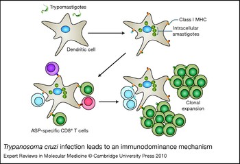
The T. cruzi genome encodes many antigens that contain amino acid sequences that are putative binders to MHC molecules. Although T cell receptors from CD8+ T cells could potentially recognise all these peptides, the actual repertoire of CD8+ T cells is rather restricted. This phenomenon, known as immunodominance (Fig. 5), is responsible for the strong recognition of only a few T. cruzi antigens (Ref. Reference Tzelepis79), and for immune protection during T. cruzi infection. However, infection with T. cruzi is never completely cleared from the host, and it can be speculated that immunodominance and the limited parasite-specific CD8+ T cell repertoire may allow the parasite to evade the immune response, leading to persistent infection with T. cruzi (Ref. Reference Rodrigues80).
Trypanosoma cruzi infection leads to an immunodominance mechanism. Mice infected with T. cruzi develop an immunodominant CD8+ T cell response. Some epitopes from the transialidase and amastigote specific protein-2 (ASP) antigens were found to be specific for the CL and Y strains, respectively. The immunodominance leads to clonal expansion of CD8+ T cells specific for one or a few epitopes, which might contribute to parasite immune evasion.

Highly immunodominant proteins, such as the amastigote surface protein (ASP)-2 and the trans-sialidase (TS) antigen, which belong to the same gene family, have been identified. The ASP-2 antigen was demonstrated to contain an immunodominant epitope for T. cruzi Y strain, while TS is the immunodominant antigen for the CL strain, proving the importance of parasite strain on specificity of immunodominant antigens/epitopes (Refs Reference Tzelepis79, Reference Tzelepis81, Reference Martin82, Reference Haolla83). The TS gene family has more than 1000 variants in the parasite genome – a frequency that is high enough to generate a strong immune response (Ref. Reference El-Sayed6). Mice vaccinated with immunodominant antigens generate memory CD8+ T cells specific for immunodominant epitopes, leading to clearance of parasites after a challenge with T. cruzi and preventing pathology (Ref. Reference Tzelepis81).
What have we learned from immunological studies on Chagas disease?
Several aspects have been identified to be important in the development of T-cell-mediated immune responses and protective immunity against infection with T. cruzi in vertebrate hosts: (1) the ability of T. cruzi parasites to elicit a strong pro-inflammatory response (including IL-12 production by dendritic cells) via activation of TLR7/TLR9 and the MYD88 adaptor (Refs Reference Brener and Gazzinelli4, Reference Golgher and Gazzinelli27, Reference Gazzinelli and Denkers44); (2) induction of MYD88/IL-12-dependent Th1 cells, which persist during chronic infection (Refs Reference Brener and Gazzinelli4, Reference Golgher and Gazzinelli27, Reference Gazzinelli and Denkers44); (3) induction of immunodominant and persistent CD8+ T cell responses (Ref. Reference Tarleton5); and (4) induction of effector mechanisms by IFN-γ (Refs Reference Brener and Gazzinelli4, Reference Golgher and Gazzinelli27). Together, these responses are essential for host resistance to infection with T. cruzi. These are all essential features that a priori need to be incorporated in a vaccine against Chagas disease. However, it is clear that many of these components are also associated with the development of myocarditis and pathogenesis of Chagas disease, and caution is needed, especially in immunotherapeutic protocols, in order to avoid potentiation of the inflammatory process elicited by T. cruzi and thereby increased severity of Chagas disease.
Immunopathogenesis of Chagas disease
The inflammatory reaction elicited by T. cruzi is responsible for the local control of parasite replication and parasitism, but also causes tissue damage, enlargement of cardiomyocytes and development of chagasic cardiomyopathy (Refs Reference Andrade26, Reference Golgher and Gazzinelli27). From studies performed in T-cell-deficient mice, it is clear that T cells are the main inflammatory cells present in the myocarditis elicited by T. cruzi. The activation of innate immunity is crucial for initiating a parasite-specific T cell response, as well as establishing an environment that is conducive to recruitment of T cells to the parasitised tissue. Different studies show that activation of nucleic-acid-sensing TLRs is an important step in activation of dendritic cells, which will favour the differentiation of CD4+ Th cells into Th1 cells. In turn, the Th1 cells contribute as a major source for IFN-γ, a cytokine that is essential for host resistance to infection but also to the genesis of myocarditis elicited by T. cruzi parasites.
Activation of TLRs is critical in bridging innate and acquired immunity during T. cruzi infection (Refs Reference Brener and Gazzinelli4, Reference Gazzinelli and Denkers44). Importantly, evidence for the role of the TLR signalling pathway in the genesis of inflammatory process elicited by T. cruzi is rapidly accumulating (Refs Reference Campos45, Reference Coelho84, Reference Petersen and Burleigh85). Translocation of NF-κB and phosphorylation of mitogen-activated protein kinases (MAPKs) are hallmarks of cellular activation by T. cruzi (Refs Reference Campos42, Reference Ropert43, Reference Hall86, Reference Huang87, Reference Huang88, Reference Ropert89, Reference Machado90). In addition to activating macrophages, infection with T. cruzi triggers signalling pathways in fibroblasts, endothelial cells and cardiomyocytes (Refs Reference Tardieux, Nathanson and Andrews70, Reference Petersen and Burleigh85, Reference Hall86, Reference Huang87, Reference Huang88, Reference Burleigh and Woolsey91, Reference Scharfstein92), leading to the production of cytokines, chemoattractants and adhesion molecules (Ref. Reference Michailowsky93). In some instances, this cellular activation occurs indirectly via immunologically active molecules (most notably IFN-γ) elicited during T. cruzi infection (Refs Reference Michailowsky93, Reference Talvani94, Reference dos Santos95, Reference Michailowsky96, Reference Aliberti97). Indeed, it has been shown that the GPI-mucins induce both local production of monocyte chemoattractant protein (MCP)-1 [CCL2; chemokine (C-C motif) ligand 2] and leukocyte recruitment, in a TLR2/IFN-γ-dependent manner (Ref. Reference Coelho84). An additional study suggested the involvement of IL-1R in hypertrophy of cardiomyocytes exposed to trypomastigotes. This cellular enlargement was found to be dependent on TLR2-induced IL-1β secretion by cardiomyocytes exposed to T. cruzi (Ref. Reference Petersen and Burleigh85).
During T. cruzi infection, IFN-γ also favours migration of T cells and establishment of myocarditis by inducing expression of chemokines, such as CCL5 (also known as RANTES), CXCL10 [chemokine (C-X-C motif) ligand 10; also known as IFN-γ-inducible protein 10 (IP10)] and CXCL9 (also known as monokine induced by IFN-γ; MIG), and adhesion molecules, such as intercellular adhesion molecule (ICAM) and vascular cell adhesion molecule (VCAM) (Refs Reference Talvani94, Reference dos Santos95). Importantly, therapy with a CCR5 (C-C chemokine receptor 5) antagonist has a beneficial effect, controlling T. cruzi-elicited myocarditis (Refs Reference Marino98, Reference Medeiros99). The presence of IFN-γ-producing cells is clearly diminished in the heart tissue of mice deficient in IL-12, a cytokine triggered by T. cruzi infection in a TLR/MYD88-dependent manner (Refs Reference Almeida41, Reference Campos45, Reference Aliberti97, Reference Camargo100). Similarly, the production of IL-12 and IFN-γ is impaired in Myd88 −/− as well as in TLR9 −/− mice (Refs Reference Campos45, Reference Bartholomeu50). Together these studies suggest that TLRs and MYD88 are important for inducing the synthesis of IL-12 and IFN-γ, which are essential in host resistance to infection but also promote myocarditis during Chagas disease.
Evaluation of human cytokine responses during Chagas disease has demonstrated that asymptomatic patients have a high ratio of lL-10 to TNF-α compared with cardiac patients (Ref. Reference Souza101). Furthermore, high levels of pro-inflammatory cytokines such as IFN-γ and TNF-α are associated with severe pathology (Ref. Reference Cunha-Neto102), establishing that the balance between anti-inflammatory and inflammatory cytokines is a key determinant in the pathogenesis of Chagas disease. Polymorphisms in cytokine genes such as IL10, TGFB1 and TNF, in the chemokine receptor gene CCR5 and in the TLR adaptor gene TIRAP are also associated with the outcome of Chagas disease in humans (Refs Reference Ramasawmy62, Reference Calzada103, Reference Calzada104, Reference Costa105, Reference Drigo106).
Immune response and efficacy of chemotherapy for Chagas disease
Benznidazol interferes with oxidative stress and is currently the only drug used to treat chagasic patients (Ref. Reference Urbina8). Unfortunately, low efficacy and failure on therapy, especially in the case of chronically infected individuals, is a common scenario. A variable degree of susceptibility of different T. cruzi strains to benznidazole therapy has been described (Refs Reference Filardi and Brener107, Reference Murta108, Reference Veloso109). In addition, when used to treat acute and chronically infected patients the efficacy of treatment with benznidazole varies from 70–80% to 30–50%, respectively (Refs Reference Galvao110, Reference Krettli, Cancado and Brener111). This difference in efficacy of chemotherapy during acute and chronic infection is reproducible in mice infected with the same parasite isolate. We speculated that the higher efficacy of treatment during the early stage of infection is related to the strong activation of the immune system and production of pro-inflammatory cytokines observed during acute infection with T. cruzi (Refs Reference Murta112, Reference Sathler-Avelar113). In fact, adjunct therapy with IL-12 (a potent inducer of IFN-γ) enhances the efficacy of chemotherapy for experimental T. cruzi infection in mice (Ref. Reference Aliberti97). Furthermore, a very low efficacy of benznidazole is observed in mice deficient in endogenous IFN-γ, and to a lesser extent IL-12, inducible nitric oxide synthase, or CD8+ T cells (Refs Reference Romanha114, Reference Michailowsky115, Reference Ferraz116, Reference Ferraz117). Together, these studies support the importance of the immune system in mediating the efficacy of chemotherapy to Chagas disease, and suggest that adjunct immunological therapy, including therapeutic vaccines, may be beneficial to treat chronic chagasic patients.
Development of vaccines for Chagas disease
Unfortunately, most (if not all) commercial vaccines available for human diseases mediate protective immunity by inducing neutralising antibodies (Ref. Reference Rodrigues118). Thus, a great challenge to immunologists and microbiologists is to develop vaccines – that is, identify antigens and define vaccine formulations – that induce T-cell-mediated protective immunity against intracellular microorganisms and tumour cells. The development of recombinant technology, and more recently proteomic approaches associated with immunological assays for high-throughput screening, has made it easier to identify new and often better antigens to be used in vaccine formulations. Live vectors and chemically defined TLR agonists as immunological adjuvants able to induce strong T-cell-mediated immunity are now readily available (Refs Reference Vollmer and Krieg119, Reference Tagliabue and Rappuoli120, Reference Moyle and Toth121). However, the lack of understanding of immunological mechanisms involved in protection and lack of identification of biomarkers of protective immunity are huge limitations for the rational development and testing of effective vaccines for human diseases.
A large number of T. cruzi antigens have been shown to induce protection against experimental challenge in mice (Ref. Reference Rodrigues118). In particular, during recent years two proteins – TS and ASP-2 – that elicit a strong response of CD8+ T cells have emerged as important candidates for an anti-T. cruzi vaccine (Ref. Reference Araujo122). Protective immunity is induced when these antigens (Refs Reference Machado123, Reference Vasconcelos124, Reference Hoft125) are produced via viral vectors (e.g. adenovirus) and DNA vaccines, or as recombinant proteins together with TLR agonists, such as CpG, monophosphoryl lipid A (MPLA) or flagellin. In mice, protective immunity elicited by different vaccination protocols is highly dependent on induction of CD4+ T cells as well as CD8+ T cells, and to a lesser extent on antibodies elicited by parasite antigens (Refs Reference Machado123, Reference Vasconcelos124, Reference Hoft125). The translation of these studies to humans remains to be discussed, since natural transmission of the disease is largely controlled in some countries where the disease is endemic. Nevertheless, the use of therapeutic vaccines must be considered, especially if we take into account the number of individuals who are chronically infected with T. cruzi and the lack of an effective therapy for chronic disease. Importantly, a few studies have demonstrated the effectiveness of therapeutic vaccines, reversing myocarditis in mice chronically infected with T. cruzi (Refs Reference Zapata-Estrella126, Reference Sanchez-Burgos127).
Beyond Chagas disease: T. cruzi and the fight against cancer
With regard to the immune response, cancer cells and tumours have three main stages during their development (Ref. Reference Dunn, Old and Schreiber128). In the first stage, the elimination phase, tumours that arise are recognised, and this is followed by induction of acquired immunity and elimination of cancer cells. The elimination phase is followed by the equilibrium phase, when tumor growth is continuously controlled by acquired immunity. Stages one and two are often subclinical and part of the immunological surveillance. The third and last stage is the escape phase, which is characterised by a rupture of the equilibrium phase, leading to uncontrolled tumour growth, which is often lethal, particularly if left untreated. It is assumed that in the third stage, due to immunological pressure, mutant cancer cells bypass immune recognition, leading to uncontrolled growth of tumour cells.
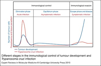
A parallel between the natural course of T. cruzi infection and the stages of tumour development is shown in Figure 6. The elimination and the equilibrium phases of T. cruzi infection are similar to the ones described for tumour development, although the elimination phase of T. cruzi infection is often clinical and associated with an exuberant activation of the innate and acquired immune system. The main difference is observed during the escape phase. The symptomatic forms of Chagas disease occur at the late stage of infection and are associated with the presence of parasites. The tissue damage is thought to be caused by an intense inflammatory reaction, associated with relatively low parasitism and parasitaemia. Uncontrolled replication of T. cruzi in chronically infected hosts is never seen, unless the host becomes immunosuppressed/immunodeficient, as is the case during immunosuppressive therapy or in acquired immune deficiency syndrome (AIDS) (Ref. Reference Vaidian, Weiss and Tanowitz129). More important, most chagasic patients will remain asymptomatic for life. Thus, the parasite seems to be able to keep boosting an effective immune response during the long-lasting parasitism.
Different stages in the immunological control of tumour development and Trypanosoma cruzi infection. As both pathologies progress they have three critical stages. The first stage is characterised by the immunological recognition and control of tumour cells or parasites, protecting against disease. The second stage, namely the equilibrium phase, is characterised by an efficient control of tumour growth and parasite replication and is normally asymptomatic. The third phase is characterised by immunological evasion and development of pathology, when the immune system can no longer maintain homeostasis and consequently prevent the clinical forms. A hallmark of T. cruzi infection is the strong activation of both innate and acquired immune system leading to intense inflammation, both in the recognition phase and at late stages during the ‘escape’ phase, when there is an increase in parasite replication but this is still kept in check by the host immune system. By contrast, tumour cells are less effective in stimulating the innate and acquired immune system. Consequently, the elimination phase of tumour development is subclinical, and during the escape phase tumour cells are left completely unchecked, growing very rapidly. Red and blue lines represent the intensity of parasitaemia/parasitism and tumour growth during the elimination, equilibrium and escape phases (Ref. Reference Dunn, Old and Schreiber128).

In the 1920s, the Russian scientist Grigorii Roskin conducted a broad study using various trypanosomes (T. pecaudi, T. equiperd, T. suauri, T. gambiense and T. cruzi) to evaluate their ability to inhibit the development of tumours implanted in mice (Ref. Reference Krementsov130). Inspired by the ‘malariotherapy’ developed by the Nobel Prize winner Julius Wagner-Jauregg, which used Plasmodium to treat paralysis caused by syphilis, Roskin thought that these parasites that cause serious diseases in their hosts produced a ‘toxin’ that could also act on and kill tumour cells (Ref. Reference Krementsov130). Almost ten years later, he published an article demonstrating tumour regression after infection with T. cruzi in mice (Ref. Reference Roskin and Exempliarskaia131). This was the first of a series of studies using live T. cruzi and some parasite extracts in the treatment of tumours (Refs Reference Roskin and Romanova132, Reference Roskin and Romanova133). In 1939 he published the first article demonstrating tumour regression in human patients (Ref. Reference Bongard and Roskin134).
During the following years, the US National Cancer Institute (NCI) demonstrated interest in this new approach to treat cancer, supporting several research projects in the USA around the subject (Refs Reference Malisoff135, Reference Hauschka and Goodwin136). At that time, the NCI also supported Carlos Chagas Filho, the youngest son of Carlos Chagas, who tested the effect of infection with different T. cruzi strains on tissue cultures of embryonic cells (Ref. Reference Meyer and Chagas137). A growing interest in using T. cruzi to treat cancer was observed around the world and, in the 1960s, T. cruzi preparations for cancer therapy were launched by a French pharmaceutical company, followed by a Russian company (Ref. Reference Krementsov130). Although similar results were found (Ref. Reference Galliard, Brumpt and Martines138), the ambiguity of the findings obtained by various investigators regarding efficacy against experimental tumours (Ref. Reference Belkin139) led to the discontinuation of this line of investigation and of commercial distribution of this anticancer component.
Based on the ability of T. cruzi parasites to strongly activate both the innate and acquired immune response in the vertebrate host, we decided to investigate the mechanism by which T. cruzi infection controls tumour development from a different angle. Our initial hypothesis was that, by triggering innate immunity and a strong pro-inflammatory response, T. cruzi infection would lead to activation of immunological effector mechanisms that are also effective in killing or controlling the growth of tumour cells. In particular, the ability of T. cruzi parasites to stimulate host innate immunity, resulting in the induction of strong and long-lasting antigen-specific polarised CD4+ Th1 as well as CD8+ T cell responses, led us to explore a genetically engineered attenuated T. cruzi strain expressing tumour antigens to develop an anticancer vaccine. Importantly, protective immunity seems to be dependent on persistent infection and, as for other parasite strains, parasitological cure results in loss of parasite-specific immune responses and protection.
Of course, we have no difficulties envisaging the major ethical barriers of employing T. cruzi parasites to treat cancer. Nevertheless, we understand that by exploring this road and achieving the proof of concept, we may highlight new strategies that effectively induce a strong and long-lasting T-cell-mediated immune response, which is the key aim, although often poorly achieved, in vaccine protocols against different type of tumours. To conduct our studies, we used the clone CL14, isolated decades earlier from the CL strain and shown to be consistently avirulent, even when infecting newborn mice that are otherwise highly susceptible to T. cruzi infection (Ref. Reference Lima140).
Parasitaemia and parasitism are absent or scarce in adult mice infected with CL14, and thus the systemic and aberrant activation of the immune system is not observed, as one would predict from studies with virulent strains of T. cruzi (Ref. Reference Lima, Lenzi and Gattass141). Consequently, we have not seen any effect of vaccination/infection with CL14 in the B16 experimental model for melanoma. Nevertheless, vaccination with live CL14 was repeatedly shown to induce a potent and long-lasting parasite-specific antibody and T-cell-mediated immunity, characterised by high levels of immunoglobulin (Ig) G2, IFN-γ and CD8+ T cell responses and strong protection against challenge with highly virulent strains of T. cruzi (Refs Reference Pyrrho142, Reference Paiva143, Reference Paiva144). Thus, we assume that the immunological adjuvant properties of CL14 are still effective, and that this particular clone should be explored as a possible vaccine vector for an antitumour vaccine.
We next engineered a transgenic CL14 (CL14-NY-ESO-1) to express a member of the cancer testis antigen (CTA) family, named NY-ESO-1 (CTAG1B) (Ref. Reference Chen145). NY-ESO-1 is a cytoplasmic protein expressed only in germinal and tumour cells, and, in contrast to most tumour antigens, it is described as highly immunogenic (Ref. Reference Gnjatic146). Importantly, vaccination with CL14-NY-ESO-1 induced complete protection against a melanoma expressing NY-ESO-1, but not against the counterpart control cell line, which did not express NY-ESO-1. By contrast, we observed only partial protection when we used recombinant NY-ESO-1 associated with TLR agonists, and we are currently investigating the immunological mechanisms responsible for such a solid protection elicited by CL14-NY-ESO-1 (C. Junqueira and R.T. Gazzinelli, unpublished). Currently, experiments are being performed to determine the ability CL14-NY-ESO-1 to reverse tumour growth in immunotherapeutic protocols, which would be clinically relevant.
Concluding remarks
Despite recent advances, outstanding questions still remain regarding the innate and acquired immunological mechanisms involved in host resistance and pathogenesis of Chagas disease. Are families of innate immune receptors other than TLRs involved in the initial activation of innate immunity? What are the crucial molecular and cellular events necessary for the induction of immunodominant and long-lasting protective CD8+ T cell response induced by T. cruzi? What are the key steps required for establishment of chagasic myocarditis and Chagas disease? Can we explore the potent immunological adjuvant properties from T. cruzi parasites to develop a therapeutic antitumour vaccine without deleterious effects? Hopefully, these and other questions will stimulate the field of immunology of T. cruzi infection, providing new insights for immunological intervention to prevent or treat Chagas disease.
Acknowledgements and funding
We are grateful for comments and suggestions from the peer reviewers. Our work on Trypanosoma cruzi and Chagas Disease is funded by the Program of Clinical Discoveries from the Ludwig Institute of Cancer Research (LICR) and Atlantic Philanthropies (AP); Fundação de Amparo a Pesquisa de Minas Gerais (FAPEMIG); Fundação Oswaldo Cruz (FIOCRUZ); National Institute of Science and Technology for Vaccines/Conselho Nacional de Desenvolvimento Científico e Tecnológico (CNPq); and the US National Institutes of Health (NIH). R.T.G. and M.M. are research fellows from CNPq; C.R. is a research fellow of FAPEMIG; and C.J. is a research fellow from Coordenação de Aperfeiçoamento de Pessoal de Ensino Superior (CAPES).