Implications
The progressive applications of nanosilver as commercialized products like contraceptive devices and maternal hygiene items, make its access to human reproductive system, which may result in fertility problems. In addition, the application of silver nanoparticles as a powerful disinfection agent in the animal farm has recently been recommended. Nanosilver deposit in the animal testes could be occurring if accidentally used orally, which may result in infertility. This study explained that nanosilver could imply its toxic effects on gene levels in addition of enzyme activity.
Introduction
Silver is a white metallic element along with mercury, lead, cadmium and gold. This metal has been used over 6000 years as a medicinal drug and antibacterial treatment for serious burn wounds (Troy et al., Reference Troy, Derossi, Prochiantz, Greene and Shelanski1996). Metallic silver is engineered into ultra-fine particles, which is called nanosilver. Nanosilver is silver particle with at least one dimension <100 nm (Chen and Schluesener, Reference Chen and Schluesener2008). Recently, silver nanoparticles have been in the center of attention because of their strong antimicrobial activity. The potential of nanosilver as an alternative antimicrobial growth-promoting supplement for broiler chickens has been considered. In this regard, several studies have been designed to investigate the effects of nanosilver on vital and growth parameters of poultry such as performance, metabolism, gastrointestinal microflora, morphology of intestine, blood parameters, egg and meat qualities and development of chicken embryos. In these studies, it has been confirmed that defined concentrations of nanosilver was not harmful in the chickens in vivo (Sawosza et al., Reference Sawosza, Bineka, Grodzika, Zieliñskaa, Sysaa, Szmidt, Niemiec and Chwalibog2007; Mahmoud, Reference Mahmoud2012; Pineda et al., Reference Pineda, Sawosz, Lauridsen, Engberg, Elnif, Hotowy, Sawosz and Chwalibog2012a and Reference Pineda, Chwalibog, Sawosz, Lauridsen, Engberg, Elnif, Hotowy, Sawosz, Gao and Ali2012b; Farzinpour and Karashi, Reference Farzinpour and Karashi2013). However, in spite of the wide application of nanoparticles, there are few studies about the effect of these elements on animal and human health; and the mechanism of their toxicity is not completely clear. Small sizes of nanoparticles allow their penetration of cell membranes, making distribution easy in the body. Distribution of nanoparticles could lead to increased interactions with proteins, leading to biological and physiological changes of cells (Chen and Schluesener, Reference Chen and Schluesener2008). An important fact is that silver has an oxidation state that makes it useful as a catalyst (Wijnhoven et al., Reference Wijnhoven, WJGM, Herberts, Hagens, Oomen, Heugens, Roszek, Bisschops, Gosens and Van De Meent2009). Toxicity and cellular responses to nanosilver particles are a serious concern. It has been reported that nanoparticles mediate their toxicity by induction of reactive oxygen species (ROS; Foucaud et al., Reference Foucaud, Goulaouic, Bennasroune, Laval-Gilly, Brown, Stone and Falla2010; Chairuangkitti et al., Reference Chairuangkitti, Lawanprasert, Roytrakul, Aueviriyavit, Phummiratch, Kulthong, Chanvorachote and Maniratanachote2013). This increase in the cellular ROS levels was associated with induction of reduced cell growth, apoptotic and necrotic cell death (Zanette et al., Reference Zanette, Pelin, Crosera, Adami, Bovenzi, Larese and Florio2011).
Nitric oxide (NO) is a molecule, which has variety of biological functions. NO is known as an important modulator of cellular function and involves numerous physiological and pathological processes (Guzik et al., Reference Guzik, Korbut and Adamek-Guzik2003). NO is generated from conversion of amino acid l-arginine to l-citrulline and this reaction is catalyzed by a family of isoenzymes from three distinct genes. Endothelial NO synthase (eNOS, Type III) and neuronal NO synthase (nNOS, type I) are calcium dependent and usually considered to be involved in physiological homeostasis generating basal level of NO. The third isoform, inducible NO synthase (iNOS, type II) is thought to be calcium independent (Coleman, Reference Coleman2001). iNOS is activated in the pathological situation, although its presence also has been confirmed under physiological condition (Teshfam et al., Reference Teshfam, Nikbakht Brujeni and Hassanpour2006). iNOS produces very large, toxic amounts of NO in a sustained manner, whereas other NOS isoforms produce NO rapidly and transiently. Low amounts of NO activate cell signaling, and within a few seconds, is oxidized to its metabolites, that is, nitrite or nitrate; while iNOS-derived NO (large amounts) forms oxidant agents that mediate cytotoxic effects of NO such as DNA damage, low-density lipoprotein oxidation, tyrosine nitration and inhibition of mitochondrial respiration (Coleman, Reference Coleman2001; Guzik et al., Reference Guzik, Korbut and Adamek-Guzik2003).
Superoxide dismutases (SOD) are enzymes that belong to antioxidant defense system of cell and catalyze the dismutation of superoxide radical through a redox reaction. Eukaryotic cells express two forms of SOD: copper/zinc SOD (SOD1) and manganese SOD (SOD2). SOD1 is found primarily in the cytosol of cell and SOD2 in the mitochondria (Mruk et al., Reference Mruk, Silvestrini, Mo and Cheng2002). These enzymes are one of the first-line defenses against ROS damage, and convert superoxide anion to hydrogen peroxide, which in turn, is converted to water by glutathione peroxidase and catalase. Therefore, these antioxidant enzymes keep cellular levels of ROS low. Mitochondria are particularly susceptible to damage induced by ROS, which are generated continuously by the mitochondrial respiratory chain; this is the reason for the existence of a specific SOD in this organelle (Mruk et al., Reference Mruk, Silvestrini, Mo and Cheng2002).
This study was undertaken to examine the potential toxicological effect of nanosilver on chicken Sertoli cells, a critical cell type of the male reproductive system. In this experiment, NOS and SOD genes expression, lipid peroxidation, antioxidant capacity and NO production of the cultured Sertoli cells were evaluated.
Material and methods
Nanosilver colloid characterization
The colloidal nanosilver, type L (commercial name: Nanocid), purchased from Nano Nasb Pars Co. (Tehran, Iran), were synthesized using a novel process involving the photo-assisted reduction of Ag+ to metallic nanoparticles, registered under United States Patent Application no.: 20090013825. Briefly, 4.5 g of linear alkyl benzene suffocate were dissolved in 95 ml of distilled water and then added to a solution containing 0.32 g of silver nitrate. After mixing thoroughly, the addition of 0.2 g of a hydrazine solution (0.03 M) formed a yellowish silver colloidal solution. According to information provided by the manufacturer, the product was a water-based colloidal suspension containing 4000 mg/l spherical silver nanoparticles (average size 16.6 nm). The zeta potential of a 100-mg/l suspension, measured by dynamic light scattering using a Malvern Zetasizer model 3000 HSa (Malvern Instruments Ltd, Worcestershire, UK), was −53.33±7.86 mV. Distribution of the hydrodynamic diameter of the particles in the diluted nanosilver suspension (100 mg/l) was 54.8 nm in average.
Cell cultures and treatments
Sertoli cells were obtained from 50 to 54 weeks old (mature) Cobb 500 roosters. All birds were housed in individual battery cages under a 14-h light/10-h dark photoperiod; feed and water were provided ad libitum. For each culture, six chickens were killed by cervical dislocation. Testes were immediately recovered and pooled in cold culture medium. For cell preparation, HEPES-buffered F12/Dulbecco’s modified eagle’s medium (DMEM; Sigma-Aldrich) supplemented with 10% fetal calf serum (FCS), 100 U/ml of penicillin and 100 µg/ml of streptomycin (Sigma-Aldrich) was used. Testes were decapsulated, slightly minced, and incubated for 45 min at 37°C with DNase (20 μg/ml) and type IV collagenase (0.4 mg/ml; Sigma-Aldrich). Sertoli cells were isolated from chicken testes according to previous studies with little modifying (Guibert et al., Reference Guibert, Briere, Pelletier, Brillard and Froment2011, Park et al., Reference Park, Lee, Oh, Shim, Nah, Choi and Gye2011). Briefly, cells were centrifuged to remove collagenase and were allowed to sediment by gravity to separate seminiferous tubules in the pellet. The pelleted seminiferous tubules were resuspended and digested by collagenase (1 mg/ml), DNase (20 µg/ml; Sigma-Aldrich) and trypsin (25 mg/ml; Sigma-Aldrich) for 20 min in 5% CO2 in air at 37°C to reduce peritubular cell contamination. Digested materials were centrifuged and resulting cells pellet were suspended in DMEM/HEPES-buffered F12 (with FCS, penicillin and streptomycin) and washed twice by centrifugation. Final cell pellet was suspended in fresh medium, plated on the six-well dish in 1.5×10⁵ cells/ml, and incubated in 5% CO2 in air at 37°C. After 48 h, cells were treated with 20 mM Tris (pH 7.3) for 2 min to lyse residual germ cells. This was followed by two washes with fresh medium to remove germ cell debris. To characterize Sertoli cells, Vimentin was detected in cultured Sertoli cells by immunocytochemistry (Minaee Zanganeh et al., Reference Minaee Zanganeh, Roudkenar and Kashani2013). Nanosilver colloid was added to culture medium of Sertoli cells at concentrations of 0 (as control), 25, 75 and 125 ppm (selected from a primary dose–response study with a range of 1 to 500 ppm nanosilver). After the 48 h of culture at 37°C in a humidified atmosphere of 5% CO2 in air, sample of cultured Sertoli cells from each group was stained with 0.4% trypan blue (Sigma-Aldrich, St. Louis, MO, USA) for 3 min to test cell viability. Dead cells accumulated the dye and were stained blue, whereas living cells eliminated it and remained clear. Finally, Sertoli cells were stored at −70°C for subsequent measurements.
RNA extraction and cDNA synthesis of Sertoli cells
Sertoli cells were processed for total RNA isolation according to the acid guanidinium thiocyanate-phenol-chloroform single-step extraction protocol (Hassanpour et al., Reference Hassanpour, Yazdani, Soreshjani and Asgharzadeh2009). Total RNA was treated with RNase-free DNase (Sinaclon Bioscience, Karaj, Iran) to avoid amplification of contaminating genomic DNA. The integrity and quality of the purified RNA were assessed by agarose gel electrophoresis and by measurement of the A260/A280 nm ratio. Only RNA samples exhibiting an A260/A280 ratio >1.9 and showing integrity of the RNA by electrophoresis were used for synthesis of cDNA.
Total RNA was reverse transcribed into cDNA using M-MLV reverse transcriptase (Sinaclon Bioscience, Karaj, Iran) as previously described (Hassanpour et al., Reference Hassanpour, Yazdani, Soreshjani and Asgharzadeh2009). The reverse transcription mix was heated to 75°C for 15 min to denature the RNA and then stored at –20°C.
Quantitative real-time PCR Analysis
The levels of eNOS, iNOS, SOD1, SOD2 and β-actin transcripts were determined by real-time reverse transcriptase (RT)-PCR using Eva-Green chemistry (Sinaclon Bioscience). To normalize input load of cDNA between samples, β-actin was used as an endogenous standard. Specific primers of SOD1, SOD2 and β-actin were designed with Primer-Blast (www.ncbi.nlm.nih.gov/tools/primer-blast/index.cgi?LINK_LOC=BlastHome). For eNOS and iNOS previously described primer pairs were used (Hassanpour et al., Reference Hassanpour, Yazdani, Soreshjani and Asgharzadeh2009). Primers are listed in Supplementary Table S1. PCRs were carried out in a real time PCR cycler (Rotor Gene Q 6000, Corbett Robotics Pty Ltd, Brisbane, Australia). 1 µl cDNA was added to the Titan Hot Taq Eva-Green Ready Mix (0.5 µM of each specific primer, and 4 µl of Titan Hot Taq Eva-Green Ready Mix) in a total volume of 20 µl. The thermal profile was 95°C for 10 min, 40 cycles of 95°C for 40 s, 63°C for 45 s and 72°C for 30 s. At the end of each phase, fluorescence was measured and used for quantitative purposes. Data of genes expression were normalized to β-actin. The formula ΔΔC t (Livak and Schmittgen, Reference Livak and Schmittgen2001) was used to calculate relative transcript levels using Rotor Gene Q software, version 2.0.2 (build 4).
Thiobarbituric acid reactive substances (TBARS) and ferric ion reducing antioxidant power (FRAP) assays
All reagents for these assays were purchased from Sigma-Aldrich. The cultured Sertoli cells were sonicated for cell lysate. Using the TBARS, malondialdehyde, an indicator of lipid peroxidation, was measured according to Yagi (Reference Yagi1998). Briefly, trichloroacetic acid was added to Sertoli cell lysate to precipitate proteins. After centrifuging, the supernatants were recovered. Then an equal volume of TBA was added and the samples incubated in boiling water bath for 10 min. After cooling of samples, absorbance was measured at 532 nm by a spectrophotometer (Corning Inc., New York, USA).
FRAP assay was used to measure the antioxidant capacity. FRAP assay was performed as previously described (Aiassa et al., Reference Aiassa, Baronetti, Paez, Barnes, Albrecht, Pellarin, Eraso and Albesa2011) (modified for cell culture). This method is based on the reduction of ferric-tripyridyl triazine (TPTZ) to ferrous colored form in the presence of antioxidants. Briefly, the FRAP reagent contained TPTZ, FeCl3 and acetate buffer (sodium acetate and acetic acid) was added to each sample and incubated at 37ºC for 4 min. Absorbance was measured at 593 nm by a spectrophotometer.
Measurement of nitrite
Nitrite as a metabolite of NO, could be good candidate of NO production in the cell culture. All reagents for this test were purchased from Sigma-Aldrich. The amount of nitrite was measured by Griess reaction (Goudarzi and Hassanpour, Reference Goudarzi and Hassanpour2007). Briefly, after sonication of Sertoli cell cultures, ZnSO4 and NaOH solutions were added to deproteinize samples. Upon centrifuging, the supernatants were recovered and diluted by glycine buffer. Sulfanilamide solution was added to the sample tubes and incubated for 10 min at room temperature while protected from light. N-napthylethylenediamine dihydrochloride was then dispensed to all samples and absorbance was measured at 540 nm.
TBARS, FRAP and NO data were calculated from standard curves and expressed as micromoles (µmol) in mg−1 total protein of cell cultures. Mass protein of cells was measured by Bradford method.
Statistical treatment of results
All data points are presented as means±s.e. Differences between experimental group means were analyzed through one-way ANOVA followed by Duncan’s multiple range tests. Differences were considered significant at P<0.05.
Results
Sertoli cells isolation and characterization
The presence of Vimentin protein on Sertoli cells was confirmed by immunocytochemistry, demonstrating that the majority (up 90%; data not shown) of freshly isolated cells were Vimentin positive (Supplementary Figure S1). All these results indicated that the Sertoli cells were properly isolated.
Cell viability
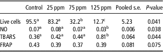
Viability of the cultured Sertoli cells exposed to nanosilver was evaluated by trypan blue exclusion and is presented in Table 1. The percentage of live cells progressively decreased with increasing nanosilver concentration. This reduction was significant (P<0.05) in 75 and 125 ppm of nanosilver that was 0.37- and 0.17-folds, respectively, compared with control.
Effect of nanosilver on live cells, NO, TBARS and FRAB in the cultured Sertoli cells of chickens

NO=nitric oxide; TBARS=thiobarbituric acid reactive substances; FRAP=ferric-ion reducing antioxidant power.
Values are means.
a,b,cMeans with the different letters between groups are significantly different (P<0.05).
NO, TBARS and FRAP are based on µmol/mg total cell protein. Live cells are based on percent (%).
SOD and NOS genes expression
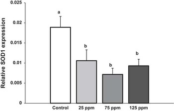
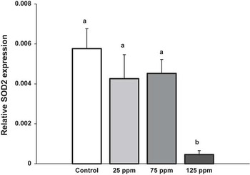
Expression of SOD1, SOD2, eNOS and iNOS genes was studied using quantitative RT-PCR in the cultured Sertoli cells exposing to different concentrations of nanosilver. Real time PCR results are shown in Figures 1 to 4. Expression of β-actin was detected in all experimental groups and was not changed by the different treatments. Expression of SOD1 gene in all nanosilver-treated groups was significantly (P<0.05) lower than control that was 0.55-, 0.38- and 0.49-folds in the nanosilver concentrations of 25, 75 and 125 ppm, respectively (Figure 1). Between treated groups, the mRNA amounts of this gene were unchanged (P>0.05). Expression of SOD2 gene expression was reduced only by 125 ppm nanosilver (about 92%) compared with control (P<0.05; Figure 2).
Relative expression of superoxide dismutase 1 (SOD1) gene in the cultured sertoli cells of chicken exposing to three concentrations of nanosilver. Results of real-time PCR of SOD1/β-actin were compared between different groups. Values are means±s.e. a,bMeans with the different letters between groups are significantly different (P<0.05).

Relative expression of superoxide dismutase 2 (SOD2) gene in the cultured sertoli cells of chicken exposing to three concentrations of nanosilver. Results of real-time PCR of SOD1/β-actin were compared between different groups. Values are means±s.e. a,bMeans with the different letters between groups are significantly different (P<0.05).

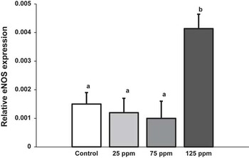
Relative expression of endothelial nitric oxide synthase (eNOS) gene in the cultured sertoli cells of chicken exposing to three concentrations of nanosilver. Results of real-time PCR of eNOS/β-actin were compared between different groups. Values are means±s.e. a,bMeans with the different letters between groups are significantly different (P<0.05).

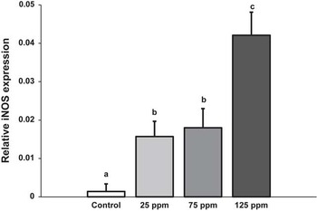
Relative expression of inducible nitric oxide synthase (iNOS) gene in the cultured sertoli cells of chicken exposing to three concentrations of nanosilver. Results of real-time PCR of iNOS/β-actin were compared between different groups. Values are means±s.e. a,b,cMeans with the different letters between groups are significantly different (P<0.05).

Expression of eNOS gene was significantly (P<0.05) increased about 2.8-fold in 125 ppm of nanosilver compared with control, but unchanged by 25 and 50 ppm of nanosilver (Figure 3).
Expression of iNOS gene was detectable in all groups. This expression was progressively increased with increasing concentrations of nanosilver compared with control (P<0.05). These elevations were about 10.7-, 12.9- and 30.1-folds for 25, 75 and 125 ppm of nanosilver, respectively (Figure 4).
Lipid peroxidation, antioxidant capacity and NO production
Results of lipid peroxidation (TBARS assay), antioxidant capacity (FRAP assay) and NO production (Griess reaction) in the nanosilver-treated Sertoli cells are presented in Table 1. Lipid peroxidation was significantly increased in cells exposed to 125 ppm of nanosilver compared with control (P<0.05). However, antioxidant capacity of nanosilver-treated cells did not change as measured by FRAP assay (P>0.05). Nitrite as index of NO production, was only reduced in 125 ppm of nanosilver compared with control (P<0.05), whereas variations of this parameter in the other nanosilver-treated groups were not significant (P>0.05).
Discussion
This research was designed to investigate adverse effects of nanosilver colloid on chicken Sertoli cells. The morphological properties of chicken Sertoli cells have high similarities with Sertoli cells in mammals, and it has been demonstrated that culture of Sertoli cells could be suitable for reprotoxicity studies (Guibert et al., Reference Guibert, Briere, Pelletier, Brillard and Froment2011).
Use of three concentrations of nanosilver in the present study confirmed that increasing exposure to nanosilver could enhance its toxic effects, consistent with other published studies (Grosse et al., Reference Grosse, Evje and Syversen2013). It has been reported that kinematic parameters of sperm was progressively reduced by higher concentrations of nanosilver in vitro (Mirshokraei et al., Reference Mirshokraei, Hassanpour, Akhavan Taheri, Riyahi and Shams-Esfandabadi2011). Following exposure to silver nanoparticles, liver, neuroendocrine and germ cell lines have shown impaired function of mitochondria (Braydich-Stolle et al., Reference Braydich-Stolle, Hussain, Schlager and Hofmann2005; Hussain et al., Reference Hussain, Hess, Gearhart, Geiss and Schlager2005; Hussain et al., Reference Hussain, Javorina, Schrand, Duhart, Ali and Schlager2006). This characteristic of response was similar in all cells. Thus, it is thought that mitochondria might be important target of nanosilver cytotoxicity (Teodoro et al., Reference Teodoro, Simoµes, Duarte, Rolo, Murdoch, Hussain and Palmeira2011). It seems that diminished SOD2 gene expression, which was confirmed in the present study, involve in dysfunction of mitochondria. SOD2 is an important antioxidant enzyme functioning in the mitochondria. Reduction of this enzyme could increase ROS leading to impairment of mitochondria. Studies have also shown that depletion of glutathione level and increased ROS were found in association with mitochondrial perturbation, suggesting that oxidative stress might mediate the cytotoxicity of silver nanoparticles (Chen and Schluesener, Reference Chen and Schluesener2008; Kim et al., Reference Kim, Choi, Choi, Chung, Park, Yi and Ryu2009; Mukherjee et al., Reference Mukherjee, O'Claonadh, Casey and Chambers2012). These studies are in agreement with our findings. The obtained results of TBARS indicated high lipid peroxidation due to high concentration of nanosilver. On the other hand, reduced SOD1 and SOD2 mRNAs could enhance this oxidation activity of ROS. Reduced SOD caused superoxide level to be high in the cell, which might react with NO and produce proxinitrite. This oxidant agent supports trigger apoptosis and cell death (Troy et al., Reference Troy, Derossi, Prochiantz, Greene and Shelanski1996). Silver nanoparticles may deplete the antioxidant defense mechanism, leading to ROS accumulation (Costa et al., Reference Costa, Ronconi, Daufenbach, Goncalves, Rezin, Streck and da Silva Paula2010). The accumulation of ROS can cause perturbation, and destruction of the mitochondria. Proteins and enzymes with thiol groups within cells like glutathione, thioredoxin, SOD and thioredoxin peroxidase, are most important components of antioxidant defense mechanism, which is responsible for neutralizing the oxidative stress of ROS (Chen and Schluesener, Reference Chen and Schluesener2008; McShan et al., Reference McShan, Ray and Yu2014). However, reduced amounts of such proteins (SOD as an example of this group) and other groups of antioxidants (see results of FRAP assay) provide evidence that the antioxidant defense mechanism of Sertoli cells has been weakened by exposure to high levels of silver nanoparticles (125 ppm). This condition could underlie toxic manifestations of nanosilver, which consist of mitochondrial dysfunction, increased membrane leakage, apoptosis and necrosis (Braydich-Stolle et al., Reference Braydich-Stolle, Hussain, Schlager and Hofmann2005; Rogers et al., Reference Rogers, Hsieh, Organti, Schmidt and Bello2008).
In the present study, gene expression of two enzymes (i.e. iNOS and eNOS) regarding to NO production was evaluated. Our data showed that despite increased transcription of eNOS and iNOS genes, the amount of NO in the Sertoli cell culture was decreased. It was predictable that oxidative stress stimulated transcription of these enzymes especially iNOS (Coleman, Reference Coleman2001). It had been determined that high concentrations of NO generated by iNOS were oxidized under aerobic conditions to reactive nitrogen oxide species (RNOS). RNOS nitrosated the thiol group to produce S-nitrosoglutathione in glutathione or to generate protein-S-NO in proteins. Such nitrosations inhibited the activity of many proteins including mitochondrial enzymes and transcription factors, and produced long-term cellular effects. Under conditions of high NO combined with high oxidative stress, superoxide interacted with NO to produce the highly toxic peroxynitrite anion (Coleman, Reference Coleman2001; Valko et al., Reference Valko, Leibfritz, Moncol, Cronin, Mazur and Telser2007). Thus, the lower level of nitrite (as an index of NO production) in nanosilver-treated culture of Sertoli cells might be due to diversion and reaction of NO into the mentioned pathways.
Taken together, the mRNA expression of chosen genes showed significant responses with increasing doses, however, not exactly reflected in the amount of TBARS, FRAP and NO especially in low concentration of nanosilver. Thus, indicating that nanosilver is not toxic in low concentration, although it affects gene expression in its all concentrations. Certainly, the activation of an enzyme was influenced by several factors in the cell in that the amount of its gene expression would be the only factor (Yamakura and Kawasaki, Reference Yamakura and Kawasaki2010; Forstermann and Sessa, Reference Forstermann and Sessa2012). In addition, the final function of an enzyme could not be resulted only from variations in its gene expression.
It is concluded that a high concentration of nanosilver can influence expression of genes associated with antioxidant mechanisms. This nanoparticle probably implies parts of its cytotoxicity with lipid peroxidation and suppression of antioxidant mechanisms via reduced expression of SOD genes and increased expression of NOS genes.
Acknowledgments
This work was supported by the funds granted by the Applied Research Centre, Vice Chancellor for Research of Shahrekord University.
Supplementary material
To view supplementary material for this article, please visit http://dx.doi.org/doi: 10.1017/S1751731114002262