Introduction
Ventricular assist devices are often utilised to support the failing systemic right ventricle as a bridge to heart transplantation in patients with d-TGA who have undergone an atrial switch operation. Reference Maly, Netuka, Besik, Dorazilova, Pirk and Szarszoi1 Although few case reports are available, the HeartMate 3 (Abbott Inc., Abbott Park, IL) has been used in such cases.
One complication associated with HeartMate 3, extrinsic obstruction of the outflow graft, was recently highlighted by a February 2024 FDA recall, 2 increasing clinicians’ awareness of this issue. This case report describes the complex catheter-based intervention to manage extrinsic obstruction of the outflow graft in a 53-year-old male with d-TGA post-Mustard procedure, who had been successfully supported by an HeartMate 3 for six years. This case underscores the importance of a multidisciplinary approach and the need for tailored technical considerations to address the unique anatomical and device-related challenges.
Case report
A 53-year-old male with d-TGA, post-Mustard procedure, had received an HeartMate 3 device in 2018 to support his systemic right ventricle as a bridge-to-decision for transplantation. Six years post-implantation, he presented with progressively reduced HeartMate 3 flow, suggesting compromised device function. Cardiac CT confirmed extrinsic obstruction of the outflow graft at the bend relief and other areas along the graft, consistent with recent FDA recall concerns. Given the high risk of surgical revision, a catheter-based intervention was chosen after thorough discussion. The multidisciplinary team included congenital and adult ventricular assist devices specialists, ventricular assist devices coordinators, imaging experts, and interventional cardiologists to ensure comprehensive pre-procedural planning.
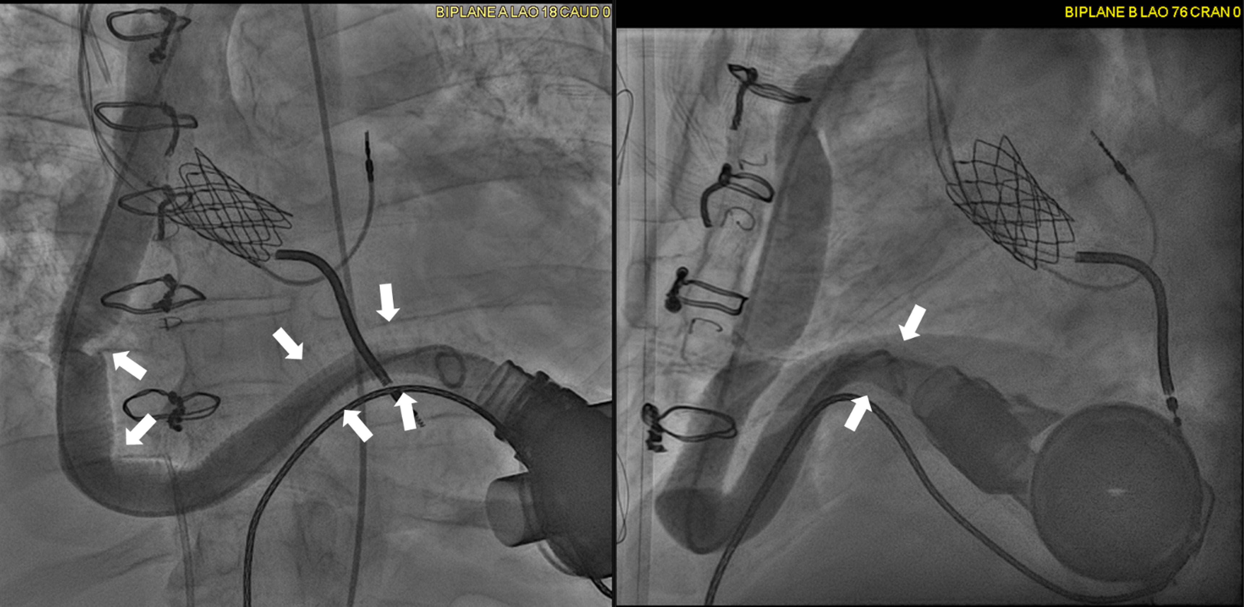
The procedure took place in the paediatric cardiac catheterisation lab, with both paediatric and adult VAD teams present. Access was obtained via the right femoral artery and vein, and the patient was anti-coagulated with heparin. Angiography confirmed multiple obstructive sites along the outflow graft (Figure 1), which had been routed in a relatively long and tortuous course through the anterior mediastinum due to the limited retrosternal space and the anterior position of the systemic right ventricle following the Mustard procedure. This anatomical constraint contributed to multiple angulations and kinks along the graft, including extrinsic obstruction of the outflow graft at the bend relief. HeartMate 3 flow was temporarily reduced to 4000 RPM (from a high of 6200 RPM) during contrast injections to facilitate imaging. The initial strategy was to place stents from the bend relief, with the plan to cover approximately two-thirds of the outflow graft.
Pre-stenting angiography of the HM3 outflow graft (left: AP view; right: lateral view). A 5 French pigtail catheter was advanced retrograde through the ascending aorta into the outflow graft of the HM3. The tip of the pigtail catheter is positioned near the bend relief. Angiography reveals stenotic areas consistent with extrinsic obstruction of the outflow graft (EOGO) and some regions of kinking (arrows).

A long sheath was carefully advanced retrograde over a guidewire from the aorta into the outflow cannula, ensuring the guidewire tip remained clear of the pump and cannula. One assistant was tasked with continually monitoring and ensuring that the tip of the guidewire was never advanced into the actual pump mechanism. Stents with an initial diameter of 11 mm were chosen and deployed in an overlapping manner (with the narrowest segment measured at 7 mm), with brief reduction of the HeartMate 3 flow for each deployment. Despite angiographic improvement, HeartMate 3 flow remained insufficient, prompting serial balloon angioplasty of the stents using a 16 mm balloon. Shortening of the stents with dilation led to separation and intervening gaps, necessitating additional stent placement.
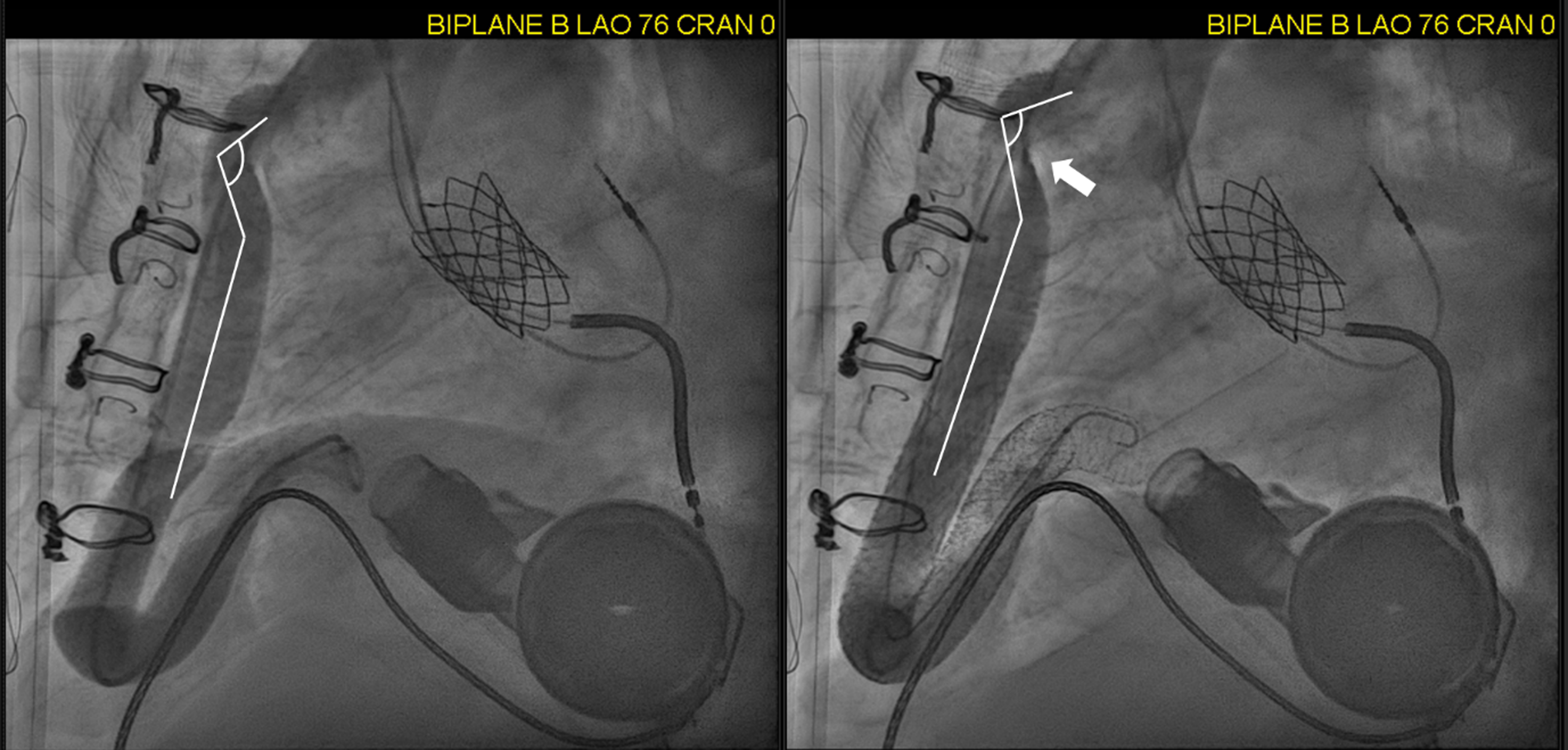
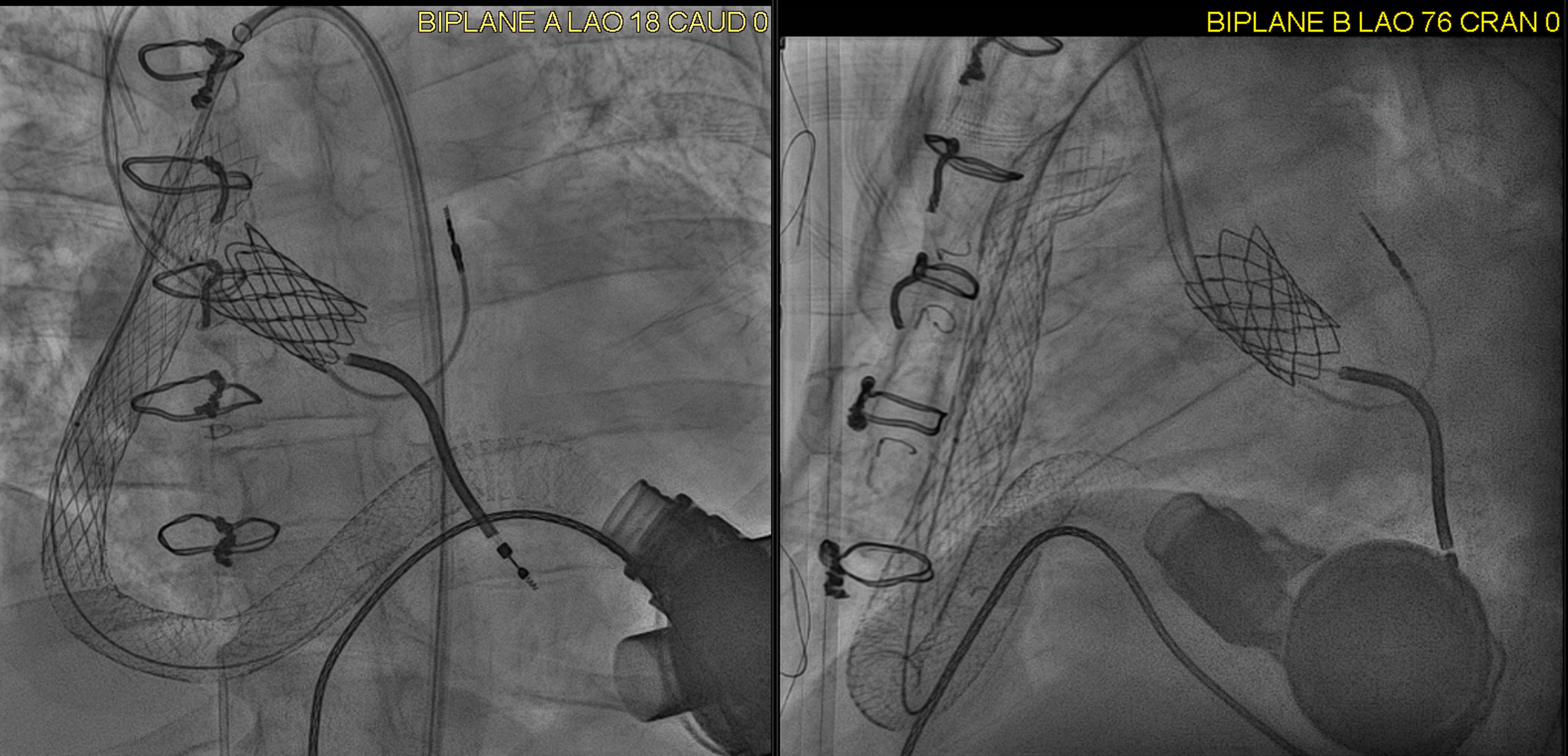
After a total of eight stents were placed, HeartMate 3 flow dropped significantly. Repeat angiography showed a sharper angle between the anastomosis of the outflow cannula and the ascending aorta, along with a contrast-opaque area suggestive of potential thrombus in that location (Figure 2, supplementary video). Stenting was thus extended up to the graft-aorta anastomosis with immediate improvement in HeartMate 3 flow (approximately 1 L/min increase). A total of 10 stents were deployed (GORE® VIABAHN®, Valeo® Biliary Stent, IntraStent® LD biliary stent, Palmaz® XL) (Figure 3). The patient experienced no complications and was discharged the following day. At the time of writing, the patient is 6 months post-procedure and remains clinically stable. He is maintained on warfarin therapy with a target PT-INR of 2.0–3.0. Follow-up imaging and clinical assessments have demonstrated preserved HeartMate 3 flow without evidence of recurrent extrinsic obstruction of the outflow graft, haemolysis, or thromboembolic events.
Lateral view angiography comparison of the outflow graft before and after initial stent placement. With the straightening of the outflow cannula, a sharper angle at the graft-aortic anastomosis site became apparent (arrow).

Final fluoroscopy at termination of the procedure. The stents show no significant gaps, with adequate overlap. A modest residual waist is present at the graft-aortic anastomosis.

Discussion
This case illustrates the technical feasibility and complexities involved in stent placement within the HM3 outflow graft to address multilevel extrinsic obstruction of the outflow graft. Key considerations included: (1) balancing HM3 flow to maintain adequate systemic blood pressure while minimising flow during balloon inflation for stent stability; (2) ensuring complete cannula coverage with adequate stent overlap, necessitating multiple stents; (3) avoiding guidewire compromise of pump function; and (4) managing the rigidity introduced by extensive stenting, altering angulation in the graft within the confined space.
Outflow graft obstruction can result from endoluminal factors (e.g., thrombus, graft-aorta anastomosis stenosis, fungal infections) or extrinsic factors (e.g., twisting, debris buildup, kinking, and mediastinal compression). Reference Goldstein, Kanwar and Cowger3 In this case, extrinsic obstruction of the outflow graft occurred at the bend relief and additional areas along the graft, along with new kinks. Extensive stenting was ultimately required from the bend relief to the graft-aorta anastomosis to relieve these obstructions. While surgical intervention, including outflow graft revision or replacement, was initially considered, it was deemed high-risk given the patient’s prior stroke and history of sternotomy. In retrospect, surgical shortening or rerouting of the outflow graft—such as directing it to the descending rather than the ascending aorta—might have mitigated the risk of anterior compression and kinking. Reference Kar, Delgado, rd and OH4 However, this alternative would have only been feasible in the context of surgical reintervention.
In d-TGA, the short distance and limited space between the anterior right ventricle and the sternum creates the substrate for an acute bend in the cannula. As a result, careful attention to stent overlap, together with application of stents with sufficient radial strength is the key to successful decompression. Any gaps remaining between stents are liable to result in recurrent stenosis. Placing narrower stents with smaller diameters is also acceptable for initial placement, as long as stent stability is maintained. However, redilation and alleviation of all stenoses along the graft is central to relieving the obstruction.
Conclusion
Careful preprocedural planning, and close intra-procedural communication ensured maintenance of cardiac output with reduction in ventricular assist devices flow for as little as possible. Complete overlap of stents dilated to an adequate diameter allowed decompression of the extrinsic obstruction of the outflow graft and safe completion of the procedure.
Supplementary material
The supplementary material for this article can be found at https://doi.org/10.1017/S1047951125100693.
Acknowledgements
We extend our gratitude to Dr Sitaramesh Emani for his invaluable clinical support and insights into the field of adult ventricular assist devices. We also wish to thank Ms. Katrina Fields and Ms. Mary Raffel for their exceptional dedication and contributions to patient care as ventricular assist devices coordinators.
Financial support
This research received no specific grant from any funding agency, commercial, or not-for-profit sectors.
Competing interests
Angela Lorts: Research grants from A.L. Abbott, Berlin Heart, Syncardia, and Abiomed. Consulting for A.L. Abbott.