Patent foramen ovale is known as a risk factor for paradoxical embolism and cryptogenic stroke. Reference Lee, Nam, Heo and Kim1 Patent foramen ovale closure is recommended for patients who are at risk for recurrent events. Reference Pristipino, Sievert and D’Ascenzo2
The standard technique of patent foramen ovale closure is established from the femoral vein and inferior vena cava, and the suitable standard interventional materials have been designed to approach the femoral vein.
On the other side, interventions from the jugular vein have been described in cases in which it is not suitable to pass through the inferior vena cava due to thrombus or filter in inferior vena cava, or when the inferior vena cava is congenitally absent.
Here, we present a case who underwent patent foramen ovale closure from the right jugular vein in which support had to be provided by a balloon anchoring to the pulmonary vein.
Case
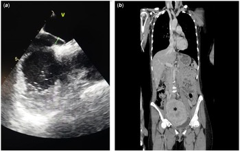
A 45-year-old female patient with an intramural 120 × 119 × 100 mm giant uterine myoma had a history of recurrent deep vein thrombosis and stroke. Echocardiography revealed a patent foramen ovale with a tunnel length of 16 mm and 1 mm left atrial and 4 mm right atrial width (Fig 1a). A bidirectional, left-to-right and right-to-left shunt, was detected on transesophageal echocardiography.
A patent foramen ovale was detected on transesophageal echocardiography with 16 mm in length and 1 mm left atrial and 4 mm right atrial width ( a ); the intramural 120 × 119 × 100 mm giant uterine myoma that caused occlusion in the inferior vena cava is marked with a black star. The inferior vena cava flow is not observed in this area on the computed tomography venous angiography ( b ).

On the computed tomography venous angiography, diffuse thrombus was detected in both iliac veins associated with inferior vena cava compression of the myoma. Furthermore, there were dominant subcutaneous visceral-portal venous collaterals which bypassed the hepatic veins and portal veins over the inferior vena cava (Fig 1b).
Hysterectomy was initially planned by gynaecology but due to the possible risk of embolisation of inferior vena cava thrombus and stroke after removal of the compressive mass during hysterectomy, first patent foramen ovale closure and then hysterectomy was decided in the Gynecology-Cardiology-Cardivascular Surgery council.
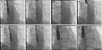
A 7F sheat was placed in the right internal jugular vein and a 6F a Medtronic Launcher 6Fr EBU catheter was used to approach the foramen ovale. Terumo wire was directed from the foramen ovale to the left atrium and approached to the pulmonary vein. Although the stiff wire passed into the left atrium, the catheter could not be passed to the left atrium, probably because of the tight left atrial ostium of the patent foramen ovale. A 0.014-inch guidewire was placed in the pulmonary vein through the 6F EBU catheter, and anchoring was performed with a 5.0 × 15 mm coronary balloon. With this anchor support, the EBU catheter could be passed to the left atrium. 0.0035-inch hydrophilic guidewire was replaced with stiff curved wire (Fig 2a-d).
The EBU catheter was directed to the patent foramen ovale (PFO) ( a ) and the Terumo wire was passed from the PFO to the left atrium (LA) ( b ) and then the wire was placed to the pulmonary vein (PV) ( c ), but the EBU catheter could not be passed to the LA because the left atrial ostium of the PFO was narrow and tight. Then, a 0.014-inch wire was sent to the PV ( c ) and the PV was anchored with a 5.0 × 15 mm coronary balloon over the wire; this procedure allowed the EBU catheter to pass to the LA ( d ). A stiff curved wire was placed through the EBU catheter to the LA ( e ), and the PFO occluder delivery system was passed through this wire ( f ). A 25-no Amplatzer PFO occluder device was brought over the PFO delivery system and first the left disc ( g ), then the right disc ( h ) was opened. All these procedures were performed under transesophageal echocardiography and fluoroscopy guidance before the device was released.

Delivery system of patent foramen ovale was advanced through the stiff curved wire. After the device’s left and right occluders were opened, it was observed that the flow via patent foramen ovale was stopped on the pre-operative transesophageal echocardiography (Fig 2e-h).
The occluder device position was confirmed on transesophageal echocardiography and fluoroscopy before the device was released. The occluder device was released and patent foramen ovale was successfully closed with the 25 no Amplatzer patent foramen ovale occluder (Abbott Medical, IL, United States) (Video 1).
The patient underwent the hysterectomy, after patent foramen ovale closure, during the same hospitalisation. After hysterectomy, the patient was followed up in the ICU due to the risk of any pulmonary thromboembolisation that could occur after the procedure. The patient was discharged with recovery without any problem.
Although the literature data are not clear on this subject, according to our clinical experience, the patient was planned to be treated with warfarin + clopidogrel + acetyl salicylic acid for 1 week after 1 week of warfarin + clopidogrel 6 months and warfarin for a total of 1 year.
The patient was asymptomatic at 6 months of control.
Discussion
Patients with patent foramen ovale and paradoxical embolism have a high prevalence of recurrent paradoxical embolism, ranging from 3.2 to 3.8% per year. Reference Mas and Zuber3 Transcatheter patent foramen ovale closure has been shown to be superior to medical therapy in patients with paradoxical embolism. Reference Martin, Sanchez and Doherty4,Reference Kitsios, Dahabreh, Abu Dabrh, Thaler and Kent5
The femoral vein approach for transcatheter patent foramen ovale closure is the most frequent way used by invasive cardiologists. Reference Martin, Sanchez and Doherty4 Deoxygenated blood flow from the inferior vena cava is directed through the foramen ovale by the Eustachian valve. There is an appropriate angle for a guide wire to cross into the left atrium between inferior vena cava and patent foramen ovale which is around 45 degrees. By this way, the patent foramen ovale delivery system inserted along the guidewire can usually reach the left atrium without difficulty, allowing deployment of the device. Reference Hascoet, Fraisse and Elbaz6
As in our patient, in cases where standard femoral vein access cannot be used due to the occlusion of inferior vena cava with thrombus, filter, compression, or congenital absence of inferior vena cava, the current literature mainly indicates the use of right jugular vein, but there are also rare cases where the hepatic vein was used. This approach is defined by paediatric interventional cardiologists. Reference Shim, Lloyd, Cho, Moorehead and Beekman7 The patent foramen ovale closure is performed without specific technical difficulty via transhepatic approach. But this technique needs gel foam or coils in the tract of approach through the liver to achieve haemostasis or it can cause peritoneal bleeding. Reference Hussain, Strumpf, Ghandforoush, Jamal and Diethrich8
Sader et al. presented a patent foramen ovale closure from the right jugular vein via a Boston Scientific Voda Left catheter. Reference Sader, De Moor, Pomerantsev and Palacios9 He et al. performed a patent foramen ovale closure from the right internal jugular vein via a Medtronic Launcher 6Fr EBU catheter. Reference He, Xue, Du, Xie, Wang and Zhang10 Hascoet et al. and Qintar et al. also presented cases in which patent foramen ovale closure was performed from RIJ using steerable catheters. Reference Hascoet, Fraisse and Elbaz6,Reference Qintar, Villablanca and Lee11
In light of the literature, intervention via right internal jugular vein has been described with different catheters, including steerable catheters for patients whose inferior vena cava intervention is not possible for different reasons, and there are case reports about transhepatic intervention even in adults. Reference Hussain, Strumpf, Ghandforoush, Jamal and Diethrich8 Due to the different anatomical structures of the patent foramen ovale, it is not always possible to pass the patent foramen ovale with VL or EBU catheters.
While steerable catheters that provide better support across the patent foramen ovale may be beneficial during the procedure, these catheters may not always be feasible due to difficulties in reaching these catheters and thick structures that are not available for small vascular or atrial sizes.
We successfully closed the patent foramen ovale via the right internal jugular vein access by balloon anchoring to pulmonary vein and passing through the patent foramen ovale to left atrium.
Conclusion
This is the first case that presented a balloon anchor support to the pulmonary vein method presented during the percutaneous closure of patent foramen ovale to deal with the venous access problem. Balloon anchoring is an easy and practical method, usually used by most invasive cardiologists during coronary interventions and may also be considered in patent foramen ovale closure when additional support is needed despite the use of steerable and more supported catheters.
Supplementary material
To view supplementary material for this article, please visit https://doi.org/10.1017/S104795112300063X
Acknowledgements
None.
Financial support
This research received no specific grant from any funding agency, commercial, or not-for-profit sectors.
Conflicts of interest
None.
Ethical standards
The authors assert that all procedures contributing to this work comply with the ethical standards of the relevant national guidelines on human experimentation (please name) and with the Helsinki Declaration of 1975, as revised in 2008, and all procedures were performed under the patient’s verbal and written approval.