Implications
Different stressors result in different responses from the hypothalamus–pituitary–adrenal (HPA) axis and stress can reduce reproductive performance in mammals. Considering the different roles of males and females in reproduction, it is reasonable to think that each sex has its own strategy for dealing with the effects of stress. There is abundant evidence that the gonads affect the way the HPA axis responds to stress. Understanding the regulatory pathways of sex steroid modulation of HPA activity will help to develop methods to reduce the impact of stress in animal husbandry and improve animal welfare.
Introduction
There is strong evidence that the gonads modulate the hypothalamus–pituitary–adrenal (HPA) axis. Sex steroids affect the HPA axis centrally, affecting the secretion of corticotropin-releasing hormone, arginine vasopressin and ACTH in rodents and primates (Burgess and Handa, Reference Burgess and Handa1992; Handa et al., Reference Handa, Burgess, Kerr and O’Keefe1994; Young, Reference Young1995). The stress response of the HPA axis of female rats is greater than that of male rats and gonadal steroid levels are in part responsible for these differences (Handa et al., Reference Handa, Burgess, Kerr and O’Keefe1994). This has been observed both under basal and stressful conditions in rats (Kitay, Reference Kitay1961; Viau and Meaney, Reference Viau and Meaney1991) and in primates (Uhart et al., Reference Uhart, Chong, Oswald, Lin and Wand2006). Oestrogens enhance ACTH secretion and testicular androgens suppress corticosterone secretion in the rat (Ogilvie and Rivier, Reference Ogilvie and Rivier1997). However, most reports have focused on the central effect of steroids (central nervous system, Burgess and Handa, Reference Burgess and Handa1992; Young, Reference Young1995; Viau and Meaney, Reference Viau and Meaney1996). Direct effects of oestrogens on adrenal corticosterone secretion have been observed in ovariectomised female rats (Figueiredo et al., Reference Figueiredo, Ulrich-Lai, Choi and Herman2007). In sheep, the response of the adrenal gland to ACTH administration – in terms of cortisol secretion – is also affected by sex: ewes secrete more cortisol than rams (Van Lier et al., Reference Van Lier, Pérez-Clariget and Forsberg2003a), which has also been found in vitro (Canny et al., Reference Canny, O’Farrell, Clarke and Tilbrook1999). Gonadectomy seems to have an inverse effect on cortisol levels after ACTH treatment in both sexes; differences were observed in intact rams and ewes, while the cortisol response in gonadectomised rams and ewes was similar (Van Lier et al., Reference Van Lier, Pérez-Clariget and Forsberg2003a), although this is not always observed (Tilbrook et al., Reference Tilbrook, Canny, Serapiglia, Ambrose and Clarke1999; Turner et al., Reference Turner, Canny, Hobbs, Bond, Clarke and Tilbrook2002). Therefore, sex differences in the stress response appear also to be expressed peripherally at the adrenal, and are probably partly due to the gonadal steroids. However, to our knowledge, no hormone replacement studies have been performed in sheep in order to test their effects on the adrenal gland.
Synthesis of cortisol in the adrenal cortex is induced by the binding of ACTH to its membrane receptor (melanocortin 2 receptor, MC2R), which activates adenylate cyclase, resulting in increased intracellular levels of cAMP, which in turn activates protein kinase. The activated protein kinase stimulates transcription of the steroid acute regulatory protein (STAR), which participates in the transport of cholesterol towards the inner mitochondrial membrane where the first enzymatic reaction of steroidogenesis occurs. This transfer of cholesterol from the outer to the inner mitochondrial membrane is the rate-limiting step in steroidogenesis (Manna et al., Reference Manna, Dyson and Stocco2009). Modulation of cortisol secretion by sex steroids would be exerted through their binding to a specific receptor in adrenocortical cells. Oestrogen receptors (ER) and androgen receptors (AR) have been found in the rat adrenal gland (ER: Cutler et al., Reference Cutler, Barnes, Sauer and Loriaux1978; AR: Calandra et al., Reference Calandra, Purvis, Naess, Attramadal, Djøseland and Hansson1978) and in the sheep adrenal gland (ER: Van Lier et al., Reference Van Lier, Meikle, Bielli, Akerberg, Forsberg and Sahlin2003b; AR: Gay et al., Reference Gay, Campbell, McNeilly and Duncan2007). Sex steroids do affect adrenocortical sensitivity to ACTH in rats (Atkinson and Waddell, Reference Atkinson and Waddell1997), but no reports in ruminants were found. We hypothesise that steroid treatment modulates adrenal response – in terms of cortisol secretion – to an exogenous ACTH treatment, and that the differences between cortisol secretion in male and female sheep are partly due to sex steroids (oestrogens and testosterone) acting directly on the gland. Furthermore, the sex steroids might affect the sensitivity of the adrenal gland to ACTH by modulating the concentration of MC2R, or they might affect precursor availability for cortisol production by modulating STAR abundance (Van Lier et al., Reference Van Lier, Meikle, Bielli, Akerberg, Forsberg and Sahlin2003b). Thus, in the first study we compared the cortisol response to ACTH in gonadectomised sheep with or without sex steroid replacement. In the second study we measured the relative expression of ERS1, AR, MC2R and STAR mRNAs in adrenal glands of gonadectomised male and female sheep either with or without sex steroid replacement (Testosterone Cyclopentilpropionate (TC) and Oestradiol Benzoate (EB), respectively).
Material and methods
Working conditions
All of the sheep were of the Corriedale breed and were accustomed to frequent handling. Before the experiments the animals were kept at pasture with free access to water. The experiments were carried out in autumn (May, breeding season, Southern Hemisphere). Both experimental protocols were approved by the Honorary Committee of Animal Experimentation of the University of Uruguay (Comisión Honoraria de Experimentación Animal, CHEA, protocols No. 7101/05 and 7101/07).
Experiment 1
Six adult rams (mean BW±s.e.m.: 70.6±1.52 kg) and four adult ewes (mean BW±s.e.m.: 42.5±1.53 kg) were used. Gonadectomy was performed 4 weeks before the experiment. On the day before each of two frequent blood-sampling periods, each animal’s neck was shorn and fitted with an indwelling jugular catheter for the collection of blood samples into heparinised tubes. The samplings took place in a shed with the animals in small pens in pairs of the same sex. The animals were all in visual contact with each other and were fed with hay and had free access to water. During these periods, blood samples were taken every 15 min for 4 h.
On the day of the first frequent blood sampling each animal received 0.5 mg of an ACTH analogue (a polypeptide based on the first 24 amino acids of the 39 occurring in the natural molecule: Tetracosactid, Synacthen Depot®, 1 mg/ml; Ciba Geigy, Basel, Switzerland) i.v., immediately after the sample at 1 h from the beginning of the trial, which was considered time 0 (Van Lier et al., Reference Van Lier, Pérez-Clariget and Forsberg2003a). Four days later the males received 100 mg of TC i.m. (in oil vehicle, 100 mg/ml, Laboratorios Dispert S.A., Montevideo, Uruguay; recommended dose for androgenisation of wethers for oestrus detection) and the females received 2.5 mg of EB i.m. (in oil vehicle, 5 mg/ml, Laboratorios Dispert S.A.; half of the minimum therapeutic dose recommended). One week after the first frequent blood sampling a second identical sampling was performed (72 h after TC or EB administration) with ACTH administration at 0 h (1 h after the beginning of the trial). Sampling began at 1000 h on both occasions. In order to establish the effectiveness of the TC and EB treatment, samples for analysis of testosterone or 17β-oestradiol were taken at 0, 24, 48 and 72 h relative to steroid replacement.
Experiment 2
Eight adult rams (mean BW±s.e.m.: 70.6±2.80 kg) and eight adult ewes (mean BW±s.e.m.: 43.1±2.70 kg) were used. Gonadectomy was performed at least 3 weeks before the experiment. Four rams received 100 mg of TC i.m. (in oil vehicle, 100 mg/ml) and four females received 0.5 mg of EB i.m. (in oil vehicle, 5 mg/ml; 1/10 of the minimum therapeutic dose recommended). Blood samples were taken at 0, 12, 24, 48 and 72 h relative to steroid replacement in order to establish the effectiveness of the TC and EB treatments and the animals were thereafter slaughtered in order to obtain their adrenal glands. Slaughter was performed under barbiturate general anaesthesia (1 g of thiopental sodium) and the adrenal glands were sampled within 15 min. Adrenal glands were weighed and then cut in half on ice. The tissues were packed and immediately frozen in liquid nitrogen and then stored at −80°C.
Hormone assays
Plasma was obtained by centrifugation of the samples immediately after collection. Approximately 3 ml of plasma was taken from each sample and then stored at −20°C within 2 h after collection. All hormone concentrations were determined using previously validated radioimmunoassays (Coat-A-Count radioimmunoassay kits; Diagnostic Products Corporation, Los Angeles, CA, USA).
Cortisol was analysed in every half an hour sample for the first hour and then in every 15 min sample for 2 h and then each half hour until the end of experiment 1. All of the samples were analysed in the same assay. Quality control samples containing endogenous cortisol (low 36.9 nmol/l, medium 154.9 nmol/l and high 296.0 nmol/l) were assayed in duplicates at both the beginning and end of the assay. The intra-assay CV were 8.6%, 3.7% and 5.2%, respectively. The analytical detection limit of the assay was 10.8 nmol/l.
Testosterone was determined in the samples of the rams in order to evaluate the effectiveness of the replacement treatment. For experiment 1 the samples at 0, 24 and 48 h relative to steroid replacement, as well as one sample before and after ACTH administration were analysed, and for experiment 2 all of the samples of the rams were analysed. The intra-assay CV for two control samples (4.0 and 24.1 nmol/l), assayed in duplicates at the beginning and end of each assay, were 9.9% and 6.1%, respectively. The corresponding inter-assay CV were 12.9% and 5.7%, respectively. The analytical detection limit of the assay was 0.4 nmol/l.
In the samples of the ewes 17β-oestradiol was determined in order to evaluate the effectiveness of the replacement treatment. For experiment 1 the samples at 0, 24 and 48 h relative to steroid replacement, as well as one sample before and after ACTH administration were analysed, and for experiment 2 all of the samples of the ewes were analysed. The samples of each experiment were analysed in the same assay. The intra-assay CV for two control samples (low 14.4 pmol/l and medium 45.3 pmol/l) were 23.6% and 6.2%, respectively. The analytical detection limit of the assay was 2.6 pmol/l.
Real-time reverse transcription PCR (RT-PCR)
In order to quantify mRNA expression of ERS1, AR, MC2R and STAR using real-time RT-PCR, total RNA was extracted using TRIZOL (Invitrogen, Life Technologies, Carlsbad, CA, USA), precipitated with lithium chloride to remove cDNA synthesis inhibitors, and treated with DNAse to remove contaminating DNA (DNA-Free kit; Applied Biosystems/Ambion, Austin, TX, USA). The concentration of RNA was determined by measuring absorbance at 260 nm, the purity of all RNA isolates was assessed from 260/280 absorbance ratio and the integrity by electrophoresis (1% agarose gel). All samples presented A260/280 ratios between 1.8 and 2.0.
For each sample, cDNA was synthesised by reverse transcription using the SuperScript III First-Strand Synthesis System Kit (Invitrogen) with random primers and 1 μg of total RNA added as a template. Primers to specifically amplify cDNA of MC2R, ERS1 and STAR, as well as the housekeeping genes hypoxanthine phosphoribosyltranspherase (HPRT) and ribosomal protein L19 (RPL19) were obtained from the literature (Schams et al., Reference Schams, Kohlenberg, Amselgruber, Berisha, Pfaffl and Sinowatz2003; Myers et al., Reference Myers, Hyatt, Mlynarczyk, Bird and Ducsay2005; Chen et al., Reference Chen, Green, Antoniou, Ealy, Mathialagan, Walker, Avalle, Rosenfeld, Hearne and Roberts2006; Jaquiery et al., Reference Jaquiery, Oliver, Bloomfield, Connor, Challis and Harding2006; Carriquiry et al., Reference Carriquiry, Weber, Fahrenkrug and Crooker2009) and further primer information is given in Supplementary Table S1. Primers to amplify cDNA of AR were designed based on ovine nucleotide sequences available from NCBI (http://www.ncbi.nlm.nih.gov/) using Primer Express 3.0 software from Applied Biosystems. Before use, primer product size (1% agarose gel separation) was determined to ensure that primers produced the desired amplification products (results not reported). Real-time PCR reactions were performed using 10 µl SYBR green (Quantimix; Biotools, Madrid, Spain) equal amounts (200 nM) of forward and reverse and 3 µl diluted cDNA (1 : 7.5 in RNase/DNase free water) in a final volume of 20 µl. Samples were analysed in duplicate in a 72-disk Rotor-GeneTM 6000 (Corbett Life Sciences, Sydney, Australia). Standard amplification conditions were 5 min at 95°C and 40 cycles of 10 s at 95°C, 45 s at 60°C and 20 s at 72°C. Each disk included a pool of total RNA from adrenal samples analysed in triplicate to be used as the basis for the comparative expression results (exogenous control) and duplicate tubes of water (non-template control). Gene expression was measured by relative quantification to the exogenous control and normalised to the mean expression (geometric mean) of the endogenous control genes (HPRT and RPL19). Dilution curves were developed for each primer from the quantification of 6.25, 12.5, 25, 100 and 200 ng of transcribed RNA and amplification efficiency was estimated by linear regression (Supplementary Table S1). The intra- and inter-assays CV were <7.6% and 13.1%, respectively.
Statistical analysis
Data were analysed using the SAS Systems programs (SAS 9.2 V; SAS Institute Inc., Cary, NC, USA). Cortisol data (experiment 1) were analysed as repeated measures, using the MIXED procedure. The model included the effect of sex (male or female) and steroid replacement (with or without) within sex, time (repeated measure within subject) and their interactions as fixed effects and animal (subject) as a random effect. The first-order autoregressive covariance structure (AR(1)) and the Kenward–Rogers procedure to adjust the degrees of freedom of denominator were specified. Gene expression data, animal BW, absolute and relative tissue weight (experiment 2) were analysed using the MIXED procedure with a model that included the fixed effects of sex (male or female) and steroid replacement (with or without) within sex. Mean separation was performed using the Tukey test. Correlation coefficients used to describe relationships between gene expressions were estimated using the CORR procedure. The level of significance was considered to be P⩽0.05, and tendency as 0.5<P<0.10.
Results
Testosterone and 17β-oestradiol
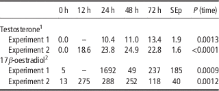
Testosterone was not detected in any of the samples of the non-treated castrated rams. Steroid replacement with TC effectively raised testosterone levels in all of the castrated rams (Table 1). Testosterone concentrations in the samples after ACTH administration were not different from the sample taken just before ACTH administration (data not shown). In all of the ovariectomised ewes 17β-oestradiol was low, but detectable. EB administration increased 17β-oestradiol levels in plasma of all of the ewes (Table 1). The concentrations of 17β-oestradiol in the samples after ACTH administration were not different from the sample taken just before ACTH administration (data not shown).
The effect of steroid replacement (immediately after the 0 h sample) on mean concentrations of testosterone (nmol/l) in Testosterone Cyclopentilpropionate (TC)-treated castrated rams and concentrations of 17β-oestradiol (pmol/l) in Oestradiol Benzoate (EB)-treated ovariectomised ewes either before ACTH administration (experiment 1) or before slaughter (experiment 2)

–=no sample; SEp=pooled standard error.
1 The same dose of TC (100 mg/ram) was used in both experiments. The analytical detection limit of the assay was 0.4 nmol/l.
2 Different doses of EB per ewe were used in experiments 1 and 2 (2.5 and 0.5 mg of EB, respectively). The analytical detection limit of the assay was 2.6 pmol/l.
Cortisol in ACTH trials
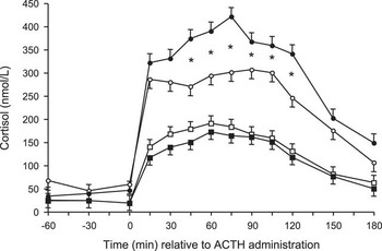
The main effects sex (P<0.0001), steroid replacement(sex) (P=0.024) and time (minutes relative to ACTH treatment) (P<0.0001), were significant, as were the interactions sex×time (P<0.0001) and time×steroid replacement(sex) (P=0.0073). Before ACTH administration cortisol levels were low, while after ACTH administration they were significantly higher in all of the animals and did not reach pre-treatment levels before the end of the experiment (Figure 1). The rams had lower cortisol levels than the ewes at time 0 (P=0.0321) and after ACTH administration (P<0.0002), but TC did not affect these cortisol levels. In the EB-treated ewes, however, the cortisol levels after ACTH administration were higher as compared with non-treated ewes (P=0.0097) (Figure 1).
The effect of sex and steroid replacement on mean (±s.e.m.) cortisol levels (nmol/l) before and after administration (i.v.) of an ACTH analogue at time 0 in gonadectomised sheep (dots are ewes and squares are rams; open symbols are non-steroid-treated sheep and solid symbols are steroid-treated sheep). The cortisol response was significantly different between rams and ewes and stars indicate significant differences between Oestradiol Benzoate treated and control ewes (P<0.05).

BW, absolute and relative adrenal weight
BW was different between sexes (P<0.0001), but steroid replacement(sex) had no effect (P=0.6777) (Table 2). The absolute adrenal weight was not different between sexes (P=0.3811) and steroid replacement(sex) had no effect (P=0.8304) (Table 2). The relative adrenal weight was different between sexes (P=0.0002), but steroid replacement(sex) had no effect (P=0.8877) (Table 2).
The effect of sex and steroid replacement on mean BW (kg), absolute adrenal weight (g) and relative adrenal weight (mg adrenal tissue/kg BW) in gonadectomised ewes (control and replaced with Oestradiol Benzoate, EB) and gonadectomised rams (control and replaced with Testosterone Cyclopentilpropionate, TC) (experiment 2)

SEp=pooled standard error.
a,bValues within a row with different superscripts differ significantly at P<0.05.
MC2R, AR, ERS1 and STAR mRNA
The effect of sex on the relative amount of MC2R transcript was not significant while the effect of steroid replacement(sex) tended to be significant (P=0.0558). Gonadectomised rams treated with TC presented greater MC2R mRNA expression than males without the steroid replacement and ewes treated with EB (Table 3). For the relative amount of the AR transcript the effect of sex tended to be significant (P=0.0844) while the effect of steroid replacement(sex) was significant (P=0.0421). Gonadectomised rams with TC had greater AR mRNA expression than non-replaced rams and ewes treated with EB. Gonadectomised ewes treated with EB tended to present lower AR mRNA than the ewes without steroid replacement (P=0.0881) (Table 3). No significant effects were found on the relative amount of ERS1 and STAR transcripts (Table 3). However, gonadectomised rams with TC had greater ERS1 mRNA expression than ewes treated with EB. The transcripts of AR and MC2R were highly, positively and significantly correlated (r=0.95, P<0.0001) and ERS1 was highly, positively and significantly correlated with AR (r=0.79, P=0.0008) and with MC2R (r=0.80, P=0.0007). The STAR transcript was also positively correlated with the receptor transcripts, although to a lesser extend (ERS1: r=0.60, P=0.0244; AR: r=0.58, P=0.0302; and MC2R: r=0.52, P=0.0548). In rams TC was positively correlated (r=0.75) with the AR and MC2R transcripts (P=0.0538 and 0.0522, respectively), and in ewes EB was negatively correlated (r=−0.78) with the AR transcript (P=0.0384).
The effect of sex and steroid replacement on relative expression of MC2R, AR, ERS1 and STAR mRNA in gonadectomised ewes (control and replaced with Oestradiol Benzoate, EB) and gonadectomised rams (control and replaced with Testosterone Cyclopentilpropionate, TC) (experiment 2)

AR=androgen receptor; ERS1=estrogen receptor α; MC2R=melanocortin 2 receptor (ACTH receptor); STAR=steroid acute regulatory protein; SEp=pooled standard error.
a,bValues within a row with different superscripts differ significantly at P<0.05, † P=0.0881.
Discussion
Steroid replacement
Before steroid replacement testosterone was not detectable in the blood samples of the rams indicating successful castration. However, the ovariectomised ewes had low levels of 17β-oestradiol, which has been observed (Adams et al., Reference Adams, Atkinson, Hoskinson, Abordi, Briegel, Jones and Sanders1990; Van Lier et al., Reference Van Lier, Meikle, Bielli, Akerberg, Forsberg and Sahlin2003b) and suggests an extragonadal source of 17β-oestradiol (or its precursors), possibly the adrenal cortex. Both EB and TC were effective in increasing 17β-oestradiol and testosterone concentrations, respectively, in all of the treated animals. The levels of 17β-oestradiol in the animals of experiment 1 were pharmacological and the dose of EB was therefore reduced in experiment 2, resulting in lower, yet still pharmacological levels of 17β-oestradiol in those animals. Testosterone levels of the animals in both experiments were within the physiological range, although by intramuscular administration the active compound is usually released into the bloodstream at a relatively steady rate and not in discrete pulses, as occurs in intact rams (Sanford et al., Reference Sanford, Palmer and Howland1977).
Cortisol response to ACTH
The differences in cortisol response to ACTH in gonadectomised sex steroid replaced sheep confirm our previous findings of sex differences in cortisol secretion in intact sheep (Van Lier et al., Reference Van Lier, Pérez-Clariget and Forsberg2003a) and expand these findings as in the present study we found differences in cortisol levels of gonadectomised sheep. This is in contrast with earlier studies (Tilbrook et al., Reference Tilbrook, Canny, Serapiglia, Ambrose and Clarke1999; Turner et al., Reference Turner, Canny, Hobbs, Bond, Clarke and Tilbrook2002) where no difference in cortisol response was observed between gonadectomised male and female sheep. In our earlier work (Van Lier et al., Reference Van Lier, Pérez-Clariget and Forsberg2003a), cortisol levels of the gonadectomised rams and ewes were in between the levels of the intact animals and tended to be different (P=0.0701). However, differences in cortisol secretion in gonadectomised sheep have been found (Broadbear et al., Reference Broadbear, Pierce, Clarke and Canny2005), which were not reverted by sex steroid replacement, although the ewes were replaced with luteal phase levels of progesterone and oestrogens, which means oestrogens were at nadir. In rats, gonadectomy reduces or eliminates sexual differences in HPA function (Nowak et al., Reference Nowak, Neri, Nussdorfer and Malendowicz1995). This discrepancy in the effect of gonadectomy on cortisol secretion in sheep suggests that other factors are involved in the cortisol response to ACTH administration. Sex steroids present during the perinatal period can organise neuronal substrates, resulting in lifelong alterations in endocrine function (McCormick et al., Reference McCormick, Furey, Child, Sawyer and Donohue1998). This might also cause permanent ‘organisational’ differences in peripheral tissues such as the adrenal cortex, although it is not clear why the differences in cortisol secretion in gonadectomised male and female sheep are not always observed. On the other hand, life cycle stage of the animals also modulates cortisol secretion (Korte et al., Reference Korte, Koolhaas, Wingfield and McEwen2005) and might conceal differences in gonadectomised ewes and rams.
Hormone replacement affected the ACTH-induced cortisol secretion only in ewes. EB increased the cortisol response to ACTH in female sheep, confirming our hypothesis that oestrogens are involved in the regulation of the response of the adrenal cortex to ACTH, at least at the dose of EB used in our experiment. The effect of oestrogens on the secretion of cortisol has been described in rodents since the 1960s (Kitay, Reference Kitay1961) and later the presence of ER in the adrenal cortex of rats and primates was demonstrated (Cutler et al., Reference Cutler, Barnes, Sauer and Loriaux1978; Hirst et al., Reference Hirst, West, Brenner and Novy1992). Since then, ER has also been described in the sheep adrenal gland (Van Lier et al., Reference Van Lier, Meikle, Bielli, Akerberg, Forsberg and Sahlin2003b), supporting a possible role for oestrogens as modulators of cortisol secretion.
Although in our experiment TC did not modify the cortisol response to ACTH in the rams, it cannot be ruled out that this might be due to the type of steroid replacement leading to sustained testosterone levels and not discrete pulses (Sanford et al., Reference Sanford, Palmer and Howland1977). However, Broadbear et al. (Reference Broadbear, Pierce, Clarke and Canny2005) did not detect differences in cortisol response after HPA stimulation of castrated rams with or without testosterone implant replacement (mean testosterone concentrations 6.11 nmol/l; lower than in the present study). Androgen receptors have been found in adrenal glands from rats (Calandra et al., Reference Calandra, Purvis, Naess, Attramadal, Djøseland and Hansson1978; Bentvelsen et al., Reference Bentvelsen, McPhaul, Wilson, Wilson and George1996), monkeys (Hirst et al., Reference Hirst, West, Brenner and Novy1992) and sheep (Gay et al., Reference Gay, Campbell, McNeilly and Duncan2007), indicating that androgens can act on the tissue. Van Lier et al. (Reference Van Lier, Pérez-Clariget and Forsberg2003a) reported that while ovariectomised ewes had lower cortisol levels than the intact ewes, castration of rams did not affect cortisol secretion compared with the intact rams, suggesting that cortisol secretion may be regulated by oestrogens – but not testosterone – at the adrenal cortex. However, in other species, androgens have been reported to inhibit corticosteroid secretion (rodents: Handa et al., Reference Handa, Burgess, Kerr and O’Keefe1994; Ogilvie and Rivier, Reference Ogilvie and Rivier1997; McCormick et al., Reference McCormick, Furey, Child, Sawyer and Donohue1998; cattle: Verkerk and Mcmillan, Reference Verkerk and Mcmillan1997), probably through action on other parts of the HPA axis.
Adrenal weight
The absolute adrenal weight did not show sex differences and confirms our earlier observations (Van Lier et al., Reference Van Lier, Meikle, Bielli, Akerberg, Forsberg and Sahlin2003b). However, when the adrenal weight is related to the live weight, ewes have more adrenal mass per unit of live weight than the rams. Absolute and relative adrenal weights seem not to be affected by gonadectomy (Canny et al., Reference Canny, O’Farrell, Clarke and Tilbrook1999; Van Lier et al., Reference Van Lier, Meikle, Bielli, Akerberg, Forsberg and Sahlin2003b), which would be confirmed by our results since treatment with EB or TC were without effect. Female rats had 30% more adrenal mass than male rats, and this difference appeared after sexual maturation (Rifka et al., Reference Rifka, Cutler, Sauer and Loriaux1978). In that study gonadectomised hypophysectomised rats were used and oestrogens did not affect the adrenal weight of female rats, while testosterone induced a decrease in the adrenal weight of male rats. However, Bentvelsen et al. (Reference Bentvelsen, McPhaul, Wilson, Wilson and George1996) did not find differences in adrenal weight of castrated androgen-replaced male rats. In sheep it seems that the sex steroids do not have trophic effects on the adrenal glands. It is not clear when the differences in relative adrenal weight appear in sheep: before birth (as an organisational effect of sex) or after puberty, as in rats, due to gonadal steroid modulation.
Adrenal ERS1, AR, MC2R and STAR mRNA
This study confirms earlier observations of ERS1 presence in the sheep adrenal gland (Van Lier et al., Reference Van Lier, Meikle, Bielli, Akerberg, Forsberg and Sahlin2003b) and confirms other reports on the presence of AR (Gay et al., Reference Gay, Campbell, McNeilly and Duncan2007). No differences were found in AR or ERS1 mRNA expression in gonadectomised rams and ewes. Similarly, Van Lier et al. (Reference Van Lier, Meikle, Bielli, Akerberg, Forsberg and Sahlin2003b) did not find differences in ERS1 transcript (measured by solution hybridisation) in the adrenals of intact and gonadectomised ewes and rams, while the ER content (measured by ligand-binding assay) differed between males and females and intact and gonadectomised sheep. Thus, data suggest that steady state levels of receptor protein are regulated at other levels of protein synthesis (e.g. efficiency of translation).
Sex steroid replacement was successful in inducing differences in adrenal ERS1 and AR mRNA expression, confirming a role for the sex steroids in regulating tissue sensitivity to hormones (e.g. hormone receptors) in the adrenal gland as has been reported for other tissues (Spencer and Bazer, Reference Spencer and Bazer1995). EB replacement tended to reduce the relative expression of AR mRNA in ewes and TC increased the relative expression of AR mRNA in rams. The increase of AR mRNA expression in the adrenal gland by androgens has also been observed in the male rat (Bentvelsen et al., Reference Bentvelsen, McPhaul, Wilson, Wilson and George1996). A down regulation of steroid receptors (ER and progesterone receptor-binding activity) by oestrogen treatment has been reported in sheep uteri (Meikle et al., Reference Meikle, Forsberg, Sahlin, Masironi, Tasende, Rodriguez-Pinon and Garófalo2000), and the authors suggested that this down regulation is a mechanism to limit hormone action in the target cells. An oestrogen down regulation of ERS1 expression was also found in rat uteri, and the regulation is dose dependent, as demonstrated by the stimulating or inhibitory effects of low or high doses of oestrogens, respectively (Medlock et al., Reference Medlock, Forrester and Sheehan1994).
EB increased the cortisol response to ACTH in female gonadectomised sheep, but no effect of TC was found in male sheep. Several mechanisms have been suggested by which sex steroids might affect adrenal steroidogenesis such as adrenocortical sensitivity, steroid precursor availability or enzyme induction (Van Lier et al., Reference Van Lier, Meikle, Bielli, Akerberg, Forsberg and Sahlin2003b). In this study we have looked into the first two of these mechanisms. The most straightforward mechanism would be for the sex steroids to affect adrenocortical sensitivity to ACTH (Atkinson and Waddell, Reference Atkinson and Waddell1997). A stimulating action of oestrogens and an inhibiting action of testosterone on the adrenal have been previously proposed in rats (Kitay, Reference Kitay1963; Nowak et al., Reference Nowak, Neri, Nussdorfer and Malendowicz1995). Therefore, one of our target genes in experiment 2 was of the ACTH receptor (MC2R). Contrary to what we expected to find based on the cortisol results of experiment 1 (increased after EB and no change after TC), we found no differences in MC2R expression in EB-treated ewes and higher MC2R expression in TC-treated rams. However, adrenal sensitivity depends on actual MC2R protein content and even if mRNA is frequently used as a predictor of protein content it may not always present a direct relationship as has been described before (Meikle et al., Reference Meikle, Forsberg, Sahlin, Masironi, Tasende, Rodriguez-Pinon and Garófalo2000), thus, post-transcriptional events might affect receptor activation and/or receptor turnover. Although the time between steroid replacement and adrenal sampling was determined to allow for protein synthesis and has been used in other studies (Clark et al., Reference Clark, Schrader and O’Malley1992; Meikle et al., Reference Meikle, Tasende, Rodríguez and Garófalo1997, Reference Meikle, Forsberg, Sahlin, Masironi, Tasende, Rodriguez-Pinon and Garófalo2000), it cannot be ruled out that a critical window was missed.
Steroidogenesis could also be modulated by influencing steroid precursor (cholesterol) availability to the mitochondrial enzymes. The STAR protein participates in the transport of cholesterol towards the inner mitochondrial membrane where the first enzymatic reaction of steroidogenesis occurs. This transfer of cholesterol from the outer to the inner mitochondrial membrane is the rate-limiting step in steroidogenesis (Manna et al., Reference Manna, Dyson and Stocco2009). Both STAR gene expression and transcription are regulated by protein kinase activation in response to ACTH stimulation of cAMP (Dyson et al., Reference Dyson, Kowalewski, Manna and Stocco2009). In our study, we have found no evidence that sex or sex steroids differentially regulate STAR expression, since no difference in relative abundance of STAR mRNA was observed. However, the amount of STAR protein is primarily regulated by transcription, which then also regulates steroidogenesis (Hiroi et al., Reference Hiroi, Christenson and Strauss2004). Therefore, the possibility exists that the observed sex differences in cortisol secretion post-ACTH administration might be due to differences in STAR translation rate (caused by the sex steroids) and thus its availability either limiting or stimulating steroidogenesis.
The high and positive correlation between the transcripts is indicative of a regulatory role of the sex steroid on the adrenal gland. TC showed a high and positive correlation with the transcripts of AR and MC2R; EB showed a high and negative correlation with the transcript of AR. This contrast between a positive correlation between TC and AR and a negative one for EB and AR further supports the concept of opposite adrenal regulation of testosterone and 17β-oestradiol. However, while the sheep adrenal gland expresses the receptor proteins for both 17β-oestradiol and testosterone, and therefore may be subject to sex steroid modulation, the underlying mechanisms remain unresolved.
Conclusions
Gonadectomised steroid replaced ewes and rams in this study showed the most divergent cortisol profiles as well as the most divergent relative ERS1, AR and MC2R mRNA expression, confirming a modulatory role of the sex steroids on cortisol secretion acting directly on the adrenal glands. In ewes, 17β-oestradiol modulated cortisol secretion by increasing the response to exogenous ACTH administration, but only tended to decrease the relative adrenal AR expression in ovariectomised ewes. A role for testosterone in influencing cortisol secretion in castrated rams was not confirmed, however, it did increase the relative adrenal MC2R and AR expression compared with non-steroid replaced castrated rams.
Acknowledgements
The authors thank Nelson Villegas, Daniel Agüero, Dana Montedónico, Alejandra Carbone, Angélica de Larrobla, Carolina Chiesa, Hugo Petrocelli, Stella Lanzzeri, Andrea Fernández, Beatríz Carracelas, Alejandro Castillo, Carlos Pérez, Andrés López for their help with the field work, Oscar Bentancur for statistical assistance, and Yerú Pardiñas and Enrique Rodríguez Grasso for donating part of the animals. Elize van Lier received a grant from the Uruguayan Ministry of Education and Culture, DINACYT, Fondo Profesor Clemente Estable (10083), Uruguay.