Achyrocline satureioides (Lam.) DC., Asteraceae, popularly known as macela, is an annual, aromatic herb native to South America (Retta et al., Reference Retta, Dellacassa, Villamil, Suárez and Bandoni2012). The biological activities of macela, such as antimicrobial, cytoprotective, antioxidant, and anti-inflammatory, have been frequently attributed to the abundant flavonoids found in its inflorescences, especially quercetin, luteolin, and 3-O-methylquercetin (Arredondo et al., Reference Arredondo, Blasina, Echeverry, Morquio, Ferreira, Abin-Carriquiry, Lafon and Dajas2004; Retta et al., Reference Retta, Dellacassa, Villamil, Suárez and Bandoni2012). Owing to its multiple biological activities, the use of macela is attracting interest across a range of research areas, including veterinary sciences (Both et al., Reference Both, Avancini, Spaniol and Petrovick2016; Pinheiro Machado et al., Reference Pinheiro Machado, Veleirinho, Honorato and Kuhnen2020). For example, the high antimicrobial activity found in macela extract makes it a promising alternative for the therapy of bovine mastitis (Pinheiro Machado et al., Reference Pinheiro Machado, Veleirinho, Honorato and Kuhnen2020).
Treatment of bovine mastitis typically requires the use of antimicrobials effective against pathogens causing the disease (Langoni et al., Reference Langoni, Salina, Oliveira, Junqueira, Menozzi and Joaquim2017). However, as mastitis is commonly treated through the intramammary application of antimicrobials, it is important to evaluate the possible toxic effects of the therapy on epithelial cells (Kalayou et al., Reference Kalayou, Haileselassie, Gebre-egziabher, Tiku, Sahle, Taddele and Ghezu2012) as well as its ability to permeate into mammary tissue. Damage to epithelial cells may induce not only the formation of connective tissue in the affected area, with associated interalveolar fibrosis of the mammary tissue, loss of secretory function and alteration in the quantity and quality of the milk produced, but also cause proinflammatory effects, which can worsen the clinical condition of the animal (Radostits et al., Reference Radostits, Gay, Hinchcliff and Constable2007; Fiordalisi et al., Reference Fiordalisi, Honorato and Kuhnen2019).
Antimicrobials with low permeation power may spread inadequately throughout the mammary gland, impeding the effects on all affected areas. This may contribute to the frequent recurrence of cases of mastitis, which can become chronic through the application of ineffective treatments. Furthermore, Staphylococcus aureus, a bacterium often isolated from bovine mastitis, is associated with the ability to form biofilms as a survival strategy. This ability hinders the effects of antimicrobials since they are unable to penetrate the biofilm matrix (Donlan and Costerton, Reference Donlan and Costerton2002). Nanoemulsion systems are, however, recognized as promoting the permeation of compounds across biological membranes by the reduced size of the droplets, fluidity, and capacity of interaction with the epithelium (Rai et al., Reference Rai, Mishra, Yadav and Yadav2018).
Considering the antimicrobial potential and evidence of low cytotoxicity of macela (Arredondo et al., Reference Arredondo, Blasina, Echeverry, Morquio, Ferreira, Abin-Carriquiry, Lafon and Dajas2004; Pinheiro Machado et al., Reference Pinheiro Machado, Veleirinho, Honorato and Kuhnen2020), along with the greater permeation power of nanoemulsified systems, the present study adressess the hypothesis that macela (Achyrocline satureioides Lam. DC.) extract nanoparticles promote protection of bovine mammary epithelial cells and increase the permeation of flavonoid compounds through the mammary glandular epithelium.
Materials and methods
Macela samples and extract preparation
Macela extract was prepared by macerating inflorescences with 80% ethanol (v/v) (1 : 60, w/v). Subsequentely, the extract was vacuum filtered and the organic solvent removed with a rotary evaporator at 60°C. Immediately after solvent removal, the aqueous phase of the extract was used to prepare the nanoemulsions. Details are provided in the online Supplementary File.
Preparation of nanoemulsions of flaxseed oil loaded with macela extract (NE-ML)
Nanoemulsions loaded with macela extract (NE-ML) and their respective blank-nanoemulsions (without extract: NE-BL) were prepared according to Pinheiro Machado et al. (Reference Pinheiro Machado, Veleirinho, Honorato and Kuhnen2020). To obtain NE-ML, the macela extractive solution, equivalent to 250 mg of extract, was slowly poured into an oily phase consisting of different concentrations of flaxseed oil (1, 5, and 10%, w/v) and Tween 80 surfactant (0.2, 1, and 5%, w/v). Table S1 of the Supplementary File shows the preparation conditions of the different formulations.
Determination of macela phenolic compounds by high-performance liquid chromatography with diode-array detection (HPLC/DAD)
Analysis of the phenolic composition of the macela extract was performed by HPLC/DAD according to Pinheiro Machado et al. (Reference Pinheiro Machado, Veleirinho, Honorato and Kuhnen2020). Details are provided in the online Supplementary File.
Viability of MAC-T cells
MAC-T cell suspension was plated in a 96-well microplate (100 μl/well), followed by incubation (24 h) in culture conditions for adherence. Subsequently, varying concentrations (10–0.3%, v/v) of NE-MLs and their respective NE-BLs were added for 24 h, and cell viability was determined based on the MTT (3-(4,5-dimethylthiazol-2-yl)-2,5-diphenyltetrazolium bromide; 0.5 mg/ml) method. Flaxseed oil and free macela extract were also evaluated in concentrations ranging from 4000 to 12 μg/ml. Cells cultivated in untreated DMEM were considered 100% viable. The experiments were performed in triplicate. Details are provided in the online Supplementary File.
Cell death analysis- annexin V staining and flow cytometric analysis
Annexin V coupled with FITC is typically used in conjunction with propidium iodide (PI) to identify different stages of apoptotic and necrotic cells using flow cytometry by FACSCalibur™ BD. MAC-T cell death was determined after exposure to different concentrations of NE-ML1:5 and NE-BL1:5 at 0.6, 1.2, and 5% (v/v) (24 h). Subsequently, the percentage of MAC-T cells in apoptosis and necrosis was determined after exposure to NE-ML1:5 (1%, v/v) for 23 h, followed by exposure to H2O2 (2 mM) for 1 h. The trypsinization and labeling with annexin V and PI were performed following the manufacturer's instructions (BD Bioscience, San Diego, CA, USA). Apoptotic and necrotic cells were counted in the upper and lower right quadrants, including the percentages of both early (annexin V-positive) and late apoptotic cells (annexin V- and PI-positive). Normal cells were counted in the lower left quadrant (annexin V- and PI-negative), and necrotic cells were counted in the upper left quadrant (PI-positive). Two independent assays were performed according to Craciunescu et al. (Reference Craciunescu, Constantin, Gaspar, Toma, Utoiu and Modovan2012) with modifications.
In vitro mammary glandular epithelial permeation and retention analysis
Permeation and retention studies of flavonoid compounds found in macela extract were performed using Franz diffusion cells with an effective diffusion area of 1.58 cm2. Epithelium from the internal part of the mammary gland was used as a membrane model in this study, aiming for the intramammary treatment of bovine mastitis. From the results, curves of quantity permeated (μg/cm2) over time (h) were constructed, and the steady-state flow (J), lag time (Tlag) and permeability coefficient (Kp) were determined. The steady-state flow (J) was obtained by the angular coefficient of the straight line that relates permeate quantity (μg/cm2) as a function of time. The lag time (Tlag) (time necessary for the constant passage of the substance through the membrane in conditions of infinite dosage) was determined by extrapolating the steady-state line in the abscissa axis of the graph of permeate quantity and time (y = 0) (Barry, Reference Barry2002; Mashru et al., Reference Mashru, Sutariya, Sankalia and Sankalia2005). The Kp (permeability coefficient in cm/h) was obtained as Kp = J/C, where J is the flow (in μg/cmh−1), and C is the concentration of the flavonoids in the donor solution (in μg/cm3). The retention results were expressed as the average amount of retained flavonoids per area of the membrane (μg/cm2). The experiments were replicated six times. Details are provided in the online Supplementary File.
Statistical analysis
Statistical analysis was performed by analysis of variance (ANOVA), followed by Tukey's test for the viability and cell death assays, and two-way ANOVA, followed by a Holm–Sidak and t-test for the tissue permeation and retention experiments, using GraphPad Prism software (GraphPad Software Inc., San Diego, CA). IC50 values were determined using nonlinear regression. Values of P < 0.05 were considered significant.
Results and discussion
The macela-nanoemulsion used in the present study represents a potential and innovative antimicrobial product developed by our research group in a previous study (Pinheiro Machado et al., Reference Pinheiro Machado, Veleirinho, Honorato and Kuhnen2020). To ensure safety in studies of its in vivo evaluation for microbial control of bovine mastitis, we believe that studies of its in vitro cell viability and penetration are an important preliminary step. Therefore, the present study focused on supporting future studies of the use of macela nanoparticles by intramammary route as a safe and innovative delivery system of herbal bioactives.
Chemical characterization of macela extract
Three major flavonoids, 3-O-methylquercetin, quercetin, and luteolin, were identified via HPLC in the macela extract in concentrations of 187.3 ± 0.1, 76.3 ± 0.1 and 30.4 ± 0.0 μg/ml, respectively. These flavonoids, which occur in macela inflorescences, are recognized as providing the biological activities of the plant, such as antimicrobial, cytoprotective, antioxidant, and anti-inflammatory (Arredondo et al., Reference Arredondo, Blasina, Echeverry, Morquio, Ferreira, Abin-Carriquiry, Lafon and Dajas2004; Retta et al., Reference Retta, Dellacassa, Villamil, Suárez and Bandoni2012).
Characterization of nanoemulsions
The parameters obtained from macela nanoemulsions (NE-ML) assessment are the same as those evaluated in a previous study by Pinheiro Machado et al. (Reference Pinheiro Machado, Veleirinho, Honorato and Kuhnen2020). The characteristics of formulations are shown in Table S1 of the Supplementary File, highlighting the average particle size around 200 nm. The encapsulation efficiency of quercetin, 3-O-methylquercetin, and luteolin was over 94%. More detail on formulation development can be found in the study described by Pinheiro Machado et al. (Reference Pinheiro Machado, Veleirinho, Honorato and Kuhnen2020).
Viability of MAC-T cells
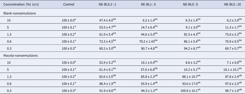
The viability of MAC-T cells was affected by exposure to NE-BLs and NE-ML formulations (Table 1). For blank-formulations, the cell viability was reduced by exposure to NE-BL0.2:1 and NE-BL5:10 at all tested concentrations compared to the control group. For NE-BL1:5 and NE-BL5:5, the viability of MAC-T cells was affected from 0.6% (v/v). For NE-MLs, the cell viability was reduced from 1.3% (v/v) for NE-ML0.2:1 and 5% (v/v) for the others compared to the control group (Table 1). Interestingly, the cell viability was higher after exposure to formulations NE-ML1:5 and NE-ML1:10 compared to its respective NE-BL at the same concentration (Table 1). For NE-ML1 : 5, this effect was found between 0.6 and 5% (v/v) and for NE-ML1 : 10 between 0.1 and 1.3% (v/v). These results suggest the ability of NE-MLs to counteract the negative effects of the blank nanoemulsions (NE-BL).
Percentage of viable bovine epithelial cells from the MAC-T lineage (mean ± sem) after exposure to different concentrations of macela-nanoemulsions (NE-ML) and blank-nanoemulsions (NE-BL)

The results shown are the average of three independent experiments. Different lowercase letters in the same row represents significant differences between treatments (NE-BL or NE-ML) and control. Different uppercase letters in the same column represents significant differences between NE-BL and its respective NE-ML at the same concentration (P < 0.05).
The IC50 values found for the NE-BL0.2:1, 1:5, 5:5 and 5:10 were 5.3 ± 0.0, 1.3 ± 0.0, 3.1 ± 0.0 and 1.4 ± 0.0, respectively. The IC50 values found for the NE-ML0.2:1, 1:5, 5:5 and 5:10 were 9.7 ± 0.0, 4.4 ± 0.0, 2.6 ± 0.0 and 2.8 ± 0.0, respectively. For NE-ML, the IC50 values found are equivalent to 242, 110, 65 and 70 μg/ml, respectively, of macela extract in nanoemulsions. Among the formulations, NE-ML0.2:1 and NE-ML1:5 showed IC50 values 2–3.4 times higher, respectively, than their corresponding NE-BL.
Similar results were found when comparing the cell viability of NE-BL and NE-ML to flaxseed oil and macela extract (Table S2 of the Supplementary File). For flaxseed oil the inhibitory concentration capable of reducing cell viability by 50% (IC50) was 3.8 mg/ml (Table S2 of the Supplementary File). On the other hand, when nanoemulsified (NE-BL), the IC50 values varied from 50 to 150 mg/ml (values corresponding to flaxseed oil concentration in formulations: Table S1 of the Supplementary File). Of the analyzed formulations, NE-BL1:5 showed lower cell viability with an IC50 of 1.3% (v/v), which corresponds to approximately 50 mg/ml of flaxseed oil in the formulation. Macela nanoemulsions did not significantly change the IC50 value, especially for the NE-ML0.2:1 and NE-ML1:5 formulations. While the IC50 of the extract was 0.14 mg/ml, in nanoemulsions it ranged from 2.6 to 9.7% (v/v), equivalent to 0.07 and 0.24 mg/ml of macela extract in nanoparticles (Table 1 and Table S2 of the Supplementary File).
The maintenance of cellular viability may be related to the high encapsulation efficiency found for these nanoemulsions (>94%) (Pinheiro Machado et al., Reference Pinheiro Machado, Veleirinho, Honorato and Kuhnen2020). In the study that developed these formulations, the high encapsulation efficiency values for flavonoids of macela extract are probably related to their affinity to flaxseed oil or the surfactant used in the formulation (Pinheiro Machado et al., Reference Pinheiro Machado, Veleirinho, Honorato and Kuhnen2020). Similarly, a reduced level of toxicity was found for nanoparticles of macela essential oil (Do Carmo et al., Reference Do Carmo, Baldissera, Vaucher, Rech, Oliveira, Sagrillo, Boligon, Athayde, Alves, França, Lopes, Schwertz, Mendes, Monteiro and Silva2015).
For the current study, the increased antimicrobial activity of innovative formulations containing macela extract (Pinheiro Machado et al., Reference Pinheiro Machado, Veleirinho, Honorato and Kuhnen2020) should be associated with reduced toxicity to bovine mammary gland cells. The antimicrobial evaluation of the macela was previously investigated against microbial strains of mastitic milk (Pinheiro Machado et al., Reference Pinheiro Machado, Veleirinho, Honorato and Kuhnen2020) showing a minimum inhibitory concentration value (MIC) lower than the IC50 found in the present study. In this context, when comparing our results with those for antimicrobial activity, the potential of these formulations is clear, particularly for NE-ML1:5. NE-ML1:5 showed a MIC50 value of 1.2% (v/v) against methicillin resistant S. aureus (MRSA) bacteria strains from mastitic milk (Pinheiro Machado et al., Reference Pinheiro Machado, Veleirinho, Honorato and Kuhnen2020), that is to say almost four times less than the IC50 found herein (4.4%, v/v). Such results suggest a promising potential for this formulation for analysis in future in vivo studies. When in contact with the NE-ML1:5, it should be noted that cell viability greater than 90% was observed in MAC-T cells at the concentration capable of inhibiting microbial growth (MIC50 1.2%, v/v), which corresponds to 30 μg/ml of crude macela extract (Pinheiro Machado et al., Reference Pinheiro Machado, Veleirinho, Honorato and Kuhnen2020). The NE-ML1:5 formulation highlighted in the previous forced stability analysis showed high stability (Pinheiro Machado et al., Reference Pinheiro Machado, Veleirinho, Honorato and Kuhnen2020). Therefore, the remaining analyses performed in the current study were conducted using the NE-ML1:5 formulation.
Cellular death analysis – apoptosis and necrosis
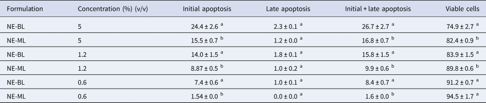
After exposure to NE-ML1:5, an analysis of the type of cellular death induction revealed a reduction in the percentage of cell apoptosis compared to the NE-BL1:5 formulation at all tested concentrations (5, 1.2 and 0.6%; v/v). For example, approximately twice as many apoptotic cells were found after exposure to the highest concentration of the NE-BL1:5 formulation (5%, v/v) (Table 2). For all tested concentrations (5, 1.2 and 0.6%, v/v), cell viability of > 75% was observed (Table 2). After exposure to NE-ML, a higher percentage of viable cells were found at concentrations of 5 and 1.2% compared to NE-BL (Table 2). In addition, after exposure to these same concentrations, a lower percentage of apoptotic cells were found.
Percentage of viable MAC-T cells and in early apoptosis, late apoptosis, and early + late apoptosis after exposure to different concentrations of macela-nanoemulsion (NE-ML1:5) and blank-nanoemulsion (NE-BL1:5)

Values expressed as an average ± sem of experiments performed in triplicate. Different lowercase letters in the column represent significant pairwise differences between NE-BL and NE-ML1 : 5 for each of the tested concentrations (P < 0.05).
The MTT test corroborated these results (Table 1). Since exposure of MAC-T cells to the macela nanoemulsion (NE-ML1:5) resulted in a reduction in the percentage of apoptotic cells compared to the respective blank-nanoemulsion (NE-BL1:5), we can suggest that macela extract may be involved in triggering a possible mechanism of apoptosis inhibition, thus conferring cytoprotection. It is important to note that we did not observe any cells to be marked with propidium iodide, that is to say necrotic as a result of losing plasma membrane integrity after exposure to both formulations. Even at the highest concentration (5%, v/v), the type of cell death induced by both formulations was predominantly apoptotic (Figure S1 of the Supplementary File).
Previous studies have already suggested the induction of anti-apoptotic mechanisms with macela extract, which is also related to its cytoprotective potential in that flavonoids of this plant have been able to prevent cellular death after oxidative stress (Arredondo et al., Reference Arredondo, Blasina, Echeverry, Morquio, Ferreira, Abin-Carriquiry, Lafon and Dajas2004; Sabini et al., Reference Sabini, Cariddi, Escobar, Mañas, Comini, Reinoso, Sutil, Acosta, Montoya, Contigiani, Zanon and Sabini2013). In the present study, the maintenance of cellular viability and the anti-apoptotic effects presented by the macela extract formulation (NE-ML1:5) represent an important advantage in the therapy of bovine mastitis, as cytoprotective effects would be triggered.
Subsequently, we evaluated the type of cell death after prior treatment of MAC-T cells with NE-ML1:5 (1%, v/v), followed by exposure to H2O2 (2 mM) for 1 h (Table 3). In this case, initial apoptosis increased in the exposed cells pretreated with NE-ML1:5, but there was no reduction in the percentage of cells in apoptosis (initial + late). A significant reduction in necrotic cells was seen (Table 3), along with an increase in viable cells, when exposed to the pretreatment, again suggesting the cytoprotective effect of NE-ML1:5 on MAC-T cells in terms of necrotic death. This result may be related to the fact that damaged cells are being induced to die by apoptosis owing to the presence of macela extract.
Percentage of viable MAC-T cells and in early apoptosis, late apoptosis, early + late apoptosis, and necrosis after exposure to macela-nanoemulsion (NE-ML1:5) for 23 h, followed by exposure to H2O2 (2 mM) for 1 h

Values expressed as an average ± sem of experiments performed in triplicate. Different lowercase letters in the column represent significant differences between treatments (P < 0.05).
Since macela is popularly used for its cytoprotective potential, previous research has assessed such effects (Arredondo et al., Reference Arredondo, Blasina, Echeverry, Morquio, Ferreira, Abin-Carriquiry, Lafon and Dajas2004). In that study, exposure to H2O2, along with the infusion of macela (75 or 100 μg/ml), resulted in significant cytoprotection when compared to PC12 cells treated only with H2O2. On the other hand, 5 to 50 μg/ml of the infusion of this plant showed no cytoprotective activity (Arredondo et al., Reference Arredondo, Blasina, Echeverry, Morquio, Ferreira, Abin-Carriquiry, Lafon and Dajas2004).
The cytoprotective potential of macela has been attributed to the high content of flavonoids found in this plant in which aglycones could be the main components responsible for the cytoprotective activity (Arredondo et al., Reference Arredondo, Blasina, Echeverry, Morquio, Ferreira, Abin-Carriquiry, Lafon and Dajas2004). The same authors showed that two free flavonoids present in the plant (quercetin and luteolin) had cytoprotective activity when added to cells exposed to H2O2.
The apoptotic cell death of MAC-T cells may call for further research since S. aureus, a bacterium that causes mastitis, presents intracellular survival capacity as a protective mechanism against the effects of antimicrobials (Hébert et al., Reference Hébert, Sayasith, Sénéchal, Dubreuil and Lagacé2000; Gruet et al., Reference Gruet, Maincent, Berthelot and Kaltsatos2001; Erskine et al., Reference Erskine, Wagner and DeGraves2003). The programmed death of some infected cells could release the pathogen, meaning that this possible association with the type of cellular death is important information in the evaluation of a product's potential for the intramammary therapy of mastitis, as necrotic cell death can aggravate the clinical condition of the animal by the triggering of pro-inflammatory cytokines and chemokines.
In vitro mammary glandular epithelial permeation and retention studies
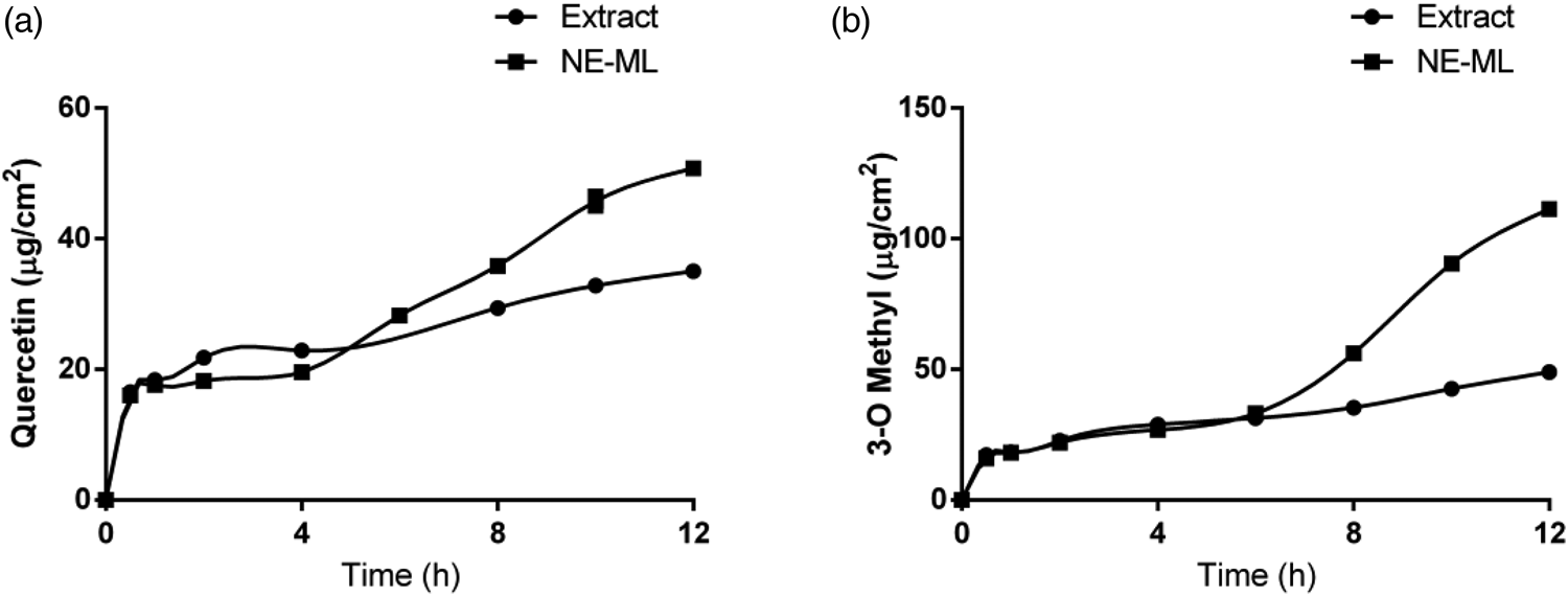
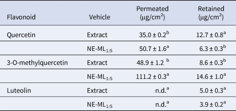
The epithelial tissue from the internal part of the mammary gland was used as a membrane model in this study. To date, no publications have been identified that use epithelium from the internal section of the bovine mammary gland with diffusion cells. The cumulative permeation profiles of the chemical markers of macela extract (quercetin and 3-O-methylquercetin), after application of NE-ML1:5 nanoemulsion and macela extract to the mammary gland explants can be seen in Figure 1. The results of the permeated and retained amounts of the compounds quercetin, 3-O-methylquercetin, and luteolin after 12 h can be seen in Table 4.
Cumulative permeation profiles of quercetin and 3-O-methylquercetin after application of macela nanoemulsion (NE-ML) and macela extract to the mammary glandular epithelium.

Amounts of flavonoids quercetin, 3-O-methylquercetin, and luteolin permeated and retained by area (μg/cm2) in mammary glandular epithelium over 12 h.

n.d.: not detected in samples as a result of concentrations below the detection/quantification limit of the method. Values expressed as an average ± sd. Different lowercase letters in the column represent significant pairwise differences (P < 0.05).
Considering the analysis of chemical markers of this extract, luteolin could not be quantified in the receptor medium because it was at concentrations below the detection limit of the method of analysis. It is important to note that the permeation profiles of quercetin and 3-O-methylquercetin were similar after initial application of the nanoemulsion and the extract to the explant. After 6 h of testing, the permeation rate of the flavonoids from the free extract was slower, when compared with the permeation profile obtained for the nanoemulsion. In part, this result can be attributed to the ability of the nanoemulsion to protect flavonoids from degradation caused by external factors, such as light, oxygen, and temperature (Pinheiro Machado et al., Reference Pinheiro Machado, Veleirinho, Honorato and Kuhnen2020). In this case, the protection conferred by the nanoemulsion may have enabled a gradual release and permeation of active compounds over time. Also, similar to skin and other epithelial mucosa, the nanoemulsion components (oil and surfactants) can act as permeation enhancers by interacting with the mammary glandular epithelium, increasing flavonoid permeability. The flow parameters (J) and permeability coefficient of quercetin and 3-O-methylquercetin were calculated from the inclination of the apparent linear portion of the cumulative permeation curves, including only data obtained at times greater than the T-lag. Both the flow (J) and the permeability coefficient (Kp) differed when quercetin and 3-O-methylquercetin in the nanoemulsion (4.6 ± 0.2 and 8.3 ± 0.2 μg/cm2/h, respectively) was compared to the extract (3.4 ± 1.2 and 4.5 ± 0.1 μg/cm2/h, respectively: details are shown in the Table S3 of the Supplementary File).
We observed that both permeated and retained total amounts were affected after the association of flavonoid compounds with the macela-nanoemulsion (NE-ML1:5). At the end of the experiments, the amount of quercetin and 3-O-methylquercetin retained in the mammary explants was approximately two times greater for the extract (12.7 and 14.6 μg/cm2, respectively) compared to the nanoemulsion (6.3 and 8.6 μg/cm−2, respectively), while the amount of luteolin retained was not affected. In contrast, NE-ML1:5 significantly increased the amount of permeated quercetin and 3-O-methylercetin in relation to the extract with permeate amounts of 50.7 and 111.2 μg/cm−2, compared to 35.0 and 48.9 μg/cm−2, respectively (P < 0.05). These results are probably related to the ability of nanoemulsions to promote the permeation of active compounds through biological barriers, which has been widely described in skin permeation studies (Franken et al., Reference Franken, Eloff, Du Plessis and Du Plessis2015). However, this is the first time the result has been reported for bovine mammary explants.
In light of these results and considering the therapy of bovine mastitis, the intramammary application of NE-ML1:5 nanoemulsion would allow for greater permeation and spread of the active compounds beyond the gland cistern, possibly reaching ducts and alveoli and ensuring greater performance in terms of antimicrobial potential. It is important to highlight that S. aureus is a particularly virulent bacterial cause of bovine mastitis that is difficult to control based on its ability to survive in cells and form abscesses and biofilms that protect it from the effects of antimicrobials and the host's immune system (Hébert et al., Reference Hébert, Sayasith, Sénéchal, Dubreuil and Lagacé2000; Gruet et al., Reference Gruet, Maincent, Berthelot and Kaltsatos2001; Erskine et al., Reference Erskine, Wagner and DeGraves2003). Therefore, an antimicrobial with greater penetration capacity is relevant for therapeutic treatments of the disease. According to Radostits et al. (Reference Radostits, Gay, Hinchcliff and Constable2007), the most successful antimicrobial agent for the treatment of mastitis during the dry period has characteristics that depend on the release time and transport agent in the formulation, as well as the size of the particles and the capacity of diffusion of the antimicrobial through lipophilic biological membranes.
In conclusion, we have shown a protective effect on MAC-T cells of a formulation comprising a nanoemulsion of the medicinal herb known as macela. This arises as a consequence of a higher IC50 and an ability to inhibit cell apoptosis and necrosis. Cell integrity is maintained at values above 82% and there is a higher permeation capacity of phenolics through the mammary glandular epithelium, showing that this formulation has potential as an innovative product for the treatment of bovine mastitis. The need for future in vivo studies to evaluate its efficacy is clear, especially in dry cow therapy.
Supplementary material
The supplementary material for this article can be found at https://doi.org/10.1017/S0022029922000139.
Acknowledgements
This study was funded by CNPq (Edital 39/2013, 403415/2013-6). The authors thank the LMBV (Laboratory of Plant Morphogenesis and Biochemistry, UFSC) and LAMEB (Multi-User Laboratory of Studies in Biology, UFSC).