Introduction
The efficiency of somatic cell nuclear transfer (SCNT) is affected by many factors, for example the quality of recipient oocytes, stage of donor cell cycle, activation method, exposure duration of the donor nucleus into inactivated oocytes, epigenetic and genetic status of the donor cell genome (Campbell et al., Reference Campbell, McWhir, Ritchie and Wilmut1996; Wakayama & Yanagimachi, Reference Wakayama and Yanagimachi2001; De Sousa et al., Reference De Sousa, Dobrinsky, Zhu, Archibald, Ainslie, Bosma, Bowering, Bracken, Ferrier, Fletcher, Gasparrini, Harkness, Johnston, Ritchie, Ritchie, Travers, Albertini, Dinnyes, King and Wilmut2002; Kawahara et al., Reference Kawahara, Wakai, Yamanaka, Kobayashi, Sugimura, Shimizu, Matsumoto, Kim, Sasada and Sato2005; Sung et al., Reference Sung, Gao, Shen, Yu, Song, Smith, Chang, Inoue, Kuo, Lian, Li, Tian, Tuck, Weissman, Yang and Cheng2006; Inoue et al., Reference Inoue, Noda, Ogonuki, Miki, Inoue, Katayama, Mekada, Miyoshi and Ogura2007). In particular, the diploid complement retention is one of the most important factors that directly affects the developmental competence of SCNT embryos receiving donor cells in the G0/G1 phase; this is because the DNA content during the first cell cycle affects the subsequent development of SCNT embryos (Wakayama et al., Reference Wakayama, Perry, Zuccotti, Johnson and Yanagimachi1998). Unless SCNT embryos injected with donor nuclei in the G0/G1 phase are treated with cytoskeletal inhibitors such as cytochalasins, they exclude some chromosomes as a pseudo-polar body (pPB) after artificial activation, resulting in aneuploidy (Lai et al., Reference Lai, Tao, Machaty, Kuhholzer, Sun, Park, Day and Prather2001).
Cytochalasins, a group of fungal metabolites, can bind to actin and block the barbed end of actin filaments (F-actin), which inhibits both the association and dissociation of subunits at that end (Brown & Spudich, Reference Brown and Spudich1981). It is known that the effect of cytochalasin D (CD) on the growth or shortening of the barbed end of F-actin is approximately 10 times more potent than that of cytochalasin B (CB) (Cooper, Reference Cooper1987). In addition to binding actin, cytochalasins A and B inhibit monosaccharide transport across the plasma membrane, whereas cytochalasins C, D, E and H do not (Rampal et al., Reference Rampal, Pinkofsky and Jung1980).
In the SCNT of many animals, CB has generally been used to control pPB extrusion (Wakayama et al., Reference Wakayama, Perry, Zuccotti, Johnson and Yanagimachi1998; Miyoshi et al., Reference Miyoshi, Saeki and Sato2000). As described above, it is known that CB possess cytotoxicity, whereas CD does not (Cooper, Reference Cooper1987). This cytotoxicity of CB might affect the subsequent development of SCNT embryos. In pigs, however, there has been no report that compares the effects of CB and CD on the subsequent development of SCNT embryos. In addition, disruption of the cortical F-actin after activation has a detrimental effect on the development of porcine SCNT embryos (Kawahara et al., Reference Kawahara, Mori, Tanaka and Shimizu2002; Yamanaka et al., Reference Yamanaka, Sugimura, Wakai, Shoji, Kobayashi, Sasada and Sato2007).
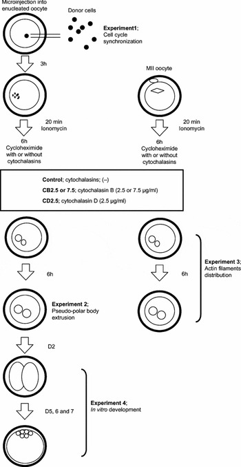
The purpose of the present study was to compare the effects of CB and CD treatments on SCNT embryos that were reconstructed using miniature pig foetal fibroblasts (mFFs) in the G0/G1 phase. As shown in Fig. 1, we first analysed the phase of the cell cycle of mFF cultured under various conditions and determined a more effective synchronization method in the G0/G1 phase. Next, we compared the effects of CB and CD on the pPB extrusion of SCNT embryos and reorganization of the cortical F-actin of porcine parthenogenetic oocytes after artificial activation. Finally, the effects of CB and CD treatment on the blastocyst formation rate of SCNT miniature pig embryos were investigated on days 5, 6 and 7 after in vitro cultivation.
Schematic representation of the experimental studies. The numbering of the experimental studies is retained from Experimental studies in the Materials and methods section.

Materials and Methods
Donor cells
Miniature pig foetuses (CSK, Suwa, Japan) were collected from the sow on day 56 of pregnancy. Each foetus was decapitated and eviscerated. The remaining tissues were washed in Dulbecco's phosphate-buffered saline (PBS, Sigma Chemical Co.) and then digested with 0.1% trypsin–ethylenediaminetetraacetic acid (EDTA), (Sigma) for 45 min at 38.5 °C. After digestion, the cells collected as mFFs were cultured in Dulbecco's modified Eagle medium (DMEM, Sigma) supplemented with 10% foetal bovine serum (FBS, Sigma). The culture medium was replaced every 2 days until the cells proliferated to confluency. After reaching confluence, the cells were harvested with 0.1% trypsin in PBS containing 0.5 mm EDTA for 5 min at 38.5 °C, frozen with a cryoprotectant (Cellbanker, Zenyaku, Tokyo) and stored in liquid nitrogen (passage 0). Prior to the start of experiments, the cells derived from a single foetus were thawed and cultured in DMEM plus 10% FBS and used between passages four and nine.
Cell cycle analysis
Cell cycle analysis was performed using a flow cytometer (Becton Dickinson) by measuring the DNA content of individual cells. After fixation, the cells were centrifuged, washed in PBS and then resuspended in a 500 μl droplet of PBS containing 10 μg/ml of propidium iodide (Sigma) and 30 μg/ml RNase (Sigma), followed by incubation for 30 min at 37 °C. The samples were filtered through a nylon mesh with 50 μM pores and maintained on ice until analysis. The phase of the cell cycle in individual cells was analysed by flow cytometry at 488 nm excitation and gated using forward light scatter. The percentages of G0/G1, S and G2/M phases were calculated using the CellQuest software programme (Becton Dickinson).
In vitro maturation of oocytes
Ovaries from prepubertal gilts were collected at a local slaughterhouse and transported to the laboratory within 2 h in a container containing warm saline. Porcine oocytes were aspirated from the antral follicles with 3–6 mm diameter by using a 5 ml disposable syringe attached to an 18 gauge needle. Cumulus–oocyte complexes (COCs) with uniform ooplasm and compact cumulus cell mass were selected in PB1 (Quinn et al., Reference Quinn, Barros and Whittingham1982). After washing in PB1, the COCs were cultured in bovine serum albumin (BSA, Sigma)-free NCSU-23 medium (Miyoshi et al., Reference Miyoshi, Saeki and Sato2000) for 44 h at 39 °C in a highly humidified atmosphere of 5% CO2 in air. For the first 22 h of maturation, the medium contained 10 IU/ml pregnant mare serum gonadotropin (PMSG; Serotropin; Teikokuzouki), 10 IU/ml human chorionic gonadotropin (hCG; Puberogen; Sankyo), 0.1 mg/ml cysteine (Sigma), 1 mm dibutyryl cyclic AMP (Sigma) and 10% porcine follicular fluid. The cells were cultured for the subsequent 22 h in the same medium without hormonal supplementation. After cell culture, expanded cumulus cells of the COCs were removed by vortexing in PB1 containing 1 mg/ml hyaluronidase (Sigma) and the oocytes with the first polar bodies were selected as mature oocytes under a stereomicroscope. The matured oocytes were transferred into PB1 and used for the experiments.
Nuclear transfer
The cumulus-free oocytes were stained with 5 μg/ml Hoechst 33342 (Sigma) for 5 to 10 min at 39 °C and transferred into PB1 containing 2.5 μg/ml CD (Sigma). Enucleation was performed by aspirating the first polar body and a small volume of the adjacent cytoplasm with a bevelled pipette using a piezo-drive unit (Primtech) and was confirmed by visualizing the presence of metaphase plates within the removed cytoplasm under ultraviolet (UV) light. The enucleated oocytes were washed three times and transferred to a 100 μl droplet of porcine zygote medium-3 (PZM-3) for the subsequent microinjection of the donor nucleus (Yoshioka et al., Reference Yoshioka, Suzuki, Tanaka, Anas and Iwamura2002). The donor cells prepared were transferred to a 50 μl droplet of PB1, and their plasma membranes were broken by gently aspirating them in and out of the bevelled pipette. The denuded nucleus was microinjected into the cytoplasm of the enucleated oocyte.
Activation and culture of SCNT embryos
The activation protocol was performed as described by Yamanaka et al. (Reference Yamanaka, Sugimura, Wakai, Shoji, Kobayashi, Sasada and Sato2007). In order to activate the SCNT embryos, they were treated with 15 μM ionomycin (Sigma) in PZM-3 containing fatty acid-free BSA (Yoshioka et al., Reference Yoshioka, Suzuki, Tanaka, Anas and Iwamura2002) for 20 min at 38.5 °C in 5% CO2 in humidified air and then washed with PZM-3. Next, SCNT embryos were cultured in PZM-3 containing 5 μg/ml cycloheximide (Sigma) with or without cytochalasins (see the section ‘Experimental Studies’) for 5 h at 38.5 °C in 5% CO2 in humidified air and then washed five times with cycloheximide-free PZM-3. Lastly, the embryos were transferred to PZM-3 and cultured at 38.5 °C in an atmosphere of 5% CO2 in air.
Immunostaining
To assess the distribution of F-actin, activated porcine oocytes were stained after in vitro cultivation by a modified technique described by Yamanaka et al. (Reference Yamanaka, Sugimura, Wakai, Shoji, Kobayashi, Sasada and Sato2007). F-actin was detected by immunostaining. After three washes with PBS containing 0.1% polyvinyl alcohol (PBS–PVA, Sigma), the activated oocytes were fixed in PBS–PVA containing 2% paraformaldehyde (Sigma) and 0.2% Triton X (Wako) for 40 min. The fixed oocytes were washed three times in PBS–PVA and then cultured in blocking solution (PBS–PVA supplemented with 1% BSA) for at least 1 h at room temperature. The activated oocytes were stained with 5 μg/ml phalloidin conjugated with fluorescein isothiocyanate (FITC–phalloidin, Sigma) for 1 h at 39 °C in PBS containing 0.1% Tween 20. The slides were examined using a laser-scanning confocal microscope. Confocal microscopy was performed using a Bio-Rad MRC-1024 confocal microscope system equipped with LaserSharp Processing software. Images were obtained by repeated laser scanning (10 times in 10 s) to improve the signal-to-noise ratio.
Experimental studies
In Experiment 1, four different types of synchronization of the cell cycle in foetal fibroblasts were studied: (i) mitosis (cell-cycling); (ii) serum-starved; (iii) just grown to confluence (just-confluent); and (iv) 7 days after confluence (long contact-inhibited). The cells during mitosis were collected within 48 h after culture. The serum-starved cells were prepared by culturing for 48 h in DMEM containing 0.5% FBS (Fig. 1).
In Experiment 2, the effect of CB or CD treatment on pPB extrusion of SCNT embryos was studied (Fig. 1). SCNT embryos were cultured in PZM-3 containing cycloheximide with or without cytochalasins for 6 h. The following concentrations of CB and CD were used: 2.5 μg/ml CB (CB2.5), 7.5 μg/ml CB (CB7.5) and 2.5 μg/ml CD (CD2.5). After cycloheximide treatment with or without CB or CD, the SCNT embryos were cultured in PZM-3 and fixed in acetic alcohol (1:3 v:v at 6 h after in vitro culture), followed by staining with 1% orcein. The pPB extrusion and the pseudo-pronucleus formation of the SCNT embryos were observed under the microscope.
In Experiment 3, the effect of CB or CD treatment on the distribution of F-actin in porcine parthenogenetic oocytes was assessed (Fig. 1), because in our previous study, we have confirmed that oocyte manipulation such as enucleation caused no effects on the distribution of F-actin in porcine oocytes (Kawahara et al., Reference Kawahara, Mori, Tanaka and Shimizu2002). Porcine oocytes matured in vitro were activated and treated with or without CB2.5, CB7.5 and CD2.5 by using the same method as that for SCNT embryos. After cycloheximide treatment with or without cytochalasins, porcine parthenogenetic oocytes were cultured in PZM-3. After fixation at 0 and 6 h of in vitro cultivation, cortical F-actin of porcine parthenogenetic oocytes was stained with phalloidin.
In Experiment 4, the effect of CD and CB treatments on the in vitro development of SCNT embryos was studied (Fig. 1). SCNT embryos were cultured in PZM-3 containing cycloheximide with or without CB2.5, CB7.5 and CD2.5 for 6 h. After cycloheximide treatment with or without cytochalasin, the number of cleaved SCNT embryos was examined on day 2 of in vitro cultivation. The number of SCNT embryos that developed to the blastocyst stage was examined on days 5, 6 and 7 of in vitro cultivation. The cell number of all blastocysts stained with 0.1 mg/ml Hoechst 33342 was counted on day 7.
Statistical analysis
The cell number of SCNT embryos developed to the blastocyst stage and cell cycle phases were analysed with ANOVA, followed by Tukey's test for determining significant difference (p < 0.05). Other data were analysed by the chi-square test (p < 0.05).
Results
Experiment 1. Synchronization of the cell cycle of mFFs
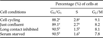
Table 1 shows the population of mFFs in each of the mitotic phases of G0/G1, S and G2/M, as determined by flow cytometry. The percentage of cells in G0/G1 was significantly lower (p < 0.05) in the cell-cycling (88.2%) and just-confluent (89.1%) groups than in the long contact-inhibited (90.5%) and serum-starved (90.5%) groups. The proportion of cells in the S phase of the cell cycle was greater (p < 0.05) in the cell-cycling (2.8%) and just-confluent (2.7%) groups than in the long contact-inhibited (1.5%) and serum-starved (1.6%) groups. This result indicated that the cell cycle of mFFs as well as of foetal fibroblasts derived from domestic pigs could be synchronized in the G0/G1 phase by long contact inhibition.
Cell cycle phases in mFFs under various conditions

Experiments were repeated three times.
a,b Values with different superscript letters within the same column are significantly different (p < 0.05).
Experiment 2. Effect of CB and CD treatments on pPB extrusion and pseudo-pronucleus formation
We investigated the effect of CB2.5, CB7.5 and CD2.5 treatments on pPB extrusion in SCNT embryos after artificial activation (Table 2). Pseudo-pronucleus formation was observed at rates ranging from 68.8 to 83.3%, with no significant difference among the groups. However, in the control group without cytochalasin treatment, the rate of SCNT embryos with a pPB was 43.3%; this rate was significantly higher (p < 0.05) than that in the other groups treated with cytochalasin. In the groups treated with CB7.5 and CD2.5, the rates of SCNT embryos with a pPB decreased significantly (p < 0.05) to 14.2 and 4.0%, respectively, as compared with those of the control group without cytochalasin treatment and the CB2.5 group. This result indicates that, at similarly low concentrations, CD treatment can completely inhibit pPB extrusion of SCNT embryos, while CB treatment cannot.
Effects of CB and CD treatments on pPB extrusion in SCNT embryos microinjected with mFF, followed by artificial activation

Experiments were repeated three times. PN, pronucleus; PB, polar body.
a, bValues with different superscript letters within the same column are significantly different (p < 0.05).
Experiment 3. Effect of CB and CD treatments on the distribution of F-actin in porcine parthenogenetic oocytes
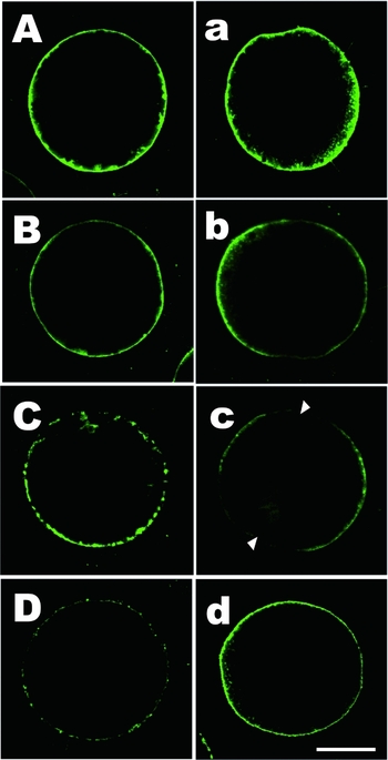
We observed the distribution of F-actin in porcine parthenogenetic oocytes at 0 and 6 h after in vitro cultivation with CB2.5, CB7.5 or CD2.5 (Fig. 2). Cortical F-actin in oocytes treated with CB7.5 and CD2.5 is distributed non-uniformly immediately following artificial activation (0 h) (Fig. 2C,D), whereas oocytes treated with CB2.5 showed insufficiently destabilized F-actin. After in vitro cultivation for 6 h, oocytes treated with CD and CB2.5 showed reorganization of cortical F-actin and their uniform distribution at the cytoplasmic membrane surface; this finding was similar to that of control oocytes without cytochalasin treatment (Fig. 2a,d). On the other hand, non-uniform distribution of F-actin at the pronuclear stage was observed after CB7.5 treatment (Fig. 2c). This result demonstrated that CD treatment was sufficient for the destabilization of cortical F-actin and did not prevent their reorganization.
Confocal micrographs of the cortical F-actin distribution in porcine parthenogenetic oocytes treated with or without CB and CD. A,a: control oocytes without cytochalasin treatment; B,b: oocytes treated with 2.5 μg/ml CB; C,c: oocytes treated with 7.5 μg/ml CB; D,d: oocytes treated with 2.5 μg/ml CD. Letters (A–D) and (a–d) correspond to two time points at 0 and 6 h after cytochalasin treatment, respectively. Arrows indicate the ruptured domain. Bar = 60 μm.

Experiment 4. Effects of CB and CD treatments on the in vitro development of miniature pig embryos
As shown in Table 3, CD treatment after activation significantly increased the cleavage rate (77.9%) and the blastocyst formation rate on day 5 (13.7%) as compared with other treatments. On days 6 and 7, cytochalasin treatment did not significantly increase the blastocyst formation rates of SCNT embryos. Furthermore, the total cell number (37.1 ± 8.0) of blastocysts derived from CD-treated SCNT embryos was the highest, but not significant. Thus, CB, but not CD, treatment resulted in the delay in the in vitro development of SCNT embryos to the blastocyst stage.
Effect of CB or CD treatment on the development of SCNT embryos following artificial activation

Experiments were repeated three times.
a–cValues with different superscript letters within the same column are significantly different (p < 0.05).
Discussion
In many animals, the application of somatic cells in the G0/G1 phase as donor sources for SCNT embryo reconstruction involves the use of cytochalasins to inhibit the extrusion of the pPB (Campbell et al., Reference Campbell, McWhir, Ritchie and Wilmut1996; Wakayama et al., Reference Wakayama, Perry, Zuccotti, Johnson and Yanagimachi1998; Onishi et al., Reference Onishi, Iwamoto, Akita, Mikawa, Takeda, Awata, Hanada and Perry2000; Beyhan et al., Reference Beyhan, Ross, Iager, Kocabas, Cunniff, Rosa and Cibelli2007). However, limited information is available on the effects of cytochalasins on the in vitro development of SCNT embryos. In the present study, we first determined an effective synchronization method for donor cells derived from miniature pigs and then compared the effects of CB and CD treatments on pPB extrusion, cortical F-actin distribution and the in vitro development of SCNT embryos.
Donor nuclei must be in the G0/G1 phase during their transfer to recipient oocytes with a high level of maturation-promoting factor (MPF) (Tani et al., 2001). Hence, we have described the preparation of donor cells in detail. In the present study, to determine a suitable method for the synchronization of the cell cycle of donor nuclei before nuclear transfer, we evaluated mFF cultured under various conditions by analysing their stages of the cell cycle. The long contact-inhibited and serum-starved groups contained significantly higher and lower percentages of G0/G1 and S cells, respectively, than the cell-cycling and just-confluent groups. No significant difference was observed between the long contact-inhibited and serum-starved groups. It has been suggested that serum-starved culture might induce the degeneration of the cell surface, with the resultant effect that the cells with a rough surface might have damaged chromosomes, which cannot be remodelled by the ooplasm of matured oocytes (Tao et al., 1999). Furthermore, it has been reported that donor cells that reach confluence are positive for proliferating cell nuclear antigen (PCNA), but lose this PCNA immunoreactivity after further 10 days of culture (long contact inhibition) (Onishi et al., Reference Onishi, Iwamoto, Akita, Mikawa, Takeda, Awata, Hanada and Perry2000). Taken together with the present results, we selected cells from the long contact-inhibited group to produce the SCNT embryos.
To gain further understanding on the effect of both CB and CD on the cytoskeleton of SCNT embryos, we observed cortical F-actin of porcine parthenogenetic oocytes by using the same activation method as that for SCNT embryos. We compared the effect of both CB and CD on the reorganization of cortical F-actin in parthenogenetic oocytes. The result showed that, as a cytoskeletal inhibitor, CD was more effective than CB even at lower concentrations; this is because CD-treated parthenogenetic oocytes showed faster reorganization of cortical F-actin at 6 h after artificial activation than CB-treated parthenogenetic oocytes. It was reported that the actin cytoskeleton is critical for normal mitochondrial distribution (Barnett et al., 1997) and for transfer of the mitochondria to daughter cells during cell division (Boldogh et al., 1998). The inhibition of F-actin polymerization prevents cytokinesis in porcine embryos, at least in one to two cell cycles (Wang et al., 1999). In our recent study, the disruption of cortical F-actin after activation also had a detrimental effect on the development of porcine SCNT embryos (Kawahara et al., Reference Kawahara, Mori, Tanaka and Shimizu2002; Yamanaka et al., Reference Yamanaka, Sugimura, Wakai, Shoji, Kobayashi, Sasada and Sato2007).
Next, we investigated the in vitro development of SCNT embryos that were cultured in the medium supplemented with either CB or CD. Interestingly, both the cleavage and the blastocyst formation rate in CD-treated SCNT embryos were significantly higher than those in CB-treated SCNT embryos. Furthermore, the cell number of CD-treated SCNT embryos was higher than that of CB-treated SCNT embryos. Treatment with CB2.5 could not sufficiently inhibit pPB extrusion and showed a lower cleavage and blastocyst formation rate. These results indicate that both the appropriate inhibition of pPB extrusion and faster reorganization of cortical F-actin are essential for the normal development of porcine SCNT embryos.
The affinity of CD for barbed ends is considerably higher than that of CB (Cooper, Reference Cooper1987). This is due either to the specificity of CD for actin or due to the fact that, unlike CB, CD does not bind to the glucose transporter (Rampal et al., Reference Rampal, Pinkofsky and Jung1980). Additionally, binding of CD to other targets has not been reported. CD could be used in low concentrations to minimize non-specific interactions; hence, cortical F-actin in CD-treated SCNT embryos might have rapidly reorganized as compared with those in CB-treated SCNT embryos. This rapid reorganization of cortical F-actin appeared to contribute to the improvement of both the cleavage rate on day 2 and the blastocyst formation rate on day 5, as shown in Table 3; this is because the rapid reorganization of cortical F-actin does not affect the first cell division of SCNT embryos into two cells, thereby resulting in the improved in vitro development of SCNT miniature pig embryos.
In conclusion, the present findings suggest that a thus far unidentified interference of CB is responsible for the delayed reorganization of cortical F-actin and that CD is more effective than CB as the cytoskeletal inhibitor for the production of SCNT embryos.
Acknowledgements
We thank the staff of the meat inspection office, Sendai City, for supplying porcine ovaries. This study was supported by Research Fellowships for Young Scientists from the Japan Society for the Promotion of Science to S.S. and M.K.