Introduction
Computed tomography (CT) images for radiotherapy planning are generally used as reference for image guidance. Reference Arimura1 To ensure image guidance accuracy, the reference image should not be distorted, and the tumor location should be accurate. Very few studies have reported on image distortion for planning of CT images. According to the guidelines for clinical implementation of image-guided radiotherapy (IGRT), 2 ‘Positioning uncertainty is impacted by CT imaging conditions, such as slice thickness or pitch, and the characteristics of position matching software.’
Previous studies have reported that the detector collimation Reference Katsuhiro and Yoshihisa3 and pitch factor Reference Wang and Vannier4 of imaging conditions impacted the distortion of helical CT images. The distortion of helical CT image causes image registration errors and mismatches between the reference image and the cone-beam CT (CBCT) image. Moreover, to our knowledge, no report to date has examined the impact of the distortion of helical CT image on IGRT. Determining the impact of image distortion on IGRT based on the imaging conditions of helical CT can be useful for radiotherapy planning. Therefore, this study aimed to propose a method of verifying image distortion for IGRT.
Methods
Verification method
The proposed verification method for image distortion involved the use of Duracon balls of 50·8 mm in diameter (AZUMA, Osaka, Japan). Duracon is a non-metal material and does not introduce metal artifacts into CT images; its specific gravity, 1·41, is similar to that of tough bone phantom, 1·50, which is assumed to be human bone Reference Magara and Kikumura5 and it has excellent dimensional stability. In addition, the spherical shape can eliminate rotational setup errors. In this method, four balls were placed on the couch at 0, 7·5, 15 and 22 cm from the centre of the CT image. To show the result of the verification method, the four Duracon balls were scanned with a six-detector-row CT scanner (SOMATOM Emotion 6; Siemens, Munich, Germany).
CT images were obtained using a helical scan under the following conditions: detector collimations, 0·5, 1 and 2 mm; and pitch factors, 0·40, 0·75, 1·10, 1·45 and 1·80. The following conditions were not changed: reconstruction slice thickness, 2·5 mm; field of view (FOV), 500 mm; tube voltage, 130 kV; minimum configurable tube current; gantry rotation time, 0·6 s; image reconstruction filtered back projection and convolution kernel B41s.
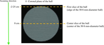
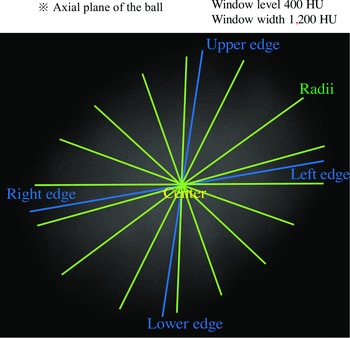
For each ball, the radii in the axial plane in the first and centre slices were measured to quantify image distortion. The slice at −22·5 mm was determined as the first slice of the balls and that at 0 mm was determined as the centre (Figure 1). The radii of the ball in a slice were measured at 22·5° intervals using image analysis software (syngo fastView; Siemens). The centre of each ball was determined by the intersection of the line connecting the top and bottom edges and the line connecting the left and right edges (Figure 2).
Ball radii measurement positions.

Measurement of the radii of the ball. The radii of the ball in the CT image are measured at every 22·5° angular interval. Window level, 400 HU and window width, 1,200 HU. CT, computed tomography, HU, Hounsfield unit.

The radii of the four balls placed at 0, 7·5, 15 and 22 cm were measured for 15 parameter pairs of three detector collimations and five pitch factors. The distortion of each ball was defined by the coefficient of variation (CV) of the radii. The CV was used to suppress the impact of the positional difference of the balls.
Impact of image distortion on image guidance
The series of 15 CT scans of the balls were transferred to the radiation therapy planning system (XiO®; Elekta AB, Stockholm, Sweden) and were used to build a pseudo-plan for image guidance. Auto skin (window level, 400 Hounsfield unit (HU); window width, 1,200 HU) was used to contour the four balls in these images. A total of 60 plans were built as the reference for image guidance; the isocentre was placed at the centre of each ball in each series.
One of the Duracon balls was placed at the isocentre of the linear accelerator (Synergy; Elekta AB) and was scanned using a CBCT system, X-ray Volume Imaging (XVI; Elekta AB). The CBCT image of the ball was registered with the corresponding reference image. The imaging conditions of CBCT were as follows: acquisition fast full scan; image reconstruction of medium resolution with matrix size of 512 × 512; FOV, 270 mm, with an image range of 270 mm in the body axis direction; start angle, 180°; stop angle, 180°; gantry speed, 360°/min; tube voltage, 100 kV; 330 frames; nominal scan dose, 220 mGy; total, 84·5 mAs and no bowtie filter. A full-angle scan was selected instead of a half-angle scan, as it can provide better spatial and contrast resolution. Reference Lu, Lu and Palta6 A small FOV of 270 mm was also selected to improve spatial resolution. To minimise potential CBCT errors, each CBCT examination was performed without changing the position of the Duracon ball. The image registration range of the reference image was limited to one ball to ensure that the image registration was not affected by other balls. A grey-value automatic registration was applied for the image registration between one of the reference images and the CBCT image. Reference Roche, Malandain, Pennec, Ayache, Wells, Colchester and Delp7,Reference Hristov and Fallone8
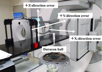
The registration results were assessed. The position with the standard condition that produced the smallest distortion of the ball was considered the true position. The registration results were compared with this true position. The American Association of Physicists in Medicine task group recommended IGRT position accuracy to be within 1 mm Reference Bissonnette, Balter and Dong9 ; therefore, the tolerance for the positional error was set at 1 mm in the X (lateral error), Y (longitudinal error) and Z (vertical error) directions (Figure 3).
Direction of image guidance.

Results
CT image distortion
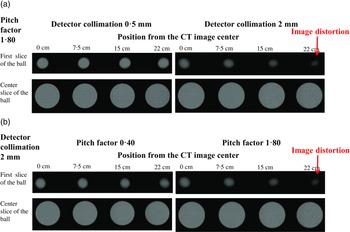
In the first slices of the ball, the distortion of the ball increased depending on the ball positions from the CT image centre and the parameter pair of detector collimation and pitch factor (Figure 4). However, the distortion of the ball did not change in the centre slice, regardless of the ball position and parameter pairs (Figure 4).
Image distortion differed between the first and centre slices of the ball. In the first slice of the ball, the distortion of the ball increased depending on the (a) detector collimation and (b) pitch factor. However, the distortion did not change in the centre slice of the ball. CT, computed tomography.

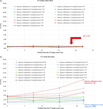
The CVs of the radii in the centre slice of the ball were < 1·5% regardless of the ball position and parameter pairs (Figure 5(a)); therefore, a CV < 1·5% was used as the threshold for distortion in the following analysis. CVs < 1·5% in the first slice were observed when CT was performed with a detector collimation of 0·5 or 1 mm and pitch factor of 0·4 (Figure 5(b)). The parameter pair of detector collimation of 0·5 mm and pitch factor of 0·40 showed the least image distortion. The narrowest configurable detector collimation was 0·5 mm, and the smallest configurable pitch factor was 0·40.
The CV of the radii differed in the first and centre slices of the ball. (a) The CV of the radii in the centre slice of the ball was less than 1·5% regardless of the detector collimation and pitch factor. (b) The CV of radii in first slice of the ball was less than 1·5% only with detector collimation of 0·5 or 1 mm and pitch factor of 0·4. CT, computed tomography; CV, coefficient of variation.

The parameter pair of detector collimation ≥ 1 mm and pitch factor ≥ 0·75 caused a distortion > 1·5% in the CVs at the first slices of the ball. In addition, the CVs increased from the centre to the edge of the CT images. The CT image with the parameter pair of detector collimation of 2 mm and pitch factor of 1·80 showed the largest distortion in the first slices. The reference image of the ball placed at the centre of the CT image, which was obtained with the parameter pair of detector collimation of 0·5 mm and pitch factor of 0·40, was used to evaluate the impact of image distortion on image guidance.
Impact of image distortion on image guidance
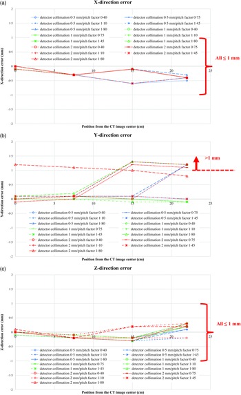
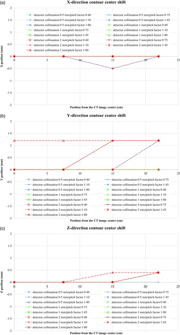
The image registration errors along each direction, compared with the position of the reference image of the ball placed at the centre of the CT image, obtained with the parameter pair of detector collimation of 0·5 mm and pitch factor of 0·40, are presented in Figure 6. The errors along the X and Z directions did not exceed 1 mm with any parameter pairs (Figure 6(a, c)). Only the error along the Y-direction exceeded 1 mm in the case of detector collimation > 1 mm, pitch factor > 1·10, or position > 15 cm away from the CT image centre (Figure 6(b)). Figure 7 presents the contour centre shifts along each direction, compared with the contour centre of the reference image of the ball placed at the centre of the CT image, obtained with the parameter pair of detector collimation of 0·5 mm and pitch factor of 0·40. Each direction error shown in Figure 6 matches each direction contour centre shift shown in Figure 7. Therefore, the main cause of the image registration error was a shift in the ball contour centre from the actual ball centre owing to image distortion, indicating that the image registration error can be predicted by the shift distance of the ball contour centre. The ball in the CT image showed high contrast and had a simple structure; therefore, image registration might be easy. The finding suggested that the minimum image registration error could be predicted from imaging conditions.
Image registration error. (a) The errors along the X-direction did not exceed 1 mm. (b) Only the error along the Y-direction exceeded 1 mm in the case of detector collimations wider than 1 mm, pitch factors larger than 1·10, or positions more than 15 cm away from the CT image centre. (c) The errors along the Z-direction did not exceed 1 mm. CT, computed tomography.

The contour centre shifts matched each direction error. (a) The X-direction contour centre shift matched the X-direction error. (b) The Y-direction contour centre shift matched the Y-direction error. (c) The Z-direction contour centre shift matched the Z-direction error. CT, computed tomography.

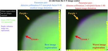
The best and worst image registration results are presented in Figure 8. The CT images scanned with the parameter pair of detector collimation of 0·5 mm and pitch factor of 0·40 showed the smallest distortion. The CVs of the ball radii in the first slices were < 1·5%. The contour of the ball in the CT image matched with that of the CBCT image regardless of the position from the CT image centre. The CT image scanned with the parameter pair of detector collimation of 2 mm and pitch factor of 1·80 showed the largest distortion. The CVs of the radii of the ball in the first slices of the scan were > 3·4%, exceeding the threshold of 1·5%. As shown in Figure 8, the reference image with the smallest image distortion matched the CBCT image and that with the largest image distortion did not match the CBCT image. In the green-purple mode, the reference image with the largest image distortion rendered images in green and purple where the CBCT and reference images protruded, respectively. Therefore, the main cause of mismatch between the contours of the reference and CBCT images was the distortion of the reference image. When the CV of the radii was smaller than the threshold in the first slice, the ball in the CT image matched that in the CBCT image. The finding suggests that imaging conditions could predict the conformity between the reference and CBCT images.
The reference image with the largest image distortion did not match the CBCT image. CBCT, cone-beam computed tomography; CT, computed tomography.

Discussion
This study proposed a validation method for the imaging conditions of helical CT for IGRT. Image distortions were considered a windmill artifact, as in a previous study. Reference Silver, Taguchi, Hein, Chiang, Kazama and Mori10 The radii CVs of the first and centre slices of the ball could quantify this windmill-like distortion. Scanning the edges of balls and dome-shaped objects has been reported to produce windmill artifacts. Reference Kanamori11 Especially, image distortion does not occur in clinical practice under image conditions, in which windmill artifacts do not occur at the edges of easily observable balls. The threshold for distortion of the CV from the radii of the centre slice of the ball should account for the uncertainty due to manual measurement. Then, the CV of the radii of the first slice of the ball could be compared with the threshold value to evaluate the image distortion under the image conditions.
This verification method was performed as follows: the CV of the radii was measured in the first and centre slices of the ball; then, the shift of the contour centre of the ball was determined. This verification method could evaluate the impact of image distortion in the imaging conditions used before helical CT for radiotherapy planning, making it possible to predict the image registration error and distortion of the reference image and thus take relevant countermeasures. It is possible to predict if the reference image will be impacted by image distortion at each position from the CT image centre and, thus, reduce the uncertainty of radiotherapy planning. The parameter pair of detector collimation of 2 mm and pitch factor of 1·80 could be scanned in 18 times less time than the parameter pair of detector collimation of 0·5 mm and pitch factor of 0·40. However, the Y-direction error for the parameter pair of detector collimation of 2 mm and pitch factor of 1·80 exceeded 1 mm even at the CT image centre (Figure 6(b)). Nevertheless, image registration errors predicted in advance can be addressed by setting appropriate planning target volume margins. The parameter pair of detector collimation of 0·5 mm and pitch factor of 0·40 had the least distortion across CT images; if geometric accuracy is the priority, this parameter pair can be selected. To suppress the impact of image distortion on IGRT, the shift between the ball contour and the actual centre should be maintained at < 1 mm and the CV of the first slice radii at < 1·5%.
The difference in effective slice thickness due to detector collimation and pitch factor might have impacted the shift of the contour centre along the scanning direction. Reference Katsuhiro and Yoshihisa3 The contouring could be affected by the partial volume effect, which differed according to the effective slice thickness. When the effective slice was thick, an object easily extended along the scanning direction, the contour also became larger than the actual structure and, then, the contour centre shifted accordingly.
Visual assessment of image guidance results is usually performed by comparing characteristic structures, such as bones and markers, between the reference and CBCT images. Therefore, evaluating the match between the reference and CBCT image in advance is useful.
The error in each direction was considered to include the mechanical variations of CBCT. A previous study has reported that the mechanical reproducibility of CBCT matching accuracy has an average standard deviation of 0·0–0·2 mm. Reference Meyer, Wilbert and Baier12 Moreover, as grey-value registration is based on voxel intensity, Reference Roche, Malandain, Pennec, Ayache, Wells, Colchester and Delp7,Reference Hristov and Fallone8 it was assumed that the distortion of the reference image also impacted the image registration error.
Catphan phantom Reference Katsuhiro and Yoshihisa3 is a tool that can evaluate CT image quality, but image distortion cannot be evaluated. Penta-guide phantom Reference Sato, Kanai and Lee13 is an IGRT evaluation tool, but its size is too large to be suitable for evaluation of image distortion at each position of CT images. Moreover, there is no method for quantifying image distortion. In the proposed method using Duracon balls, CT image distortion and the impact on IGRT could be evaluated.
This study had some limitations. Given that this experiment used balls, rotational errors in image guidance could not be evaluated. This work was conducted with a simple ball, and a complicated structure, such as that of the human body, was not examined. The CT scanner used was not capable of performing non-helical scans; therefore, no evaluation using non-helical scans was performed.
Conclusion
This study described a verification method for image distortion using four Duracon balls placed horizontally at different positions from the centre of the CT image and scanned helically under several imaging conditions. The verification method involved measuring the CV of the radii in the first and centre slices of the ball and determining the shift of the contour centre of the ball. Mismatch between the contours of the reference and CBCT images can be predicted by the CV of the radii in the first and centre slices of the ball. Moreover, the image registration error can be predicted by the contour centre shift of the ball. This verification method is useful for predicting the impact of image distortion on CT images under the imaging conditions in helical CT for radiotherapy planning, providing a reference image for image guidance. This study clarified the impact of image distortion on image guidance in clinical settings, and the results suggest that this verification method can reduce uncertainty in radiotherapy planning.
Acknowledgements
The authors thank the staff of the National Hospital Organization Kanazawa Medical Center for their cooperation in obtaining experimental data for this study.
Financial support
This research received no specific grant from any funding agency, commercial or not-for-profit sectors.
Competing interests
The authors declare none.
Authorship and contributorship
Takayuki Harada conceived, planned, and carried out the study and wrote the manuscript. Akihiro Takemura interpreted the presented evidence and made intellectual contributions, participated in the revision of the manuscript, and approved the final version.