Introduction
Triclabendazole (TCBZ) has been the mainstay for the treatment of fasciolosis in both humans and animals since its introduction in the early 1980s. Its very success has (almost inevitably) led to the emergence of TCBZ-resistant fluke populations. TCBZ resistance is now a well-established and widespread phenomenon, in many countries throughout the world (for references, see Table 6 in McMahon et al., Reference McMahon, Edgar, Hanna, Ellison, Flanagan, McCoy, Kajugu, Gordon, Irwin, Barley, Malone, Brennan and Fairweather2016). Consequently, there is a need to develop new products, especially more affordable ones for developing countries. The situation has triggered fresh interest in the evaluation of natural plant products. Traditional medicines have long been used as antiparasitic agents for the treatment of parasitic diseases in both humans and livestock (Iqbal et al., Reference Iqbal, Akhtar, Sindhu, Khan and Jabbar2003; Kayser et al., Reference Kayser, Kiderlen and Croft2003; Anthony et al., Reference Anthony, Fyfe and Smith2005; Crump, Reference Crump2006; Ndjonka et al., Reference Ndjonka, Rapado, Silber, Liebau and Wrenger2013).
A number of natural products have been reported to show activity against Fasciola hepatica, e.g. genistein, propolis and mangostin (Hegazi et al., Reference Hegazi, Abd El Hady and Shalaby2007a, Reference Hegazi, Abd El Hady and Shalabyb; Toner et al., Reference Toner, Brennan, Wells, McGeown and Fairweather2008; Keiser et al., Reference Keiser, Vargas and Winter2012; see also the review by Fairweather, Reference Fairweather2009). A separate group of compounds, the artemisinins, has attracted a considerable amount of attention (Keiser et al., Reference Keiser, Utzinger, Vennerstrom, Dong, Brennan and Fairweather2007; Kim et al., Reference Kim, Yoo, Li, Hong, Keiser and Hong2009; Kirchofer et al., Reference Kirchofer, Vargas, Braissant, Dong, Wang, Vennerstrom and Keiser2011; Keiser & Utzinger, Reference Keiser and Utzinger2012; Meister et al., Reference Meister, Duthaler, Huwyler, Rinaldi, Bosco, Cringoli and Keiser2013).
Mirazid, the subject of the present paper, is another potential compound. It contains a purified oleoresin extract of myrrh, derived from the plant Commiphora molmol, and has been developed as an antiparasitic drug. It has a broad spectrum of activity against a variety of protozoan and helminth parasites (e.g. Al-Mathal & Fouad, Reference Al-Mathal and Fouad2004; Haridy et al., Reference Haridy, Dawoud and Morsy2004, Reference Haridy, El-Sherbiny and Morsy2006; Fathy, Reference Fathy2011; Basyoni & El-Sabaa, Reference Basyoni and El-Sabaa2013). In particular, it has been shown to be effective in the treatment of fasciolosis in both humans and animals (Massoud et al., Reference Massoud, El Sisi, Salama and Massoud2001; Haridy et al., Reference Haridy, El Garhy and Morsy2003; Abo-Madyan et al., Reference Abo-Madyan, Morsy, Motawea and Morsy2004b; Soliman et al., Reference Soliman, El-Arman, Abdul-Samie, El-Nemer and Massoud2004). It is also active against the human blood fluke, Schistosoma spp. (Badria et al, Reference Badria, Abou Mohamed, El-Mowafy, Massoud and Salama2001; Sheir et al., Reference Sheir, Nasr, Massoud, Salama, Badra, El-Shennawy, Hassan and Hammad2001; Abo-Madyan et al., Reference Abo-Madyan, Morsy and Motawea2004a; Massoud et al., Reference Massoud, El Ebiary, Abou-Gamra, Mohamed and Shaker2004; Soliman et al., Reference Soliman, El-Arman, Abdul-Samie, El-Nemer and Massoud2004; Hamed & Hetta, Reference Hamed and Hetta2005); however, its efficacy against this parasite has been questioned (Botros et al., Reference Botros, William, Ebeid, Cioli, Katz, Day and Bennett2004, Reference Botros, Sayed, El-Dusoki, Sabry, Rabie, El-Ghannan, Hassanein, El-Wahab and Engels2005; El-Malky et al., Reference El-Malky, Lu, El-Beshbishi, Saudy and Ohta2013). Its activity against F. hepatica has been challenged as well (Botros et al., Reference Botros, El-Lakkany, Badawy, Mahmoud, Ebeid and Fenwick2009), although this discrepancy may be due to the treatment protocol employed, as different regimes have been used in different studies. This lack of uniformity is an issue that has plagued studies on Mirazid and has been remarked upon by Yakoot (Reference Yakoot2010); it will be returned to in the Discussion.
Previous studies on Fasciola involving Mirazid have been concerned more with efficacy than with drug action. As far as the authors are aware, only two morphological papers have been published: they focused on scanning electron microscope and histological changes in the tegument of Fasciola gigantica following in vitro treatment (Massoud et al., Reference Massoud, Shalaby, El Khateeb, Mahmoud and Kutkat2012, Reference Massoud, Shalaby, El Khateeb, Mahmoud and Kurkat2013). The present study monitored morphological changes to F. hepatica over a 7-day period following treatment in vivo with Mirazid, in a rodent (rat) model. Surface changes to the tegument were monitored by scanning electron microscopy and internal changes to the tegument and gut by transmission electron microscopy. The tegument and gut are the two surfaces across which drugs enter the fluke and so, by comparing the changes induced by Mirazid in each tissue, it should be possible to identify its main route of entry. A known TCBZ-resistant isolate was used in the investigation. To keep the design of this small trial simple, a single dose was used, rather than the repeated and variable doses often used in previous studies (see table 1). The aims of the study were fourfold: to determine whether or not Mirazid is active against F. hepatica – and active against a TCBZ-resistant fluke isolate; to clarify the route of uptake of the drug; to gain information on the time scale of drug action; and to gain information on the tissue targets of drug action.
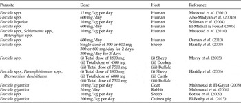
Treatment protocols for studies on Fasciola spp. involving Mirazid; doses given are for a period of 6 days unless otherwise stated.

Materials and methods
Experimental infections and in vivo drug treatment
The TCBZ-resistant Dutch isolate of F. hepatica was used for this study. For details of in vivo efficacy trials and in vitro experiments carried out with this isolate, the reader is referred to the review on fluke provenance by Fairweather (Reference Fairweather2011); see also Fairweather et al. (Reference Fairweather, McShane, Shaw, Ellison, O'Hagan, York, Trudgett and Brennan2012). Adult male Sprague–Dawley rats were each infected orally with 20 metacercarial cysts of F. hepatica under isoflurane anaesthesia by means of a stomach tube.
At 6 months post-infection, six rats were weighed individually and dosed orally with 250 mg/kg body weight Mirazid. Mirazid was used as a 10% (w/v) suspension. A similar dose (300 mg) was used in a previous sheep trial, as one of a number of treatment regimes, by Haridy et al. (Reference Haridy, El Garhy and Morsy2003). The Mirazid drench was a generous gift from Professor Osama Salama, Pharco Pharmaceuticals, Bolkely, Alexandria, Egypt. Two other F. hepatica-infected rats were not drug-treated and served as controls. From the dosed rats, flukes were recovered from the bile ducts at necropsy 2, 3 and 7 days post-treatment (pt). Each fluke was washed briefly in NCTC 135 culture medium at 37°C prior to processing. Zero-hour untreated specimens were prepared as controls: they were fixed immediately following their recovery from infected rats. For each time period pt, a minimum of four specimens were prepared for scanning electron microscopy (SEM) and three for transmission electron microscopy (TEM), as outlined below.
Tissue preparation
For SEM, 20 treated and 4 control flukes were lightly flat-fixed for 1 h at room temperature in 4% (w/v) glutaraldehyde in 0.1 m sodium cacodylate buffer (pH 7.4). They were then free-fixed, for a further 3 h, using fresh fixative before being post-fixed in 1% osmium tetroxide for 1 h. Specimens were then washed several times with cacodylate buffer before being dehydrated in an ascending series of ethanol. Following dehydration, flukes were dried using hexamethyldisilazane and mounted on aluminium stubs which were sputter-coated with gold–palladium. Samples were viewed using a FEI Quanta 200 scanning electron microscope (FEI, Eindhoven, The Netherlands) operating at an accelerating voltage of 10 keV.
For TEM, flukes were lightly flat-fixed for 1 h at room temperature in 4% (w/v) glutaraldehyde in 0.1 m sodium cacodylate buffer (pH 7.4). Specimens were then dissected into oral-cone, mid-body and tail regions, which were further split into transverse slices approximately 3 mm in thickness. These slices were free-fixed in fresh fixative for 3 h, before being washed several times in sodium cacodylate buffer. The samples were stored in buffer overnight. Following washing, fluke samples were post-fixed for 1 h in 1% osmium tetroxide, washed in buffer and dehydrated through an ascending series of ethanol. Fluke material was then embedded using Agar 100 resin. Ultrathin sections (~60–75 nm in thickness) were cut on a Reichert Ultracut E ultramicrotome (Leica Microsystems, Milton Keynes, UK) and mounted on uncoated 200-mesh copper grids. The sections were double-stained with alcoholic uranyl acetate (9 min) and aqueous lead citrate (6 min) and viewed in a FEI CM100 transmission electron microscope operated at 100 keV.
Results
Fluke recovery
At 2 days pt, a single fluke was recovered from one rat and nine flukes from the other rat. The single fluke was small and stunted and the bile duct was filled with a yellow fluid. Generally, the flukes appeared to be sluggish in their movement and were less active than normal; that is, there was a noticeable decrease in the frequency of the rhythmical waves of contraction and relaxation that pass backwards down the lateral margins of the flukes. They still retained their gut contents and eggs were present in the uterus.
At 3 days pt, two flukes were recovered from one rat and 11 flukes from the other rat. Approximately half the flukes seemed to be active, while the rest were moving more slowly than those flukes recovered at 2 days pt. Eggs were present in the uterus.
At 7 days pt, two flukes were recovered from one rat. Both flukes were completely inactive, very yellow and appeared to be autolytic. Thirteen flukes were recovered from the second rat. Approximately half the flukes were reasonably active, while the rest showed very limited movement.
Post-treatment under SEM
Control flukes
The surface architecture of the control flukes was normal, matching the images presented by Fairweather et al. (Reference Fairweather, Threadgold, Hanna and Dalton1999, Fig. 3.1) and McConville et al. (2009, Figs 1–6). For economy of space, only micrographs of flukes recovered 2 days and 7 days pt will be included; the changes observed at 3 days pt will be described, but not illustrated.
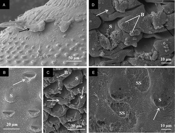
Scanning electron micrographs of the tegumental surface of the oral cone region of adult liver fluke, Fasciola hepatica (Dutch isolate) following treatment in vivo with Mirazid (250 mg/kg) for 2 days. (A) Ventral surface, with the tegument lifting away from the surface (arrow). (B) Ventral surface, with spines (S) and partial loss of spines to reveal the spine sockets (arrow). (C) Ventral surface, with submergence of the spines (S) due to severe swelling (white arrows) of the inter-spinal tegument. Blebs (B) cover the surface. There is disruption to the spine tips (black arrow). (D) Dorsal surface, with severe swelling (white arrow) and blebbing (B) of the interspinal tegument, so that the spines (S) are barely visible. There is some disruption to the spine tips (black arrow). (E) Dorsal surface, with empty spine sockets (SS) due to spine loss. The tips of spines (S) that remain are disrupted (arrow).

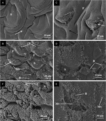
Scanning electron micrographs of the tegumental surface of the mid-body region of adult liver fluke, Fasciola hepatica (Dutch isolate) following treatment in vivo with Mirazid (250 mg/kg) for 2 days. (A) Ventral anterior mid-body region, with swelling of the inter-spinal tegument (arrow). The spines (S) are partially sunken. The general surface is smooth. (B) Ventral anterior mid-body region, with blebs (B) covering the tegumental surface. The inter-spinal tegument is roughened due to the presence of microvillus-like structures (Mv). The tegument covering the tips of the spines (S) is disrupted (arrow). F, tegumental furrows. (C) Dorsal anterior mid-body region, with swelling (black arrow) and furrowing (F) of the inter-spinal tegument. The tegument covering the spines is also swollen and disrupted and is beginning to slough away from the spine tip (white arrow). (D) Dorsal posterior mid-body region, with a spine (S) that remains in its socket, although its tegumental covering has been removed. B, bleb. (E) Dorsal posterior mid-body region, with an empty spine socket following loss of the spine. B, bleb; Mv, microvillus-like projections. (F) Ventral posterior mid-body region. The surface is almost totally covered with blebs (B). Spines (S) are barely visible, due to the severe swelling of the tegument surrounding them.

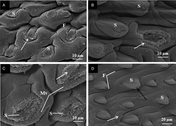
Scanning electron micrographs of the tegumental surface of the oral cone (A, B) and anterior mid-body (C, D) regions of adult liver fluke, Fasciola hepatica (Dutch isolate) following treatment in vivo with Mirazid (250 mg/kg) for 7 days. (A) Ventral surface, with disruption to the spine tips (arrow). The spines (S) are obscured by the swelling of the tegument surrounding them. F, tegumental furrow. (B) Dorsal surface, with severe disruption of the spine tips (arrow). The tegument covering the spine seems to have partially sloughed away. S, spine. (C) Ventral surface, where partial loss of the tegument (arrow) covering the spines has exposed the crystalline spine (S) beneath. Microvillus-like projections (Mv) are present on the tegumental surface. (D) Dorsal surface, with disruption to the spine tips (black arrow) and slight swelling of the tegument covering the spines (S). The inter-spinal tegument is swollen (white arrow) and furrowed (F).

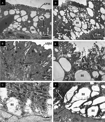
Transmission electron micrographs of the tegumental syncytium of adult liver fluke, Fasciola hepatica (Dutch isolate) following treatment in vivo with Mirazid (250 mg/kg) for 2 days (A–C) and 7 days (D–F). (A) The full depth of the tegumental syncytium, from the apical plasma membrane (APM) to the basal lamina (BL). The basal infolds (BI) are severely swollen and mitochondria (m) are accumulated in the basal region of the syncytium. Mu, sub-tegumental muscle blocks; S, spine. (B) The apical region of the syncytium, with the presence of numerous T2 secretory bodies (T2). The T1 secretory bodies (T1) are swollen. APM, apical plasma membrane; m, mitochondria. (C) The basal region of the syncytium, with swelling of the basal infolds (BI). Mitochondria (m) have accumulated just above the basal lamina (BL) and their cristae are swollen. T1, T1 secretory bodies. (D) The severe swelling of the basal infolds (BI) throughout the depth of the syncytium. APM, apical plasma membrane; BL, basal lamina; S, spine. (E) The apical region of the syncytium, with deep, convoluted invaginations of the apical plasma membrane (APM). Enlarged T1 secretory bodies (T1) have accumulated beneath the apical membrane. The swollen basal infolds (BI) reach almost to the apical membrane. (F) The basal region of the syncytium, with gross swelling of the basal infolds (BI). Mitochondria (m) and T1 secretory bodies (T1) have accumulated in the cytoplasm between the infolds. BL, basal lamina.

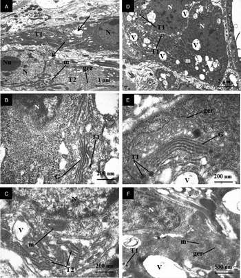
Transmission electron micrographs of the tegumental cells of adult liver fluke, Fasciola hepatica (Dutch isolate) following treatment in vivo with Mirazid (250 mg/kg) for 2 days (A–C) and 7 days (D–F). (A) Two tegumental cells, a T1 cell (T1) and a T2 cell (T2), lying beside each other. The cells are vacuolated (arrows) and contain cisternae of granular endoplasmic reticulum (ger) and mitochondria (m). N, nucleus; Nu, nucleolus. (B) A T1 tegumental cell, containing a well-developed Golgi complex (G). A T1 secretory body (T1) can be seen budding away from the complex. N, nucleus. (C) A T2 tegumental cell, with numerous T2 secretory bodies (T2). The mitochondria (m) contain swollen cristae. Vacuoles (V) are present in the cell. N, nucleus. (D) Two T1 tegumental cells, which are highly vacuolated (V). The cells contain nuclei (N), swollen T1 secretory bodies (T1) and mitochondria (m). (E) A T1 tegumental cell, containing a well-developed Golgi complex (G), which is producing T1 secretory bodies (T1). The T1 bodies are larger than normal. The cisternae of granular endoplasmic reticulum (ger) are slightly swollen. V, vacuole. (F) A T1 tegumental cell, containing T1 secretory bodies (T1), a nucleus (N), mitochondria with swollen cristae (m) and cisternae of granular endoplasmic reticulum (ger) that are slightly swollen. V, vacuole.

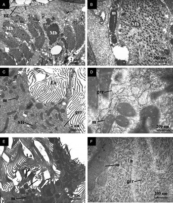
Transmission electron micrographs of the sub-tegumental musculature (A, B) and gastrodermal cells (C–F) of adult liver fluke, Fasciola hepatica (Dutch isolate) following treatment in vivo with Mirazid (250 mg/kg). (A) Seven days pt. Sub-tegumental muscle layers, showing that the muscle blocks (Mb) are more widely spaced apart than normal. BL, basal lamina. (B) Seven days pt. A muscle block (Mb), showing that the muscle fibres are fewer in number and more loosely packed than normal. The mitochondria (m) associated with the muscle blocks contained swollen cristae. (C) Two days pt. Apical region of a gastrodermal cell. The gut lamellae (La) have a normal appearance. In the cytoplasm of the cell, the cisternae of the granular endoplasmic reticulum (ger) are severely swollen, the mitochondria (m) have swollen cristae and a few secretory bodies (SB) are present. (D) Two days pt. Gastrodermal cell, showing extensive swelling and vesiculation of the cisternae of the granular endoplasmic reticulum (ger). The mitochondria (m) contain swollen cristae. (E) Seven days pt. Apical region of a gastrodermal cell, showing the presence of secretory bodies (SB) and numerous mitochondria (m). La, gut lamellae. (F) Seven days pt. Gastrodermal cell, showing swelling and fragmentation of the cisternae of the granular endoplasmic reticulum (ger). The mitochondria (m) contain swollen cristae.

Two days post-treatment with Mirazid
Severe disruption of the tegument was evident on the ventral surface in the oral-cone region, with instances of the entire syncytium lifting off just behind the oral sucker (fig. 1A). Sloughing was observed elsewhere in this region. Partial loss of spines was also seen, the tip of the spines having broken off, to expose the spine sockets (fig. 1B). In other areas, the inter-spinal tegument was swollen and was carpeted by blebs, leading to submergence of the spines (fig. 1C). Patches of swelling were observed on the dorsal oral cone surface. The swelling was limited to the inter-spinal tegument and was accompanied by surface blebbing (fig. 1D); the spines were barely visible. Elsewhere on the dorsal surface, loss of spines had taken place, leaving empty spine sockets (fig. 1E).
Swelling and furrowing of the inter-spinal tegument was observed on the ventral surface of the anterior mid-body region, which partially obscured the spines (fig. 2A). The general body surface was smooth (fig. 2A). In more severely disrupted areas, the surface was rougher, being carpeted with blebs, and the tegument covering the tip of the spine was disrupted (fig. 2B). On the dorsal surface, the inter-spinal tegument was swollen and furrowed. In these areas, the tegumental covering of the spines was also swollen and the spine tip was disrupted (fig. 2C). In other areas, the swollen tegument had a more flattened appearance, the spines barely projecting above the surface. The tegumental covering of the spine had split at the tip of the spine. In more extreme instances, the spines were missing, leaving empty spine sockets and some sloughing around these sites.
On the ventral surface of the posterior mid-body region, the inter-spinal tegument was swollen and furrowed and a small number of blebs were scattered over the surface; the general surface was smooth. The spines were still clearly visible, although their tegumental covering was swollen and the spine tips disrupted. The swelling and blebbing of the tegument were more pronounced in some areas, almost completely obscuring the spines. Swelling, furrowing and roughening of the dorsal tegumental surface was seen in the posterior mid-body region and the spine tips were disrupted. In more severely affected areas, the surface was extremely roughened and was marked by empty spine sockets. On occasion, the spine seemed to have remained in the exposed socket (fig. 2D), but generally the sockets were empty (fig. 2D and E). In extreme cases, the ventral surface was almost completely covered by blebs of various sizes (fig. 2F).
The general surface in the tail region was swollen, such that the spines appeared to be sunken and were less visible than normal.
Three days post-treatment with Mirazid
On the ventral surface of the oral-cone region, the inter-spinal tegument was swollen, so that the spines were largely submerged. The general surface was either smooth or more roughened, with microvillus-like projections extending away from the surface. There was marked disruption of the spine tips, where the tegumental covering had split to reveal the spine beneath. Widespread swelling and blebbing of the tegument was evident on the dorsal surface. Blebbing was more confined to the inter-spinal tegument than being associated with the spines. Some swelling of the tegument covering the spines was seen. More severe disruption of the covering was also seen, with partial loss of the covering, to expose the spine, or disruption and splitting at the spine tip.
The ventral tegumental surface in the anterior mid-body region was generally smooth, with mild swelling of the inter-spinal tegument but little disruption to the spines. In some areas, swelling was greater and the tegumental covering at the spine tip was disrupted. On the dorsal surface, a range of changes was observed. At times, there was limited swelling of the inter-spinal tegument, but in other areas swelling and furrowing were greater, leading to submergence of the spines. On occasion, the spines were obscured by blebs on the surface. More severe disruption to the spines was seen, with splitting of their tegumental covering and loss of the spines themselves.
The tegumental response in the posterior mid-body region was variable. In some areas, the ventral surface appeared normal, with the spines projecting clearly from the surface. In other areas, swelling and furrowing of the inter-spinal tegument obscured the spines. Blebbing on the tegumental surface was seen, but was confined to the inter-spinal tegument rather than the spines. On the dorsal surface, swelling of the tegument varied from quite mild to more severe. Some swelling of the tegument covering the spines was seen, along with splitting of the covering at the spine tip.
In the tail region, there was a general swelling and furrowing of the tegument on both the ventral and dorsal surfaces and the spines were partially or completely submerged.
Seven days post-treatment with Mirazid
Areas of swelling of the tegument were evident on the ventral surface of the oral-cone region (fig. 3A). The inter-spinal tegument was swollen and furrowed, either partially or completely submerging the spines (fig. 3A). At higher magnification, disruption to the spine tips was observed (fig. 3A). The inter-spinal tegument was slightly swollen on the dorsal surface, the spines still projecting clearly from the surface (fig. 3B). The tegument covering the spines was more severely swollen, giving the spines a ‘puffy’ appearance. An occasional bleb was seen on the spine tip. In more extreme cases, the tegumental covering of the spine had split along the spine tip and part of the covering had sloughed off the upper surface of the spines (fig. 3B).
In the anterior mid-body region, on the ventral surface, swelling of the inter-spinal tegument varied from quite mild, with the spines clearly visible and lying flat against the surface, to more severe (along with furrowing), so that the spines were almost obscured. There was some disruption to the tegumental surface around the base of the spines, but more severe disruption at the spine tip, where the tegumental covering was partially lost, exposing the crystalline spine beneath (fig. 3C). Swelling and furrowing of the inter-spinal tegument was seen on the dorsal surface, but was quite mild; some swelling of the tegument covering the spines was evident, too (fig. 3D).
In the posterior mid-body region, the swelling of the inter-spinal tegument ranged from mild to more severe, so that the spines were partially sunken, and the general surface was furrowed and roughened. Swelling and furrowing of the tegument on the dorsal surface was also mild. The tegument covering the spines was slightly swollen.
The tegument on the ventral surface of the tail region was swollen and the spines partially submerged. The swelling and furrowing on the dorsal surface were more severe, giving the surface a flattened appearance, and the spines were barely visible, being almost completely submerged by the tegument surrounding them.
Post-treatment under TEM
Tegumental system
The ultrastructure of the control specimens remained normal, as described by Meaney et al. (Reference Meaney, Haughey, Brennan and Fairweather2005, Figs 1, 5) and Halferty et al. (Reference Halferty, Brennan, Trudgett, Hoey and Fairweather2009, Figs 4A–C). Again, for economy of space, only images of flukes recovered 2 and 7 days pt will be included, although the changes observed at 3 days pt will be described.
Tegumental syncytium two days post-treatment with Mirazid
The syncytium had a vacuolated appearance, due to the swelling of the basal infolds throughout the full depth of the syncytium (fig. 4A). Type 1 and type 2 secretory bodies were present throughout the tegument, although, in the apical region, there were greater numbers of T2 than T1 secretory bodies and the T1 bodies were swollen (fig. 4B). An occasional autophagic vacuole was observed. The tegument covering the spines was thicker than normal, almost swelling up around the spine, and the apical invaginations were very deep and convoluted. At the tip of the spine, the tegumental covering was disrupted. At the base of the tegument, the mitochondria were abnormal, in that the cristae were swollen (fig. 4C).
Tegumental syncytium three days post-treatment with Mirazid
The tegument had a very vacuolated appearance, with the swollen basal infolds occupying much of the volume of the syncytium. The apical invaginations were very deep and convoluted. Few secretory bodies were present at the apex of the tegument and the type 1 bodies were swollen. T1 secretory bodies were more numerous at the base of the tegument than at the apex.
Tegumental syncytium seven days post-treatment with Mirazid
There was gross swelling of the basal infolds throughout the tegumental syncytium, and the cytoplasm around the spines was particularly disrupted (fig. 4D). At the apex of the tegument, the invaginations were deep and convoluted (fig. 4E). Type 2 secretory bodies were more numerous than T1 secretory bodies in the apical region and the T1 bodies were larger than normal, also slightly misshapen. The cytoplasm around the tip of the spines was greatly disrupted. The basal infolds were severely swollen along the length of the basal lamina and there were more T1 secretory bodies in the basal than the apical region of the syncytium (fig. 4F).
Tegumental cells two days post-treatment with Mirazid
Both T1 and T2 tegumental cells had a vacuolated appearance (fig. 5A). Distinct Golgi complexes were present in the T1 tegumental cells, with T1 secretory bodies budding away from them (fig. 5B). Numerous T2 secretory bodies were present in the type 2 tegumental cells and the mitochondria in these cells contained swollen cristae (fig. 5C).
Tegumental cells three days post-treatment with Mirazid
Some of the T1 tegumental cells were filled with slightly swollen mitochondria, while others were more vacuolated. The cisternae of granular endoplasmic reticulum (ger) in these cells were slightly swollen. Well-developed Golgi complexes were present, producing T1 secretory bodies, but these secretory bodies were larger and more swollen than normal.
Tegumental cells seven days post-treatment with Mirazid
The T1 tegumental cells were extremely vacuolated, but contained greater than normal numbers of T1 secretory bodies (fig. 5D). Golgi complexes were present and were well developed, with multiple cisternae, and the T1 secretory bodies budding away from them were swollen and larger than normal (fig. 5E). The Golgi cisternae were slightly swollen and transition vesicles were evident between the ger and the Golgi complex. The cisternae of ger were slightly swollen, as were the cristae in the mitochondria (fig. 5E and F).
Sub-tegumental musculature
The morphology of the muscle blocks in control specimens matched the descriptions given by Kumar et al. (Reference Kumar, McGeown, Reynoso-Ducoing, Ambrosio and Fairweather2003, Fig. 2A) and Toner et al. (Reference Toner, Brennan, McConvery, Meaney and Fairweather2010, Fig. 5E).
The muscle blocks in treated flukes had a similar appearance at each time-point. That is, they were more widely separated (fig. 6A) and the muscle fibres within them were fewer in number and more loosely packed than normal (fig. 6B).
Gastrodermal cells
Gastrodermal cells of the control specimens retained a normal morphology similar to that described by Robinson & Threadgold (Reference Robinson and Threadgold1975, Figs 4–8) and Meaney et al. (Reference Meaney, Haughey, Brennan and Fairweather2005, Figs 7, 8, 17, 18).
Two days post-treatment with Mirazid
The surface lamellae of the gastrodermal cells retained a normal morphology and the cells contained a number of secretory bodies (fig. 6C). The most notable feature of the cells was the gross swelling and vesiculation of the cisternae of ger; the cristae of the mitochondria were also swollen (fig. 6D).
Three days post-treatment with Mirazid
Numerous secretory bodies were present in the apical region of the gastrodermal cells. The cisternae of ger were slightly swollen, but not vesiculated.
Discussion
Mirazid caused marked changes to the external surface of F. hepatica, ranging from swelling and furrowing, to blebbing, disruption of the spine tips, splitting of the tegument covering the spines, spine loss and sloughing of the tegument. These changes were seen at 2 days pt and the general disruption did not become any worse with time. There was an anterior focus of disruption, but no clear trend as to whether the ventral or dorsal surface was the more severely affected. A similar anterior concentration of disruption has been observed following treatment with clorsulon (Meaney et al., Reference Meaney, Fairweather, Brennan, McDowell and Forbes2003) and nitroxynil (McKinstry et al., Reference McKinstry, Fairweather, Brennan and Forbes2003). As discussed below, the surface changes may be the result of internal changes in the tegumental system. One extreme event observed involved the shedding of the entire syncytium, with a new layer forming beneath. This has been seen with other drugs (e.g. Fairweather et al., Reference Fairweather, Anderson and Threadgold1986; Meaney et al., Reference Meaney, Fairweather, Brennan and Forbes2004; McKinstry et al., Reference McKinstry, Brennan, Halferty, Forbes and Fairweather2007; Halferty et al., Reference Halferty, Brennan, Hanna, Edgar, Meaney, McConville, Trudgett, Hoey and Fairweather2008). In terms of the consequences of surface disruption, any breach of the surface membrane (following, for example, splitting of the tegument at the spine tips, loss of spines and sloughing) would enhance drug penetration into the fluke and lead to further disruption of internal tissues. Also, it would expose flukes to the additional and damaging influence of environmental factors, such as bile. The spines may represent a ‘weak spot’ in the tegument, in terms of drug penetration. Similar marked surface changes (and with an anterior focus) have been observed in F. gigantica following exposure to Mirazid in vitro (Massoud et al., Reference Massoud, Shalaby, El Khateeb, Mahmoud and Kutkat2012).
In contrast to the surface changes, the changes observed in the tegumental syncytium and tegumental cells followed a clear trend, becoming progressively more severe with time pt. The most notable feature in the syncytium was the swelling of the basal infolds, which came to occupy much of the volume of the tegument. This would suggest that the osmoregulatory function of the tegument had been compromised. The internal spread of the flooding would affect the (functioning of the) deeper-lying tissues. The distension of the basal infolds may account for the swelling of the tegument seen externally. Should the swelling of the basal infolds continue, it would lead to the eventual sloughing of the tegument that was observed; this phenomenon has also been seen in previous studies on fasciolicidal drugs (e.g. Fairweather et al., Reference Fairweather, Anderson and Threadgold1986; Skuce & Fairweather, Reference Skuce and Fairweather1990; Stitt & Fairweather, Reference Stitt and Fairweather1994; Meaney et al., Reference Meaney, Fairweather, Brennan and Forbes2004; McKinstry et al., Reference McKinstry, Brennan, Halferty, Forbes and Fairweather2007; McConville et al., Reference McConville, Brennan, Flanagan, Hanna, Edgar, Castillo, Hernández-Campos and Fairweather2009; O'Neill et al., Reference O'Neill, Johnston, Halferty, Brennan and Fairweather2015).
Since the maintenance of the integrity of the apical membrane is dependent on the continued arrival and release of secretory bodies, the decreased numbers of secretory bodies (particularly the T1 type) at the apex of the syncytium would impair the fluke's ability to maintain any response to drug action. However, T1 bodies appeared to accumulate at the base of the syncytium (and in the tegumental cells), suggesting that there may be a block in the transport of the secretory bodies, from the cells to the tegumental surface (as seen with TCBZ: Stitt & Fairweather, Reference Stitt and Fairweather1994). Secretory body production (both T1 and T2 bodies) was seen to continue, although the T1 bodies were abnormally large. The cells became progressively more vacuolated with time, and organelles such as the ger and mitochondria were affected, suggesting that the normal functioning of the cells would be compromised. Similar results have been noted with other drugs (e.g. Fairweather et al., Reference Fairweather, Anderson and Threadgold1986; Stitt & Fairweather, Reference Stitt and Fairweather1994; McKinstry et al., Reference McKinstry, Brennan, Halferty, Forbes and Fairweather2007; McConville et al., Reference McConville, Brennan, Flanagan, Hanna, Edgar, Castillo, Hernández-Campos and Fairweather2009; O'Neill et al., Reference O'Neill, Johnston, Halferty, Brennan and Fairweather2015).
Changes to the sub-tegumental muscle blocks were similar at each time-point pt; namely, they were more widely spaced apart than normal and there appeared to be a decrease in the number of muscle fibres. In part, this may help to account for the (increasingly) sluggish movements exhibited by the flukes at recovery. The spacing between the muscle blocks may reflect an internal spread of the flooding observed in the syncytium. Separation of male and female schistosome (Schistosoma mansoni) worms following Mirazid treatment has been observed by Badria et al. (Reference Badria, Abou Mohamed, El-Mowafy, Massoud and Salama2001), with the female worms shifting to the liver from their usual position in the mesenteric veins draining the large intestine. Motility changes in schistosome worms induced by Mirazid in vitro have also been described by Hassan et al. (Reference Hassan, El-Motaiem, Afify, Abaza, El-Shafei and Massoud2003) and myrrh is known to possess (mammalian) smooth muscle-relaxing properties (Claeson et al., Reference Claeson, Andersson and Samuelson1991; Andersson et al., Reference Andersson, Bergendorff, Shan, Zygmunt and Sterner1997), possibly via a calcium antagonism effect (Vissiennon et al., Reference Vissiennon, Goos, Goos and Nieber2015). So, Mirazid may exert at least part of its effect on F. hepatica via a neuromuscular action on the somatic musculature.
Distinct changes to the gastrodermal cells following Mirazid treatment were evident at 2 days pt, with the swelling and vesiculation of the cisternae of ger, the reduced numbers of secretory bodies and the swollen cristae in the mitochondria. Similar changes have been seen in previous studies on F. hepatica and have been linked to a state of stress or starvation in the fluke, rendering the fluke unable to feed properly (e.g. Robinson & Threadgold, Reference Robinson and Threadgold1975; Skuce & Fairweather, Reference Skuce and Fairweather1990; Meaney et al., Reference Meaney, Fairweather, Brennan and Forbes2004, Reference Meaney, Haughey, Brennan and Fairweather2005; McKinstry et al., Reference McKinstry, Brennan, Halferty, Forbes and Fairweather2007; O'Neill et al., Reference O'Neill, Johnston, Halferty, Brennan, Keiser and Fairweather2009, Reference O'Neill, Johnston, Halferty, Brennan and Fairweather2015). In turn, this would exacerbate the changes observed in other tissues in the fluke. Surprisingly, gastrodermal cells in specimens examined at 3 and 7 days pt, did not show any greater disruption than at 2 days. This point will be discussed further below.
Comparing the changes occurring in the tegumental system and the gut pt, and their relative severity, the tegument was the more consistently and seriously affected of the two tissues, and this would suggest that trans-tegumental uptake is the predominant route of entry of Mirazid into the fluke, rather than oral uptake. A similar route is believed to be followed by other drugs, such as TCBZ (Mottier et al., Reference Mottier, Alvarez, Fairweather and Lanusse2006; Toner et al., Reference Toner, McConvery, Brennan, Meaney and Fairweather2009). In contrast, oral uptake is (known to be) responsible for the entry of clorsulon, nitroxynil and the artemisinins (Meaney et al., Reference Meaney, Haughey, Brennan and Fairweather2005; McKinstry et al., Reference McKinstry, Brennan, Halferty, Forbes and Fairweather2007; O'Neill et al., Reference O'Neill, Johnston, Halferty, Brennan, Keiser and Fairweather2009, Reference O'Neill, Johnston, Halferty, Brennan and Fairweather2015). This idea could be confirmed by the use of ligature experiments and/or by quantitative tissue analysis.
One of the features of the present investigation was the variability in the response of the flukes to Mirazid treatment. Variation was evident in the numbers of flukes recovered from different rats at the same time-point pt; in the condition of flukes in the same rat or between different rats at the same or different time-points; and in the response of different tissues at different time-points. Dead flukes were recovered at 7 days pt, but death and elimination could have occurred earlier, which might explain the consistently unequal numbers of flukes recovered from rats at the same time-point. It is possible that only those flukes that had survived drug treatment were examined. Observation of flukes recovered pt showed that they became less active over time and elimination could have occurred as early as 2 days pt. To the authors' knowledge, there is no published information on the pharmacokinetics of Mirazid, so it is difficult to correlate the morphological changes observed with the time-course of drug metabolism and action, and potential speed of elimination. The phenomenon of variability is not unique to Mirazid with regard to F. hepatica, as it has been shown to occur with the artemisinin derivative, artesunate (Keiser et al., Reference Keiser, Xiao, Xue, Chang, Odermatt, Tessana, Tanner and Utzinger2006, Reference Keiser, Veneziano, Rinaldi, Mezzino, Duthaler and Cringoli2010; Keiser & Morson, Reference Keiser and Morson2008; O'Neill et al., Reference O'Neill, Johnston, Halferty, Brennan and Fairweather2015).
Variability has also been seen in the results of Mirazid efficacy studies against both Fasciola spp. and Schistosoma spp. This inconsistency has led to controversy over its value as an anthelmintic drug. However, the variation may be due, at least in part, to the different treatment protocols used in individual studies, which makes comparison between studies almost impossible. To illustrate this point, protocols used in studies on Fasciola spp. are presented in table 1, but the concept could equally well be applied to Schistosoma spp. Indeed, Yakoot (Reference Yakoot2010) reviewed the results of trials with Mirazid, in an attempt to understand where the differences might lie, and concluded that reduced cure rates resulted from low dosages and non-recommended protocols.
In conclusion, the present study has shown that Mirazid is active against F. hepatica – and against a TCBZ-resistant fluke isolate. This is significant in terms of the future development of Mirazid as a drug for the treatment of fasciolosis. Clearly, more work needs to be done to determine the most effective concentration of drug and to standardize the dosing regimen for commercial use. Nevertheless, the results are promising and warrant further studies on drug action. This investigation is part of a wider investigation into the response of F. hepatica and F. gigantica to treatment with Mirazid, the results of which will be published separately.
Financial support
M.M.O.A. was a Visiting Research Associate at The Queen's University of Belfast. He was in receipt of an Egyptian Government Scholarship under the Channel Scheme.
Conflict of interest
None.
Ethical standards
The authors assert that all procedures contributing to this work comply with the ethical standards of the relevant national guides on the care and use of laboratory animals (rats) and have been approved by the Animal Welfare and Ethical Review Body, The Queen's University of Belfast.