

Introduction
Sugarcane (Saccharum spp.) is an economically important crop accounting for over 80% of sugar and 40% of bioethanol in the world (Kumar et al. Reference Kumar, Wang, Xu, Lu, Mao, Lin, Kong, Li, Li, Tian, Ebid, Liu and Liu2024). The Saccharum genus is currently divided into six species: S. officinarum, S. spontaneum, S. robustum, S. sinense, S. barberi and S. edule. Modern cultivated sugarcanes are derived from complex interspecific hybridisation mainly between S. officinarum and S. spontaneum, with contributions from S. robustum, S. sinense and S. barberi (D’hont et al. Reference D’hont, Paulet and Glaszmann2002; Brown et al. Reference Brown, Schnell, Power, Douglas and Kuhn2007; Pompidor et al. Reference Pompidor, Charron, Hervouet, Bocs, Droc, Rivallan, Manez, Mitros, Swaminathan, Glaszmann, Garsmeur and D’Hont2021).
S. barberi is recognised as indigenous to North India, where it has been cultivated as an ancient landrace since prehistoric times (D’hont et al. Reference D’hont, Paulet and Glaszmann2002). It is hypothesised that S. officinarum may have dispersed from New Guinea to northern India alongside human migrations over the past 10,000 years, where it likely hybridised with local S. spontaneum. This natural hybridisation is believed to be the origin of S. barberi (Daniels and Daniels, Reference Daniels and Daniels1975). Evidence suggests that S. barberi was cultivated in horticultural groups in North India during prehistoric times, with its thin and hard stalks primarily utilised for crushing rather than chewing (D’hont et al. Reference D’hont, Paulet and Glaszmann2002). Subsequently, S. barberi spread from India to other regions, including the Middle East and the Mediterranean, as one of the earliest sugarcane cultivars, significantly impacting local sugar production and consumption. S. barberi remained in use until the 16th century, when it was replaced by S. officinarum that offered higher sucrose content (D’hont et al. Reference D’hont, Paulet and Glaszmann2002). Nowadays, S. barberi is seldom regarded as a viable option for modern sugar production. However, due to its unique genetic origin, significant historical influence and several desirable agronomic traits (e.g., high fibre content, strong tillering, cold hardiness and disease resistance), S. barberi is primarily conserved as an important germ plasm of the genus Saccharum (Reed et al. Reference Reed, Ayala‐Silva, Brown, Glaz and Comstock2012).
However, due to vegetative propagation, the conservation and utilisation of S. barberi germ plasm resources are facing significant challenges, including slow multiplication rates, high pathogen transmission risks and limited genetic diversity (D’Hont et al. Reference D’Hont, Ison, Alix, Roux and Glaszmann1998; Tai and Miller, Reference Tai and Miller2001). These limitations underscore the critical need for tissue culture methodologies, which synergistically offer rapid clonal multiplication, efficient pathogen eradication and compatibility with modern genomic approaches for trait enhancement (Birch, Reference Birch, Moore and Botha2014).
Tissue culture techniques play a crucial role in innovation in sugarcane breeding. Micropropagation, the dominant application of tissue culture techniques (Yadav et al. Reference Yadav, Ahmad, Rastogi and Lal2012), accelerates variety development by rapidly multiplying elite genetic lines, thereby shortening breeding cycles (Geijskes et al. Reference Geijskes, Wang, Lakshmanan, Mckeon, Berding, Swain, Elliott, Grof, Jackson and Smith2003; Snyman et al. Reference Snyman, Meyer, Richards, Haricharan, Ramgareeb and Hucket2006). Moreover, micropropagation has demonstrated the ability to effectively eradicate systemic pathogens from regenerated plants when integrated with supplementary treatments with ultra-low temperature (Lu et al. Reference Lu, Wang, Xu, Pan, Grisham and Xu2021) or Ribavirin (Dewanti et al. Reference Dewanti, Widuri, Ainiyati, Okviandari, Maisaro and Sugiharto2016). This pathogen-free tissue culture method has been instrumental in both the commercial production of disease-free seed-cane (Kaur and Sandhu, Reference Kaur and Sandhu2015) and the in vitro preservation of germ plasm resources (Chandran, Reference Chandran2010; Zaman et al. Reference Zaman, Erum, Javed, Khan and Kanwal2014; Gupta and Sharma, Reference Gupta and Sharma2024). Additionally, tissue culture has been directly employed in sugarcane breeding endeavours. Mutagenesis induced by ethyl methylsulphone is conducted on sugarcane calli to generate genetic populations, from which mutants exhibiting enhanced drought tolerance or other desirable traits are selected (Masoabi et al. Reference Masoabi, Lloyd, Kossmann and Van der Vyver2018). Tissue culture also serves as a vital platform for genetic transformation and genome editing, facilitating the regeneration of genetically modified sugarcanes with improved traits such as herbicide resistance (Wang et al. Reference Wang, Yang, Feng, Cao, Feng, Wang, Xiong, Shen, Zeng, Zhao and Zhang2017), haploid-inducing efficiency (Guo et al. Reference Guo, Sun, Chen, Zhang, Wang, Deng, Liu, Yu, Xu, Guo, Zhang, Zhong, Zhang, Chen, Zhou, Chang, Gao and Wang2024) and optimised plant architecture (Brant et al. Reference Brant, Eid, Kannan, Baloglu and Altpeter2024).
These substantial advancements in plant biotechnology highlight the critical role of tissue culture techniques in S. barberi research. However, the full potential of these techniques remains underexploited due to insufficient systematic studies on S. barberi’s in vitro responses. Tissue culture performance is governed by multiple factors, including genotype, medium composition, explant physiological status and growth conditions. While optimised media and physical treatments can yield some improvement, in vitro regeneration of sugarcane remains constrained by genotype-specific restriction (Birch, Reference Birch, Moore and Botha2014). Although sugarcane is broadly amenable to cell culture and plant regeneration (Basnayake et al. Reference Basnayake, Moyle and Birch2011), substantial evidence demonstrates genotypic variations in tissue culture performance across Saccharum species (Abu et al. Reference Abu, Mekbib and Teklewold2014; Li et al. Reference Li, Li, Lin, Qin, Lu, Mao and Liu2021). For S. barberi, this variability may be especially critical given its extensive genetic diversity, which has been characterised through both phenotypic assessments (Ram and Hemaprabha, Reference Ram and Hemaprabha1998; Reed et al. Reference Reed, Ayala‐Silva, Brown, Glaz and Comstock2012) and genomic analyses (Li et al. Reference Li, Duan, Zhao, Jing, Feng, Kuang, Wei, Chen and Yang2022; Pan et al. Reference Pan, Todd, Lomax, White, Simpson and Scheffler2023). Notably, despite these comprehensive evaluations, the relationship between genotypic variation and tissue culture response remains inadequately defined in S. barberi, hindering the development and application of tissue culture-based biotechnological tools for this species.
In this study, we assessed the genotypic variation in callus induction and plant regeneration in S. barberi and proposed a rapid method for evaluating tissue culture performance. Our findings are expected to contribute valuable insights to tissue culture-related investigations in S. barberi.
Materials and methods
Plant materials and experimental design
The present study evaluated six S. barberi genotypes, namely Hathuni, Katha, Mungo, Nagans, Nargori and Pansahi, all of which are field-conserved accessions of S. barberi at the National Germplasm Repository of Sugarcane in Kaiyuan (NGRS-KY), Yunnan Province, China. Apical portion of healthy 8-month-old canes was collected from NGRS-KY, from which spindle leaf rolls were excised for tissue culture.
The tissue culture experiment followed a completely randomised design encompassing both callus induction and plant regeneration processes with three replicates. For callus induction, each replicate consisted of approximately 110 (ranging from 106 to 117) explants for Katha, Mungo, Nargori and Pansahi. However, due to the recalcitrance of Hathuni and Nagans towards callus induction, as identified in a preliminary experiment, their explant quantity was doubled and tripled, respectively, to ensure sufficient callus production. Subsequently, all induced calli were included in each replicate for every genotype during the regenerative competence assessment.
Media
The basal medium for this study was Murashige and Skoog (MS) medium (Murashige and Skoog, Reference Murashige and Skoog1962). In a preliminary experiment, explants of S. barberi exhibited frequent tissue browning. To mitigate this issue, citric acid, cysteine and polyvinylpyrrolidone (PVP) were added to the culture media as anti-browning agents, following methods described by Rao and Jabeen (Reference Rao and Jabeen2013) and Basso et al. (Reference Basso, da Cunha, Ribeiro, Martins, de Souza, de Oliveira Ng, Nakayama, Das Chagas Noqueli Casari, Santiago, Vinecky, Cancado, de Sousa, de Oliveira, de Souza, de Almeida Cancado, Kobayashi and Molinari2017). The concentrations of plant growth regulators in the medium were selected based on previously established protocols (Patel et al. Reference Patel, Mehta, Naik, Singh, Patel and Mali2015; Basso et al. Reference Basso, da Cunha, Ribeiro, Martins, de Souza, de Oliveira Ng, Nakayama, Das Chagas Noqueli Casari, Santiago, Vinecky, Cancado, de Sousa, de Oliveira, de Souza, de Almeida Cancado, Kobayashi and Molinari2017). The callus induction medium (CIM) comprised MS at 4.75 g/L, 2,4-dichlorophenoxyacetic acid (2,4-d) at 3.0 mg/L, casein acid hydrolysate at 0.5 g/L, citric acid at 0.15 g/L, cysteine at 0.1 g/L, PVP at 0.5 g/L, agar at 8.5 g/L and sucrose at 30 g/L. Additionally, the plant regeneration medium (PRM) contained MS at 4.75 g/L, 6-benzylaminopurine at 2.0 mg/L, naphthaleneacetic acid (NAA) at 0.5 mg/L, citric acid at 0.15 g/L, cysteine at 0.1 g/L, PVP at 0.5 g/L, agar at 8.5 g/L and sucrose at 30 g/L. Furthermore, the rooting medium (RTM) consisted of MS at 4.75 g/L, NAA at 2.0 mg/L, activated carbon at 0.75 g/L, agar at 8.5 g/L and sucrose at 30 g/L.
The pH of the medium was adjusted to 5.80–5.90 prior to autoclaving at 121°C and approximately 105 kPa for 15 min. Fresh cysteine stock solution was sterilised with syringe filters (Jet Biofil, Guangzhou, China) and added to the sterilised medium cooled to 50°C, followed by pouring the medium into sterile Petri dishes (NEST Biotechnology, Wuxi, China). All media components were purchased from Coolaber (Beijing, China).
Callus induction and regeneration
Spindle leaf rolls were stripped of their outermost leaves and then surface sterilised by thorough spraying with 75% ethanol. In a laminar airflow clean bench (Airtech, Suzhou, China), sections measuring 3–5 cm above the apical meristem of the spindle leaf rolls were excised, yielding leaf segments approximately 2 cm in length. The innermost three young leaves from each segment were extracted and cut into square slices measuring approximately 2 mm × 5 mm.
These young leaf slices were placed abaxially on CIM and incubated at 28 ± 1°C in the dark to induce callus formation. The callus induction process lasted a total of 6 weeks to ensure the complete development of embryogenic calli. During the initial 3-week period, the number of calli was recorded weekly. At the end of the third week, cultures were removed for browning or necrotic tissue and then transferred to fresh medium for a 3-week subculture. At the end of the subculture, embryogenic calli were identified by the presence of nodular structures on the surface following Taylor et al. (Reference Taylor, Ko, Adkins, Rathus and Birch1992) and Nogueira et al. (Reference Nogueira, Luis, Pasqual and Scherwinski-Pereira2019).
All calli, both embryogenic and non-embryogenic, were included for plant regeneration. The calli were cultured under light on PRM for a total of 4 weeks at a temperature of 28 ± 1°C, with a light intensity of approximately 2200 lux and a photoperiod of 16/8 h (light/dark). At the end of the second week, the calli were transferred to fresh PRM for subculture. At the conclusion of the fourth week, regenerated calli were identified by the appearance of visual green spots or shoots. The regenerated shoots were counted and subsequently transferred to RTM for rooting under the same environmental conditions as those used for regeneration.
Parameters for callus induction and plant regeneration
Seven parameters were evaluated to assess the performance of S. barberi genotypes in callus induction and plant regeneration: callus induction frequency (CIF), embryogenic callus ratio (ECR), embryogenic callus induction frequency (ECIF), callus regeneration frequency (CRF), callus regeneration coefficient (CRC), overall regeneration frequency (ORF) and overall regeneration coefficient (ORC). The equations for these parameters are as follows:
 \begin{equation*}{\text{CIF (% )}} = \frac{{{\text{Total number of calli}}}}{{{\text{Total number of explants}}}} \times 100 ;\end{equation*}
 \begin{equation*}{\text{ECR (% )}} = \frac{{{\text{Number of embryogenic calli}}}}{{{\text{Total number of calli}}}} \times 100 ;\end{equation*}
 \begin{equation*}{\text{ECIF (% )}} = \frac{{{\text{Number of embryogenic calli}}}}{{{\text{Total number of explants}}}} \times 100 ;\end{equation*}
 \begin{equation*}{\text{CRF (% )}} = \frac{{{\text{Number of differentiated calli}}}}{{{\text{Total number of calli}}}} \times 100 ;\end{equation*}
 \begin{equation*}{\text{CRC (% )}} = \frac{{{\text{Number of regenerated shoots}}}}{{{\text{Number of differentiated calli}}}} \times 100 ;\end{equation*}
 \begin{equation*}{\text{ORC (% )}} = \frac{{{\text{Number of regenerated shoots}}}}{{{\text{Total number of explants}}}} \times 100 ;\end{equation*}
 \begin{equation*}{\text{ORF (% )}} = \frac{{{\text{Number of differentiated calli}}}}{{{\text{Total number of explants}}}} \times 100 .\end{equation*}
Statistical analysis
Data analyses were performed using SPSS 24.0 software (IBM Corp, Armonk, NY, USA). Fold change was calculated as the ratio of the maximum to the minimum average value for each genotype. A one-way analysis of variance (ANOVA) was conducted to assess differences in each parameter among genotypes, followed by a multiple comparison test using Tukey’s method.
The coefficient of variation (CV, %) for each parameter was calculated as follows:
 \begin{equation*}{\text{CV}}{\text{ }}\left( \% \right){\text{ }} = {\text{ }}\frac{\sigma }{{\bar X}}{\text{ }} \times {\text{ }}100\end{equation*}
where σ is the standard deviation and  $\overline{\text{x}}$ is the mean of the population.
Broad-sense heritability (H 2, %) and the genotypic coefficient of variation (GCV, %) were estimated using variance components derived from a general linear model procedure with the ANOVA method (Falconer, Reference Falconer1989). Genotypes were treated as a random effect to estimate the genotypic variance (σ g2) and residual error variance (σ e2). Broad-sense heritability was calculated as follows:
 \begin{equation*}{{\text{H}}^{\text{2}}}{\text{ (% ) = }}\frac{{{{\sigma }}_{\text{g}}^{\text{2}}}}{{{{\sigma }}_{\text{p}}^{\text{2}}}} \times {\text{ 100}}\end{equation*}
where σp 2 = σg 2 + σe 2/n, σe 2 is the error variance and n is the total number of replicates per genotype.
The GCV (%) was determined as follows:
 \begin{equation*}{\text{GCV}}{\text{ }}\left( \% \right){\text{ }} = {\text{ }}\frac{{{\sigma _g}}}{\bar {\text{X}}}{\text{ }} \times {\text{ }}100\end{equation*}
where σg is the genotypic standard deviation (equivalent to the square root of the genotypic variance, σg 2) and  $\overline{\text{x}}$ is the population mean.
Pearson’s correlation coefficients were calculated to evaluate relationships between parameters, and their statistical significance was assessed using a two-tailed test. Scatter plots and curve fitting were performed using GraphPad Prism version 9.3.1 (GraphPad Software Inc., San Diego, CA, USA).
Results
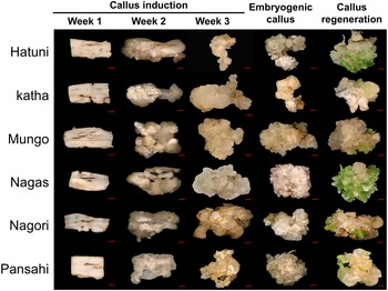
The process of callus induction and regeneration in S. barberi genotypes is shown in Fig. 1. During the first week, callus initiation was observed in leaf explants, characterised by the emergence of small, translucent calli clustered preferentially along the wounded edges. In contrast, callus formation in the central regions of the explants progressed more slowly. By the second week, the edge-derived calli exhibited accelerated growth, expanding and forming irregular structures. These calli developed superficial white, dry tissue while maintaining a moist underlying texture and translucent appearance. Concurrently, callus formation began in the central regions of the explants, forming a continuous layer over non-wounded areas. By the third week, the callus had developed into a thick tissue that fully enveloped the explants. At this stage, the Nagans genotype was distinguishable from others by its lighter callus pigmentation. During subculturing, calli from each genotype differentiated into embryogenic calli, although at varying percentages (Table 1). The embryogenic calli of different S. barberi genotypes consistently exhibited a dry and nodular surface texture. However, Mungo exhibited a more refined surface texture in the nodal structure of its callus when compared to other genotypes, including Hathuni, Katha, Nagans, Nargori and Pansahi.
Callus induction and regeneration for each of six Saccharum barberi genotypes. Hathuni, Katha, Mungo, Nagans, Nargori and Pansahi were included. Their callus induction performance in the initial 3 weeks, embryogenic callus in the sixth week and the callus regeneration are shown. Scale bar = 1 mm.

Statistical analysis of seven parameters for callus induction and plant regeneration among Saccharum barberi genotypes

Different lowercase letters indicate significant differences at P < 0.05 following Tukey’s HSD.
** and *** indicate P < 0.01 and P < 0.001, respectively. Statistical analysis was carried out as described in the ‘Materials and methods’ section.
Analysis of callus formation during induction revealed that CIF for all genotypes showed a general increasing trend over time (Fig. 2). The highest CIF values for all genotypes were observed at the third week rather than the sixth week, due to the necrosis of a small number of calli during subculture. Despite the loss of calli, there was no significant reduction in CIF, maintaining the highest level from the third week until the end of the 6-week callus induction period for each genotype.
Callus induction frequency over weeks for each Saccharum barberi genotype. Subculturing was carried out at the end of the third week. Data are presented as mean ± SD (n = 3). Tukey’s HSD was used to analyse the variation among weeks for each genotype. Different lowercase letters indicate significant differences at P < 0.05.

The response to callus induction and regeneration of S. barberi genotypes can be summarised according to the statistics of the parameters in Table 1. After the 6-week induction phase under dark conditions, 52.03% of the S. barberi explants developed callus, with 66.11% exhibiting embryogenic characteristics. This resulted in an average of 36.16% of explants producing embryogenic calli for each genotype. Following four weeks of light incubation for regeneration, 54.51% of calli exhibited differentiation, producing an average of 2.14 shoots per responsive callus. Evaluation of overall regenerative capacity revealed that 29.78% of initial explants yielded regenerated shoots, with a mean productivity of 0.483 shoots per explant. All regenerated shoots successfully developed into plantlets with well-established root systems after light incubation on RTM (Fig. S1).
Among the tested genotypes, Pansahi exhibited the best performance in callus induction and regeneration, showing the highest CIF, ECR, ECIF and CRF among all genotypes, along with a reasonable CRC. It also outperformed all genotypes in terms of ORF and ORC, two parameters for the comprehensive evaluation of regenerative competence (Table 1). In conclusion, the data indicated that Pansahi exhibited the highest regenerative capacity among all the materials tested. In contrast, Hathuni and Nagans exhibited the strongest tissue culture recalcitrance due to their poor performance in both callus induction and regeneration (Table 1).
Phenotypic diversity within the population was assessed through coefficients of variation (CVs) and fold-change analysis across seven parameters (Table 1). The observed CV ranges (22.82–68.08%) and fold-change magnitudes (1.78–7.10) revealed extensive phenotypic variation among genotypes. Variation magnitudes differed substantially across parameters, as evidenced by distinct CV and fold-change distributions. Notably, ECR and CRF exhibited the most constrained variability, with minimal CV and fold-change values compared to other parameters. Nevertheless, all tissue culture response parameters demonstrated statistically significant genotypic differences (P < 0.05; Table 1). The genotypic variance (σg 2) consistently exceeded the error variance (σ e2) for all the parameters. As a result, broad-sense heritability (H 2) values were found high across all traits, ranging from 80.77% for ECR to 93.10% for ECIF. These H 2 values suggested the effectiveness of the defined parameters in illustrating genotypic diversity and indicated that the different in vitro responses among S. barberi genotypes were primarily determined by genetic effect. The GCVs varied consistently with observation-based CVs among the parameters, with higher GCVs for ECIF, CRC and ORF, and lower for ECR and CRF, indicating different genetic diversity for the parameters in the tested S. barberi genotypes.
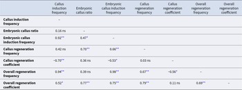
In this study, ORF and ORC serve as critical indicators of overall regenerative capacity. Statistical analysis identified three parameters exhibiting significant positive correlations with both indicators: CIF, ECIF and CRF (P < 0.05; Table 2). Among these three parameters, CIF was of particular interest due to its detectability at earlier stages compared to ECIF and CRF. To investigate whether early response to callus induction correlated with regeneration performance, the CIF data from the first three weeks were revisited (Fig. 3; Table 3). Strong positive linear correlations were found between CIFs at each of the three weeks and both ORF (Fig. 3A, C and E) and ORC (Fig. 3B, D and F). Notably, the first-week CIF demonstrated the strongest correlations with these parameters, exhibiting higher correlation coefficients (r values; Fig. 3A and B)) than those observed at the sixth week (Fig. 3G and H). During callus induction, both H 2 and GCV for CIF exhibited a progressive decline over time, with the highest values observed in the first week compared to subsequent weeks (Table 3). These patterns indicate that genotypic influences on CIF were most pronounced during the initial week of induction. Collectively, these findings demonstrate that genetically regulated early-stage performance in callus formation strongly associates with overall regenerative competence.
Relationships between callus induction frequency at different weeks and the overall regeneration frequency and the overall regeneration coefficient. (A), (C), (E) and (G) show relationships between callus induction frequency at different weeks and the overall regeneration frequency. (B), (D), (F) and (H) show the relationships between callus induction frequency at different weeks and the overall regeneration coefficient. The solid lines are the best fit linear equation for all measurements.

Correlations among parameters for callus induction and plant regeneration
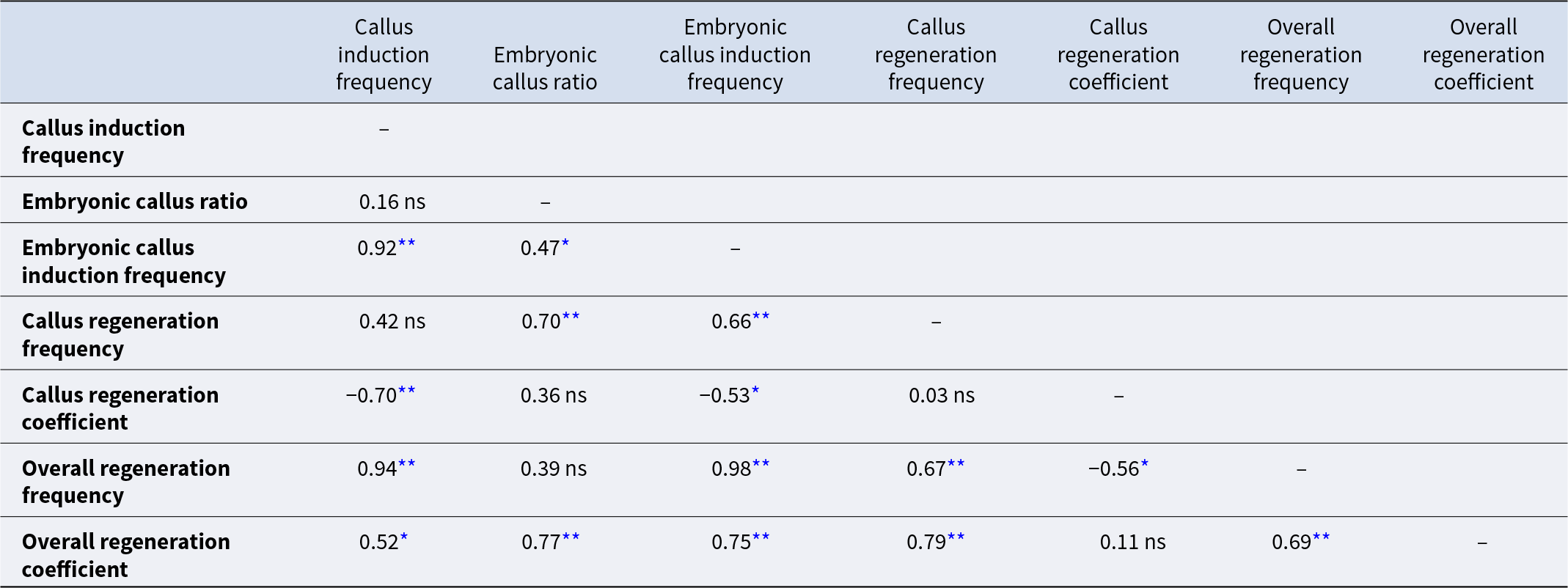
Pearson’s correlation coefficients are shown for each measurement. Statistical significance of the correlations was two-tail tested.
* P < 0.05; **P < 0.01; ns, not significant.
Variation in callus induction frequency over weeks for Saccharum barberi

* also shown in Table 1 as the result of parameter ‘callus induction frequency.’ The Calculation of GCV and H 2 is described in the ‘Materials and methods’ section.
Discussion
Tissue culture plays a pivotal role in various aspects of plant biotechnology. In this study, the callus induction response of six genotypes of S. barberi was assessed using seven parameters, revealing significant genotypic differences in each parameter.
Our findings align with previous research on tissue culture responses in sugarcane cultivars, highlighting substantial variations in tissue culture performance among different varieties (Abu et al. Reference Abu, Mekbib and Teklewold2014). However, the underlying reasons for these variations remain elusive. A quantitative analysis in S. spontaneum, a crucial wild germ plasm resource in the Saccharum genus, indicated that genetic variations predominantly drive the diverse responses to embryogenic callus induction and regeneration among genotypes (Li et al. Reference Li, Li, Lin, Qin, Lu, Mao and Liu2021). Similarly, in our study of S. barberi, broad-sense heritability (H 2) estimates revealed that genetic factors accounted for 80.77–93.10% of tissue culture traits (Table 1). These high H 2 values highlight the critical role of genotype effect in determining tissue culture response, consistent with previous findings on the genetic control of tissue culture performance (Li et al. Reference Li, Li, Lin, Qin, Lu, Mao and Liu2021).
H 2 is defined as the proportion of genetic variance contributing to overall phenotypic variance (Falconer, Reference Falconer1989). In addition to estimating the influence of genetic background on trait determination, H 2 is frequently calculated to assess the precision of experimental results (Schmidt et al. Reference Schmidt, Hartung, Rath and Piepho2019). In this study, high H 2 values were observed across all the seven parameters. These high H 2 values suggest that our data accurately reflect the impact of genetic variations in callus induction and plant regeneration. This outcome can be at least partially attributed to the experimental approach employed. Specifically, as with other tissue culture studies, our experiments were conducted under highly controlled environmental conditions. Additionally, we ensured a high level of homogeneity among the explants for each genotype by strictly limiting sampling to specific time frames and short sections of young leaf rolls. By minimising systematic error, these measures enhance the contribution of genetic variance to total phenotypic variance.
Despite the limited sample size in present study, our population exhibited substantial genetic diversity. This diversity was statistically validated by high value of CV and fold changes, as well as ANOVA result. Collectively, these metrics reflect pronounced phenotypic variation across genotypes. The high H 2values observed further indicate that genotypic variance predominantly underlies these phenotypic differences, aligning with correspondingly high GCV values. Within this compact yet diverse population, we successfully identified an elite genotype, Pansahi, with exceptional tissue culture performance. Pansahi outperformed other S. barberi genotypes in six out of the seven parameters investigated. Even when compared to highly amenable modern cultivars like RB85-5453 and IAC86-2210 (with CIF of ∼76% and callus regeneration frequency of ∼60%) (Nogueira et al. Reference Nogueira, Luis, Pasqual and Scherwinski-Pereira2019), Pansahi demonstrated comparable performance in both callus induction and plant regeneration. Based on its high performance in plant regeneration, we propose Pansahi as a model genotype for S. barberi biotechnology research, particularly in genetic engineering. Its superior in callus induction and regeneration capacity establishes Pansahi a promising transgene recipient for advancing in genetic transformation and genome editing studies. Additionally, comparative transcriptomic or proteomic studies between Pansahi and genotypes like Hathuni or Nagans, identified as having strong recalcitrance in this study, could offer insights into the molecular genetic basis of tissue culture performance in S. barberi.
Strong correlations between CIF and regeneration-related parameters were observed in S. barberi genotypes, consistent with findings in S. spontaneum (Li et al. Reference Li, Li, Lin, Qin, Lu, Mao and Liu2021). Similar associations between callus formation and regeneration capacity have been reported in other plant species like barley (Hordeum vulgare; Han et al. Reference Han, Jin, Wu and Zhang2011), duckweed (Wolffia arrhizal; Khvatkov et al. Reference Khvatkov, Chernobrovkina, Okuneva, Shvedova, Chaban and Dolgov2015), tomato (Solanum lycopersicum; Adhikari et al. Reference Adhikari, Yoon and Kang2016) and ryegrass (Lolium perenne; Grogg et al. Reference Grogg, Rohner, Yates, Manzanares, Bull, Dalton, Bosch, Studer and Broggini2022). Collectively, these studies suggest a conserved link between callus induction and plant regeneration across phylogenetically distinct species. Although these two biological processes represent distinct developmental stages, it can be hypothesised that certain pathways or mechanisms may function as a regulatory axis for both processes. Recently, Yang et al. (Reference Yang, Zhai, Wu, Deng, Chao, Meng, Chen, Liu, Bie, Sun, Yu, Zhang, Zhang, Chang, Xue, Zhao, Meng, Li, Zhang, Zhang, Zhao, Gao, Li and Li2024) identified the peptide REF1 as a local wound signal that promotes both callus formation and plant regeneration. This finding provides substantial evidence that supports the hypothesis. Exploring REF1 homologs in Saccharum complex plants could enhance our understanding of the mechanisms underlying callus induction and regeneration in sugarcane.
This study also demonstrated that CIF measured within the first week is strongly associated with regeneration-related parameters. This finding is significant for plant tissue culture research, as it implies the potential to predict overall regeneration performance through a simple and rapid callus induction test. In contrast to the comprehensive evaluation of regenerative competence, the callus induction test offers a simplified alternative that enhances both time and cost efficiency. Specifically, this method enables high-throughput screening by eliminating labour-intensive subcultures and reducing the total culture duration (e.g., processing genotype batches within 7-day cycles). Simultaneously, it lowers experimental costs by reducing chemical consumption and technician labour compared to conventional regeneration protocols. These advantages are especially relevant for programs that screening large populations for amenable genotypes in tissue culture research. In other crops, such as fodder pea (Pisum sativum var. arvense L.; Türkoğlu et al. Reference Türkoğlu, Bolouri, Haliloğlu, Eren, Demirel, Işık, Piekutowska, Wojciechowski and Niedbała2023), early callus induction performance has been recognised as a reliable predictor of subsequent plant regeneration. However, to establish robust predictive models correlating early-stage callus induction responses with regeneration performance in sugarcane, systematic expansion of tissue culture experiments is required. Future research should comprehensively evaluate the influences from critical environmental factors, including seasonal explant sourcing, media composition, photoperiod and temperature conditions. These factors have demonstrated significant impacts on callus induction and regeneration efficiency in some sugarcane genotypes (Birch, Reference Birch, Moore and Botha2014). A comprehensive analysis of their interactions with genotypes is expected to enhance the applicability of predictive models across diverse tissue culture systems in sugarcane.
In conclusion, our study reveals significant genetic variation in tissue culture performance among S. barberi genotypes. Pansahi emerged as an elite genotype due to its favourable responses in both callus induction and plant regeneration. Early callus induction performance was identified as a potential predictor of overall regenerative competence. This study represents the first report of genotypic variation in tissue culture response in S. barberi. Given the importance of tissue culture in plant research, our results could hold broad implications for applied and fundamental research in S. barberi.
Supplementary material
The supplementary material for this article can be found at https://doi.org/10.1017/S1479262125100191.
Funding statement
This study was funded by the National Key R&D Program of China (2022YFD2301100), the Science and Technology Major Project of Guangxi (Guike AA23073001), and the National Natural Science Foundation of China (32460729). S. barberi plants were provided by the NGRS-KY through the National Infrastructure for Crop Germplasm Resources (NCGRC-2024-42), the Government’s Purchase Public Services (19240681), and the Observation and Research Station of Yunnan Province (202205AM070001). The authors thank Yunnan Seed laboratory (202205AR070001-09) and the Central Government to Guide Local Science and Technology Development Funds (202407AB110007) for providing lab resources. We also thank Yanmei Dao and Hongyan Lu for assistance in tissue culture.
Competing interests
The authors declare no conflicts of interests.