Systemic sclerosis (SSc) or scleroderma is a heterogeneous autoimmune connective tissue disease of unknown aetiology that usually affects people between 30- and 50-years-old, 75% of them are female. SSc can be divided into two major subsets, limited cutaneous SSc (lcSSc) and diffuse cutaneous SSc (dcSSc), according to the extent to which the skin is affected (Ref. Reference Varga and Abraham1). The typical hallmarks of SSc are excessive accumulation of extracellular matrix (ECM) in the skin and internal organs (gastrointestinal tract, heart, lungs and kidneys), vasculopathy and immune abnormalities. SSc often leads to severe disability and premature death and among the connective tissue diseases has the highest all-cause mortality. Autoantibodies are present in more than 90% of patients. In particular, anticentromere (ACA), and anti Scl-70 (antitopoisomerase I) antibodies are closely associated with disease subset and can react with various intracellular components that bind to self DNA or RNA (Ref. Reference Hamaguchi2). These autoantibodies could potentially interact with Toll-like receptors (TLRs). Immune activation is common in SSc and although the focus has been on adaptive immunity in the form of T cells (Ref. Reference O'Reilly, Hugle and van Laar3), recent evidence suggests that innate immunity is critically important. Once histological fibrosis is established, this may be hard to reverse, however, the fact that inflammation precedes fibrosis suggests that this is playing a crucial role. The initiation phase of this disease may be mediated by innate immune signals, linking innate and adaptive immunity and fibrosis, through possible ‘danger signals’. Sterile inflammation occurring in SSc may be through such danger signals mediated via activation of TLRs. SSc is one of the most difficult rheumatic diseases to treat and although understanding of the pathogenesis of the disease has increased, there is still no proven treatment. Therefore, new approaches to SSc treatment are greatly needed. In this review, we argue that TLRs may be a novel attractive target.
Pathogenesis of SSc
SSc is a complex autoimmune connective tissue disease involving inflammation and fibrosis of the skin, lung and internal organs. There is a complex interplay between inflammation and the subsequent fibrosis although the precise molecular mechanisms remain obscure. Primarily, there is an immune activation and inflammation and then the fibroblasts differentiate into autonomous myofibroblasts secreting excessive collagens, primarily type 1. It has long been known that T cells play a critical role in SSc, possibly through the secretion of profibrotic cytokines once activated. T cells and other immune cells are found in the dermis of the skin in high numbers, especially early in the disease and are often found juxtaposed to myofibroblasts (Ref. Reference Roumm4). It has been suggested that monocytes along with fibroblasts play an important role in the production of profibrotic factors such as interleukin-6 (IL-6), collagen and tissue-inhibitor of metalloproteinase-1 (TIMP-1). TIMPs are specific inhibitors of matrix metalloproteinases (MMPs) regulating the balance of ECM turnover. Indeed, one of the hallmarks of SSc pathogenesis is pathological fibrosis development mostly because of accumulation of ECM proteins. Many different cytokines can induce regulators of ECM turnover and therefore alter the rate at which deposition of ECM occurs. Cytokines such as IL-6 can directly alter collagen I mRNA levels in targets cells. Interestingly, IL-6 is elevated in SSc serum compared with the controls and correlates both with skin thickness and has recently been identified to correlate with poor outcome (Ref. Reference Khan5). IL-6 is also induced on many ‘innate immune’ cells after activation and depending on the stimulus can result in extremely high levels. Recently, a shift in focus from adaptive to innate immunity in terms of initiation of disease has occurred across autoimmune diseases. This is further illustrated by the finding of an interferon (IFN) type gene ‘signature’ in SSc (Ref. Reference Tan6) and systemic lupus erythematosus (SLE) (Ref. Reference Baechler, Gregersen and Behrens7). Although clinically different diseases, they may share common disease mechanisms. Interestingly, dendritic cells (DCs), the sentinels of the immune system, have an elaborated response to TLR ligands producing excessive IL-6 in SSc (Ref. Reference van Bon8).
Role of TLRs in autoimmunity
TLRs are a family of evolutionarily conserved receptors that play a key role in sensing microbial or viral molecules as part of pattern recognition receptors (PRRs) (Ref. Reference Goutagny9). Indeed, TLRs are at the frontline of innate immunity protecting against invasive microorganisms playing a fundamental role in the maintenance of normal mammalian homoeostasis (Ref. Reference Hopkins and Sriskandan10). However, the members of the TLR family are also involved in the pathogenesis of autoimmune diseases, chronic inflammation and fibrosis development. In rheumatoid arthritis (RA), another chronic autoimmune disorder, TLRs have been shown to play a critical role. RA is characterised by inflammation and invasion of the synovium leading to bone erosion. Overexpression of TLRs 3 and 4 has been demonstrated in synovial tissue from RA patients (Ref. Reference Ospelt11). Indeed, injection of double stranded RNA, a TLR3 ligand, directly induces arthritis in mice and is partly mediated via IL-1 (Ref. Reference Zare12). Also, RNA released from dead or necrotic cells activates RA synovial fibroblasts to secrete a battery of cytokines and this is mediated via TLR3 (Ref. Reference Brentano13). Intriguingly, there is a clear link between smoking and development of RA and one of the mechanisms maybe through activation of TLRs, in fact, it was recently demonstrated that cigarette smoke induces the proinflammatory cytokine IL-8 by human macrophages and was TLR4-dependent (Ref. Reference Karimi14). Thus, this could link the risk of RA from smoking via TLR4 activation in macrophages. Macrophages are one of the key effector cells in the RA and their accumulation is found in the joint. Citrullinated fibrinogen has been demonstrated to stimulate macrophages to secrete tumour necrosis factor-alpha (TNF-α) and immune complexes containing citrullinated fibrinogen synergistically enhanced TNF-α secretion mediated via TLR4 and FcγR, thus citrullination may enhance the response in an inflammatory microenvironment (Ref. Reference Sokolove15). Another danger signal High mobility group box-1 (HMGB-1) has been shown to play a role in SLE pathogenesis (Ref. Reference Urbonaviciute16). SLE is another autoimmune disease in which TLRs are aberrantly activated and play a role in pathogenesis. SLE can affect the skin, joints and kidneys. SLE, such as SSc, is associated with a specific type I IFN signature. Using a well-known mouse model of SLE, Patole et al. demonstrated that RNA via TLR3 activation aggravates lupus nephritis primarily through expression on mesangial cells and antigen-presenting cells (APCs) (Ref. Reference Patole17).
The primary role of TLR signalling in connective tissue or for that matter any anatomical location is defence against pathogens. However, TLRs are also necessary for the maintenance of intestinal homoeostasis through their interaction with commensal flora and protection against gut injury (Ref. Reference Rakoff-Nahoum18). The activation is essential for innate immunity and modulating adaptive immunity but is associated with a variety of autoimmune diseases including SSc, SLE, RA, psoriasis and Sjogren's syndrome. Aberrant activation and downstream mechanisms play a prominent role in these disease settings. Thus, compounds that target such TLRs may be of therapeutic interest.
In psoriasis, an autoimmune skin condition, characterised by epidermal hyperproliferation and inflammation, TLRs aberrant activation has been identified. Keratinocytes have been reported to express TLRs 1, 2, 3, 5, 9 and 10. Whether keratinocytes express the Lipopolysaccharide (LPS) receptor TLR4 is less clear (Ref. Reference Kollisch19). Interestingly, TLR2 expression has been demonstrated to be much higher in psoriatic skin (Ref. Reference Begon20). Dectin-1, a PRR, is also differentially upregulated in psoriatic skin. Modulation of TLR1 and TLR2 has been shown to be downregulated in psoriatic skin after treatment with adalimumab (Ref. Reference De Pita21). Adalimumab leads to down regulation of TLR1 and 2 psoriatic skin, adalimumab is a fully human monoclonal antibody against TNF-α. There are also reports of induced psoriasis with the TLR7 agonist imiquimod (Refs Reference Wu, Siller and Strutton22, Reference Rajan and Langtry23). Thus, inhibition of TLR7 in psoriasis may represent an attractive therapeutic option.
SLE is an autoimmune disease characterised by chronic inflammation of the skin and joints. It is also characterised by a specific set of autoantibodies specifically antinuclear antibodies. The role of the TLR signalling system in this disease is now being elucidated. It is known in SLE that higher levels of autoantibodies against DNA and nucleoprotein circulate and that these immune complexes can be pathogenic through the deposition of these immune complexes in the kidney. There is also impaired apoptotic cell clearance in SLE patients. These DNA binding immune complexes have been shown to activate DCs, the sentinels of the immune system via ligation of the DNA-binding TLR9 system (Ref. Reference Means24). Thus, linking the DNA complexes and immune system activation lead to pathology through the secretion of soluble cytokines. Since TLR9 signalling takes place in intracellular lysosomes this can be blocked by chloroquine; chloroquine is an effective treatment in some SLE patients. The mode of action may be the blockade of intracellular signalling of TLR9-induced immune DNA complexes through the alteration of intracellular lysosomal acidification. Indeed, it has been shown that mammalian DNA and RNA are potent inducers of IFN-α mediate via TLR9 and that these can be blocked by synthetic inhibitors (Ref. Reference Barrat25). TLR9 and 7 activation triggers downregulation of blood dendritic cell antigen-2 (BDCA2) expression in SLE patients DCs suggesting a shift to a more ‘activated’ DC which is accompanied by marked increases in IFN-α (Ref. Reference Wu26). There also appears to be an expansion of B cells especially memory B cells in patients with active SLE expressing TLR9. Incubation of healthy control B cells with SLE serum leads to the upregulation of TLR9 expression on healthy B cells. Moreover, after TLR9 ligation in B cells there was an increased human leukocyte antigen-DR (HLA-DR) expression on the cells and there was a correlation between TLR9 expression and anti-DNA antibodies (Ref. Reference Papadimitraki27). However, the role of TLR9 in SLE remains controversial as TLR9 deletion in mice does not reduce pathology but rather exacerbates disease via a TLR9-independent pathway (Refs Reference Yu28, Reference Yu, Musette and Peng29). Moreover, single nucleotide polymorphisms in 3′UTR of TLR7 has been demonstrated to contribute to SLE incidence in women (Ref. Reference Kawasaki30).
Sjogrens syndrome is a systemic autoimmune disease that leads to the destruction of exocrine glands, lacrimal and salivary glands and is characterised by dry mouth and dry eyes (sicca). Although the adaptive immune system has long since been associated with SS it is now becoming clear that the innate immune system is at play. In an animal model of the SS it was shown that general inflammatory activation leads to accelerated SS-like pathology before initiation and activation of the adaptive arm of the immune system (Ref. Reference Deshmukh31). Furthermore, activation of the innate immune system via injection of TLR3 agonist Poly(I:C) caused a rapid loss of salivary hypofunction in mice and this was accompanied by a type I IFN signature. Interestingly, this was reversible after cessation of the Poly(I:C) (Ref. Reference Deshmukh31).
RA is a chronic autoimmune disease characterised by inflammation and joint destruction. It can affect multiple joints of the hands and feet and results in pain and stiffness with progressive destruction of bone and cartilage. Synovial fibroblasts are considered pivotal players in the pathogenesis of RA (Ref. Reference Neumann32). Multiple lines of evidence suggest that TLRs are involved in the disease. For example, there is a higher overexpression of TLRs3 and 4 in RA synovium and RA synovial fibroblasts even at an early stage of the disease (Ref. Reference Ospelt11). Moreover, these RA synovial fibroblasts cultured from biopsies and stimulated with the TLR3 ligand Poly(I:C) resulted in cytokine release (IL-6) and MMPs being secreted. MMPs mediate the breakdown of cartilage. Furthermore, CD16+ macrophages have been demonstrated to express higher levels of the TLR2 receptor in RA compared with healthy controls and this was also shown in synovial macrophages in the synovium. Also, in vitro studies on these macrophages from RA containing higher levels of TLR2 were found to respond to the endogenous danger signal heat shock protein 60 (HSP60) and therefore result in high release of TNF-α. TNF-α is a critical molecule in RA pathogenesis (Ref. Reference Iwahashi33). It also indicates that endogenous danger signals (damage associated molecular patterns, DAMPs) can stimulate TLRs in RA. TLRs3 and 7 have also been demonstrated to be elevated in the RA synovium and DCs derived from RA patients (monocyte-derived DCs) with combined stimulation of TLR3, 4, 7/8 resulted in marked synergy of production of proinflammatory cytokines that would perpetuate the damage in the joint (Ref. Reference Roelofs34). DCs are the sentinels of the immune system that when ‘activated’ polarise the immune response. In further animal experiments it was elegantly shown that viral double stranded RNA (vdsRNA) lead to arthritis in mice and that this was not dependent on acquired immune responses as severe combined immunodeficiency (SCID) mice, with no acquired immunity, also developed arthritis (Ref. Reference Zare12). Interestingly, by using etoposide to selectively deplete monocytes in mice, the authors also demonstrated a total abrogation of the vdsRNA-induced arthritis in mice, arguing for a central role of the monocytes (Ref. Reference Zare12). However, mice deficient for the dsRNA TLR receptor: TLR3 still developed arthritis triggered by vdsRNA, suggesting another receptor is responsible. The use of a TLR2 neutralising antibody, OPN-301, has been used in RA synovial explant cultures to attenuate TNF-α induction (Ref. Reference Ultaigh35). This is interesting as another TLR2 ligand, serum amyloid A, has recently been described and plays a role in mediating in vivo monocyte recruitment and enhanced angiogenesis in the human RA-SCID mouse xenograft model (Ref. Reference Connolly36).
Complex roles of TLRs in SSc
As outlined in the previous section the role of TLRs in mediating pathology in various autoimmune disorders is now firmly established. However, the role of TLRs in SSc pathology is only just beginning to be understood. TLRs are widely expressed by many cells of the immune system as well as nonimmune cells such as fibroblasts, epithelial or endothelial cells. Currently, 13 TLRs have been identified in mammals and 10 (TLR1-10) in humans; located on the cell surface as membrane proteins or expressed in the endocytic vesicles. Following ligand binding, TLRs activate signalling components to initiate immune responses for host defence. TLRs are type I membrane glycoproteins characterised by an extracellular domain containing leucine-rich repeats (LRRs) and a cytoplasmic tail which is shared with interleukin-1 receptors (IL-1Rs) the same highly conserved region of amino acids now called Toll/IL-1R (TIR) domain. It was shown that mice deficient in Myeloid differentiation primary response gene 88 (MyD88), a TLR-signalling adaptor protein, are less prone to fibrosis development, pathological inflammation or cardiac hypertrophy, implying a crucial role of MyD88 in fibrogenesis and thus TLRs (Ref. Reference Singh37). Interestingly, many recent studies addressed the role of endogenous ligands in TLR-mediated cell activation. Such endogenous ligands are released upon tissue damage and cell stress. Indeed, a common feature in SSc is vascular injury manifesting as Raynaud's phenomenon and ischaemia of skin and visceral organs. We recently found that both TLR4 and TLR8 play an important role in TIMP-1 and IL-6 production by SSc monocytes (Ref. Reference Ciechomska38). It is therefore tempting to speculate that more than one TLR agonist is needed to initiate and promote chronic inflammatory responses and fibrosis progression as observed in SSc. This implies that different TLR pathways might simultaneously lead not only to the breakdown of tolerance seen in autoimmunity, but also to initiate trans-differentiation of pathogenic myofibroblasts. Myofibroblasts are the highly contractile pathogenic cells found in scar tissue and facilitate the deposition of ECM. Under normal physiological conditions MMPs degrade ECM components, where as TIMPs inhibit MMPs function in a 1:1 ratio in a highly regulated manner (Ref. Reference Clark39). However, in the SSc, the equilibrium between TIMPs and MMPs favours ECM accumulation mediated by increased levels of TIMP-1. A previous study showed that TIMP-1 was increased in sera from SSc patients compared with healthy controls (Refs Reference Young-Min40, Reference Zurita-Salinas41). Also, a gene expression study demonstrated differential gene expression of MMP-1 and TIMP-1, with highly increased TIMP-1 expression observed in SSc skin (Refs Reference Rajagopal42, Reference Frost43). Such an imbalance between the inhibitor and the activator of matrix products is proposed to underlie the accumulation of ECM seen in the SSc. Furthermore, our studies demonstrate that circulating healthy CD14+ monocytes stimulated with SSc sera, but not with healthy controls (HC) or RA sera, induced strong TIMP-1 production in a MyD88-dependent fashion, whereas the level of MMP-1 remained unchanged. This demonstrates that the SSc sera contain TLR ligand(s) that trigger TIMP-1 production but not MMP-1 synthesis.
TLR4 and TLR2 in SSc
A study found that in SSc patients with interstitial lung disease (SSc-ILD), CD14+ circulating monocytes in response to TLR4 agonist LPS overexpress CD163, IL-10 and the chemokine ligand CCL18. These molecules are known to stimulate collagen secretion by fibroblasts and induce migration of T cells to inflamed lung tissue. This study also showed an increased number of circulating lymphocytes and monocytes producing collagen in SSc-ILD patients compared with age-matched controls (Ref. Reference Mathai44). Similarly, van Lieshout et al, showed that TLR4-activated monocytes and DCs from SSc patients produce high levels of CCL18 and IL-10 (Ref. Reference van Lieshout45). Indeed, previously it has been shown that CCL18 levels are elevated in SSc sera. These results suggest that TLR-stimulated monocytes along with DCs are potential sources of circulating CCL18, a T cell chemoattractant and profibrotic factor in SSc. In nephrogenic systemic fibrosis, a disease with clinical features of SSc, compounds such as gadolinium-based contrasting agents bind TLR4 and TLR7 in differentiated macrophages resulting in activation of nuclear factor kappa beta (NF-κβ) and expression of IL-4, IL-6 and transforming growth factor β (TGF-β) (Ref. Reference Wermuth and Jimenez46). All these downstream mediators are potent profibrotic molecules, thus linking external stimuli and TLR ligation in immune cells and fibrogenic factors. Interestingly, chloroquine preincubation prior to galidium application significantly attenuated the induction of the profibrotic cytokines in these macrophages. It has been shown that hepatic fibrosis can be mediated via TLR4 and its ligand LPS in vivo and that this TLR4 stimuli sensitises cells to the effects of the profibrotic cytokine TGF-β via downregulation of the TGF-β pseudoreceptor bone morphogenic protein (BMP) and activin membrane-bound inhibitor homologue (Bambi) (Ref. Reference Seki47). The increased exposure to the LPS comes from the fact that the intestinal barrier is compromised and thus hepatic exposure to bacterial products increases as in these mice administration of nonabsorbable broad spectrum antibiotics results in abolishment of the fibrosis. LPS has also been shown to mediate the upregulation of collagen facilitated by down regulation of miRNA29a and other family members who have collagen as their target genes (Ref. Reference Roderburg48), thus linking innate immune responses mediated via TLR4 and collagen secretion facilitated by an epigenetic mechanism. In addition, it was shown that the degradation of matrix proteins, including hyaluronan, fibronectin and collagen, was involved in the activation of the inflammatory response and fibrosis development. In particular, hyaluronan by binding to TLR2 and TLR4, abundantly expressed on the surface of DCs and macrophages, induced strong proinflammatory TNF-α and IL-1 production. This suggests that hyaluronan can act as a ‘danger signal’ in certain contexts, such as tissue architecture disruption as found in trauma. Also, a recent study showed that a TLR polymorphism may be correlated with SSc phenotype and disease development: SSc monocytes carrying the rare variant of TLR2 Pro631His increased the production of proinflammatory IL-6 and TNF-α upon TLR2 agonist stimulation (Ref. Reference Broen49). Furthermore, this polymorphism was associated with a higher titre of anti-topoisomerase antibody and with development of pulmonary arterial hypertension. This indicates that different TLR2 variants are associated with SSc disease severity by promotion of a proinflammatory environment.
Intracellular TLRs in SSc
Intracellular TLRs have evolved to recognise nucleic acids. A study found that healthy monocytes stimulated with TLR7, TLR9 and TLR3, but not with TLR2, induced a strong expression of Siglec-1 (CD169), which is an IFN-regulated gene. This sialoadhesin protein is highly upregulated in circulating monocytes from SSc patients. Furthermore, by blocking the type I IFN with a chemical inhibitor the expression of Siglec-1 was reduced following TLR3 agonist stimulation, suggesting that certain TLR agonists may induce monocytes differentiation towards a profibrotic phenotype observed in SSc (Ref. Reference York50). Overexpression of endothelin-1 (ET-1) has also shown to be involved in SSc development by collagen and fibronectin synthesis induction by fibroblasts. Indeed, Farina et al, showed that dsRNA (TLR3 ligand) strongly upregulated ET-1 mRNA expression by dermal fibroblasts isolated from SSc patients, whereas selective blocking of TLR3 with bafilomycin attenuated ET-1 expression (Ref. Reference Farina51). Interestingly, other TLR ligands had no effect on ET-1 expression. Furthermore, dermal fibroblasts from both SSc and healthy donors displayed an increased expression of IFN-regulated gene OAS2 and chemokines CXCL9 and CXCL10 following TLR3 stimulation (Ref. Reference Farina52). Agarwal et al, also demonstrated that SSc fibroblasts upregulated expression of TLR3 in response to IFN-α resulting in enhanced secretion of IL-6 and monocyte chemotactic protein 1 (MCP-1) (Ref. Reference Agarwal53). The elevated level of MCP-1 was seen in fibroblasts stimulated with autoantibodies that bind to fibroblasts surface by TLR4 (Ref. Reference Chizzolini54). These data suggest that TLRs activation may promote the recruitment of inflammatory cells. In addition, MyD88 deficient mice with bleomycin-induced pulmonary fibrosis had reduced recruitment of neutrophils and lymphocytes in the bronchoalveolar spaces compared with wild-type (WT) mice (Ref. Reference Gasse55). Bleomycin is a glycopeptide antibiotic used as a chemotherapeutic agent for lung and dermal fibrosis induction in animals as a model for human SSc. In particular, inhibition of fibrosis was indicated by the reduced expression of TIMP-1 and collagen in the lung tissue of MyD88-deficient mice. These data clearly underlie the essential role of MyD88 adaptor protein and TLR signalling in fibrogenesis by the promotion of monononuclear cells recruitment and matrix protein deposition in activated fibroblasts.
DAMPs in SSc
Another family of PRR ligands are DAMPs (alarmins) released by ‘stressed’ or necrotic cells or via mechanical trauma. They act as endogenous ‘danger signals’ to promote the inflammatory response mediated via TLRs, linking damage and functionally important immune changes. Numerous studies have suggested that endogenous danger signals within the inflamed joint contribute to the pathogenesis of RA (Ref. Reference Huang and Pope56) based on the observation of various endogenous ligands that are contained within the affected joints. One of the first endogenous ligands for TLRs to be described was HSP70. Indeed, TLR4 has been identified as the receptor for endogenous HSP70 (Refs Reference Asea57, Reference Ohashi58) that mediates the multiple downstream signalling effects in association with CD14. HSP70 is an intracellular protein that acts as a molecular chaperone transporting proteins within the cell that prevents protein misfolding and is also hugely upregulated under conditions of chemical, oxidative or thermal stress (Ref. Reference Ogawa59). HSP70 normally resides in the cells and lacks a leader sequence, hence it cannot be secreted in the normal way; hence release of HSP is suggested to be a danger signal to elicit an inflammatory response to restore homoeostasis. However, caution must be taken when interpreting results as we now know that a large proportion of earlier studies using recombinant proteins engineered in bacteria were contaminated with endotoxin. Therefore, spurious results and conclusions were generated. Ogawa et al demonstrated elevated levels of HSP70 from SSc patient serum compared with controls which correlated with the modified Rodnan skin score, a validated score of skin thickening (Ref. Reference Ogawa60). It is likely that damaged or stressed cells released this HSP70 into circulation to bind their cognate TLRs and induce an appropriate response, ultimately leading to an increased gene expression of proinflammatory mediators. Furthermore, it has been also shown that overexpression of HSP47 is involved in collagen accumulation that consequently leads to fibrosis development (Ref. Reference Nagata61). HMGB protein families are nuclear proteins nonhistone associated, chromatin-bound that are involved in making DNA available for the regulation of gene transcription. HMGB-1 is a 215-amino-acid soluble protein composed of two DNA-binding domains that binds to DNA in a sequence-independent manner. During injury or inflammation, HMGB-1 is released and through interactions with its receptors – TLR2, TLR4 and receptor for advanced glycation end products (RAGE), they induce production of proinflammatory mediators. HMGB-1 is elevated in the synovia of RA patients and is elevated in serum and blockade of HMGB-1 in rodent models of RA alleviates disease (Ref. Reference Kokkola62). In SLE, HMGB-1 is a component of immune complexes containing anti-DNA because of its interaction with DNA. HMGB-1 has also been proposed to play a pathogenic role in SSc (Ref. Reference Yoshizaki63). Yoshizaki et al found that both HMGB-1 and soluble sRAGE were elevated in SSc patients serum compared with healthy controls. Furthermore, elevated levels of HMGB-1 correlated with total skin thickness (Ref. Reference Yoshizaki63), suggesting a causal relationship between the two. The results from our group also showed that SSc monocytes stimulated with HMGB-1 in synergy with LPS, produce increased levels of profibrotic TIMP-1 and IL-6 compared with monocytes isolated from healthy individuals (unpublished data). Another study showed that bleomycin treated mice increased cell infiltration in the dermis and enhanced the production of hyaluronan (endogenous danger signal) in the skin, lung and sera upon TLR4 activation expressed on B-cells surface (Ref. Reference Yoshizaki64). In contrast, CD19-deficient mice suppressed hyaluronan-mediated TLR4 activation by reduced skin cell infiltration and decreased levels of IL-4, IL-6, IL-10, IFN-γ, TNF-α, TGF-β1 and MIP-2 after bleomycin treatment. These data suggest that bleomycin enhances hyaluronan production and activates B cells via TLR4 ligation to produce profibrotic cytokines and autoantibody induction that consequently promotes recruitment and retention of mononuclear cells within the tissue (Ref. Reference Yoshizaki64). Similarly, another study showed that bleomycin treated TLR2−/−TLR4−/−mice, following hyaluronan stimulation, had a reduced inflammatory response to lung injury, with a decrease in transepithelial neutrophil migration and reduced expression of MIP-2 (Ref. Reference Jiang65). A final group of endogenous danger signals are the members of the S100 protein family. The S100 proteins are calcium binding proteins and exist in the cell as dimers or multimers. These two members of the family, SA100A8 and SA100A9, have recently been described as alarmins that bind to TLR4 on monocytes (Ref. Reference Vogl66). These proteins have been shown to be strongly and independently correlated with the joint damage observed in RA (Ref. Reference Hammer67) and are highly elevated in the synovial fluid from RA patients. Furthermore, S100A7 has been shown to be elevated in SSc patients (Ref. Reference Baldini68) and S100A9 is elevated in the lungs of idiopathic pulmonary fibrosis patients (Ref. Reference Korthagen69).
Taking all the above studies together, the canonical TLR ligands and endogenous danger signals play an important role in autoimmunity and fibrosis development and remain an interesting subject for further investigation in SSc as clinical markers or even potential therapeutic targets.
TLRs signalling mechanism
Our understanding of innate immunity has led to the discovery of innate sensors or PRR which are able to recognise and eliminate highly conserved microbial components widely expressed by bacteria, fungi, protozoa and viruses. Nacht, LRR and PYD domains-containing protein-3 (NALP3), which is involved in inflammasome formation, has been shown to be important in uric acid crystals recognition during bleomycin administration resulting in IL-1β and TIMP-1 upregulation (Ref. Reference Gasse70). This suggests that nonTLR receptors are also involved in inflammation and fibrosis development (Ref. Reference Lafyatis and York71).
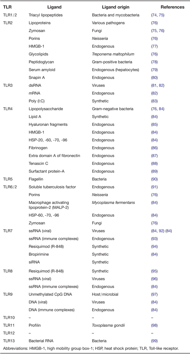
Stimulation of TLRs with their associated ligands, with the exception of TLR3, results in the association of the MyD88 adaptor protein. Homodimerisation of MyD88 is a critical step in the downstream signalling process which allows the recruitment and activation of the IL-1R-associated kinase (IRAK). This downstream MyD88 activation triggers the translocation of cytoplasmic transcription factors into the nucleus (Fig. 1). In particular, during translocation of transcription factors including NF-κβ), the activator protein-1 (AP-1) or interferon regulatory factors (IRFs) subsequently leads to activation of various genes encoding proinflammatory and profibrotic cytokines, chemokines but also collagen (Refs Reference Clancy72, Reference Couillin73). Only TLR3 uses TIR domain-containing adaptor-protein-inducing IFN-β (TRIF) as its sole adaptor molecule, whereas TLR4 activates both MyD88- and TRIF-dependent pathways. TRIF also induces the delayed activation of NF-κβ, expression IFN-β and -inducible genes through the activation of TANK-binding kinase (TBK) 1 and I-kappa-B- kinase epsilon (IKK-ε). Different TLRs recognise their corresponding PAMPs or endogenous molecules (Table 1). TLR signalling is also negatively regulated by various proteins. It has been shown that soluble decoys (sTLRs), in particular sTLR4 and sTLR2, effectively blocked LPS-induced TNF-α or IL-8 production by macrophages. Another group of negative regulators are intracellular proteins such as MyD88s or IRAK-M which disrupt the recruitment and activation of IRAK4 and IRAK1. Similarly, TRIAD3A (member of the RING-finger E3 ligases) reduce the expression of TLR by promoting the process of TLR4 and TLR9 ubiquitylation, therefore reducing the intensity and duration of TLR signalling (Ref. Reference Connolly and O'Neill100). Another important regulator of TLR signalling to dampen the TLR-induced inflammatory response is microRNAs (miRNAs). miRNAs are small 21–22 nucleotide noncoding RNAs that are intergenic or intronic. miRNAs regulate gene expression by repression of mRNA and can target multiple mRNAs (Ref. Reference O'Neill, Sheedy and McCoy101). miRNAs serve to fine tune the TLR signalling system by altering gene expression post-translationally and include miRNA 146a (Ref. Reference Taganov102). Resolution of the TLR activation and subsequent inflammation is probably mediated via miRs. Overall, the balance between activation and inhibition is the key determinant of the signal strength of TLR pathways. For that reason, the nature and specificity of the negative regulatory mechanism of TLR signalling might be a powerful therapeutic tool to prevent or modulate autoimmune diseases including SSc in the near future. Mir antagomirs have been shown to be effective in attenuating ova-induced arthritis in vivo (Ref. Reference Stittrich103).
Toll-like receptor (TLR) signalling pathways. Stimulation of TLRs can result in signalling via the myeloid differentiation primary response gene 88 (MyD88)-dependent or -independent pathways. During the MyD88-dependent pathway ligand binding to the TLR causes recruitment of the adaptor protein MyD88 to the cytoplasmic region of the receptor. MyD88 then recruits IL-1R-associated kinase 4 (IRAK4) which phosphorylates IRAK1 initiating a downstream signalling pathway leading to the phosphorylation of the inhibitor of nuclear factor κβ (NF-κB) complex (IκB complex). IκB dissociates from nuclear factor-κ light-chain-enhancer of activated B cells (NF-κB) and is degraded. NF-κB then translocates to the nucleus where it activates the transcription of inflammatory genes. MyD88-dependent signalling can also result in the nuclear translocation of the activator protein-1 (AP-1) transcription factor via c-Jun N-terminal kinase (JNK) and p38 signalling. Signalling via the MyD88-independent or TIR-domain-containing adapter-inducing interferon-β (IFN-β) (TRIF) pathway can also induce NF-κβ translocation or activate the IFN-1 transcription factor via IKKi and TANK-binding kinase 1 (TBK1) activation. MyD88-dependent signalling is targeted by negative regulators including MyD88S, a splice variant of MyD88, which can be recruited to the cytoplasmic TLR region in place of Myd88 thus inhibiting signalling, IRAK-M which prevents IRAK4 and IRAK1 recruitment, A20 which is a deubiquitylating enzyme and toll-interacting protein-1 (TOLLIP-1) which is thought to prevent phosphorylation of IRAK1. Suppressor of cytokine signalling 1 (SOCS1)

TLRs and their known identified ligands
Abbreviations: HMGB-1, high mobility group box-1; HSP, heat shock protein; TLR, Toll-like receptor.
Therapeutic targeting of TLRs in SSc
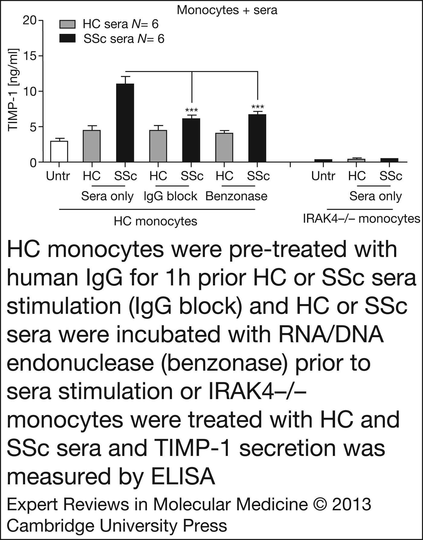
In SSc, there is inflammation leading to fibrosis, however, the link between inflammation and fibrosis is only now being elucidated and the molecular mechanisms are unknown (Ref. Reference O'Reilly104). Serum immune complexes of antibodies complexed with other protein and possibly lipids have been described in the SSc (Ref. Reference French105). It was also noted that these complexes associate with the antinuclear antibody status of the patients. Further evidence for the role of immune complexes in SSc comes from the fact that the complement that binds immune complexes is elevated in SSc patients' plasma (Ref. Reference Senaldi106). Immune complexes have been demonstrated in SSc to bind to Fcγ receptors on resident cells leading to TLR8 engagement in the endosome (Ref. Reference Kim107) with IFN-α being produced. Blockade of the Fcγ receptor on DCs with the use of a neutralising antibody anti-CD32a, reduced the immune complex-induced IFN-γ secretion demonstrating an absolute requirement for Fcγ engagement. Also, endosomal internalisation is needed to process the immune complexes as incubation with bafilomycin A1 reduced immune complex-TLR mediated IFN-γ secretion. Bafilomycin A1 is an inhibitor of lysosomal acidification which is necessary for appropriate TLR-endosomal signalling. Intracellular TLRs recognise nucleic acids. It was demonstrated that pretreatment of isolated cell extracts with DNAse or RNAse with the SSc patients' serum degraded the DNA or RNA, there was a marked reduction in IFN-γ secretion showing that nucleic acid was inducing IFN-γ secretion in combination with immune complexes. Thus, there was an immune complex in the SSc patients' serum that contains both autoantibodies, DNA or RNA, engages Fcγ receptors on appropriate target cells and binds intralysosomal TLR receptors leading to activation and secretion of IFN-γ. It was shown in SLE that the immune complexes containing small nuclear RNA in SLE patient's serum can activate TLR8 in the endosome to finally synthesise and secrete IFN-γ (Ref. Reference Vollmer108). This was also demonstrated to be mediated by the uptake through CD32/Fcγ receptors as blockade of the Fcγ receptor reduced serum-induced IFN-γ production. We have described a similar phenomenon in SSc serum where we postulate that circulating immune complexes are bound to cellular RNA and lead to secretion of TIMP-1 from immune cells (Ref. Reference Ciechomska38). These circulating factors inducing TIMP-1 could be inhibited by both blocking Fcγ receptors on CD14+ monocytes with the use of a blocking antibody and also could be inhibited with RNAse treatment, thereby degrading the cellular RNA. The mechanism is MyD88-dependent as blockade of MyD88 with a blocking peptide, but not with a scramble peptide, reduced sera-induced TIMP-1 production in monocytes. Furthermore, we used monocytes derived from a patient with a genetic defect in IRAK4, resulting in no IRAK4 protein production, a downstream signalling molecule from TLRs, to show that IRAK4 was an absolute requirement for TIMP-1 production subsequent to SSc patient serum incubation (Fig. 2). Thus, targeting TLR8 may be a therapeutic option in SSc or indeed targeting Fcγ receptors too may yield results in SSc. It is suggested that the RNA that is binding in the immune complexes prior to Fcγ receptor-mediated monocytes internalisation is derived from ‘damaged’ host cells and is acting as a danger signal to initiate wound repair. It is known that there is genetic variation in the TLRs and signalling components that result in differential responses to ligands (Ref. Reference Kiechl109), therefore, some of the clinical heterogeneity that characterises SSc could be because of genetic variation in the TLRs that mediate their effects. In other words, responses to endogenous danger signals and immune complexes may be different depending on TLR genetic variation and could account for clinical variability.
HC monocytes were pre-treated with human immunoglobulin G (IgG) for 1 h prior to HC or SSc sera stimulation (IgG block) and HC or SSc sera were incubated with RNA/DNA endonuclease (benzonase) prior to sera stimulation or interleukin-1 receptors-associated kinase 4 (IRAK4 − /−) monocytes were treated with HC and SSc sera and tissue-inhibitor of metalloproteinase-1(TIMP-1) secretion was measured by ELISA. The data show that RNA and IgG receptors play a role in induction of profibrotic TIMP-1 and that IRAK4 is critical in this process (38). *** Significantly different analysis of variance (ANOVA).
Conclusion
TLRs are a group of germline-encoded PRRs that play a role in both homoeostatic health and disease. In terms of autoimmune diseases they are prominent in disease pathogenesis including RA. In particular, in the autoimmune disease SSc they appear to be important in disease initiation and progression with multiple TLRs being implicated in provoking inflammation. The nature of the TLR ligands appears to be immune complexes of autoantibodies, complexed with RNA and nuclear material binding to Fc receptors and leading to the synthesis and subsequent secretion of profibrotic proteins including collagen and TIMP-1. TIMP-1 is a negative inhibitor of MMPs and thus if TIMP-1 is higher this will inhibit the breakdown of ECM and the net effect will be deposition of ECM: fibrosis. We have demonstrated the role of immune complexes in activation of TLR8-induced TIMP-1 production leading to matrix deposition facilitated by Fcgamma receptors on monocytes, these immune complexes contain RNA species, as incubation of an RNA degrading enzyme suppressed TIMP-1 induction. However, one question remains: where is the RNA that is bound in the immune complex coming from? What is the source? Although we could not identify the source of the cellular RNA complexed to immunoglobulins it is tempting to speculate that this may be released from dead cells. It is speculated in the SSc that the initial insult is damaged vascular tissue, thus the damaged vascular tissue may release dead cells that allow otherwise ‘hidden’ RNA to be recognised by the immune system which then binds in a complex. Targeting TLRs with specifically designed antagonists appears a real therapeutic possibility. We suggest specifically to target monocytes TLR8 based on our own observations. A TLR8 antagonist is currently under development. Redundancy in the system means that unwanted effects would be mitigated. It is worth noting that there is no effective treatment in SSc and new therapies are urgently needed.