





Introduction
In humans, tryptophan (TRP) is an essential amino acid that is exclusively obtained through dietary sources(Reference Gao, Xu, Liu, Liu, Bai and Peng1). TRP is required for the synthesis of proteins and several important bioactive molecules, such as serotonin (SER), melatonin (MEL), kynurenine (KYN), and niacin. It plays a pivotal role in gut–brain functions, bone health, immune modulation, and mitochondrial function(Reference Nayak, Singh and Buttar2). TRP and its metabolites are aryl hydrocarbon receptor (AhR) ligands(Reference Aust, Vidova, Coufalikova, Smetanova, Kozeluhova and Micenkova3), which are also discussed in this review. The gut microbiome and its produced TRP metabolites affect the host(Reference Aust, Vidova, Coufalikova, Smetanova, Kozeluhova and Micenkova3,Reference Taleb4) and the host physiology(Reference Zhang, Tang, Chen, Xie and Tao5), including stimulating the immune system development and homeostasis(Reference Kim, Galan, Hill, Wu, Fehlner-Peach and Song6,Reference Zhu, Song, Shen, Quan, Tan and Luo7) . Furthermore, they were reported to maintain intestinal barrier integrity(Reference Dodd, Spitzer, Van Treuren, Merril, Hryckowian and Higginbottom8), reaping inaccessible nutrients from the diet(Reference Koh, De Vadder, Kovatcheva-Datchary and Bäckhed9), and synthesising specific essential vitamins and neurotransmitters(Reference Lynch and Pedersen10).
In this systematic review, we provide a brief overview of TRP’s biochemical and nutritional fundamentals, and outline the essential nature of TRP, its metabolic fates (SER, MEL, KYN, its indole derivatives), and the dietary sources and requirements that determine its systemic availability as produced by the host’s gut microbiota.
Secondly, we highlight the nutritional context of TRP to disease susceptibility (e.g., immune dysregulation) and address how targeted nutritional strategies could mitigate these risks.
We also highlight the host–microbiota crosstalk mediated via the AhR and pregnane X receptor (PXR) pathways, and the role of TRP and its metabolites as modulators of the immune response of the host, intestinal barrier integrity, and neuro-immune signalling.
In addition, we focus on the relevance of TRP in diseases across multiple organ systems and address its role as a therapeutic agent in, for example, probiotic supplementation, indole-based pharmacology, and metabolic engineering of gut bacteria, thus providing a brief overview of TRP and its indole derivatives as promising modulators of health and therapeutic agents for a broad spectrum of immune-mediated, neuro-, and cardiovascular disorders.
For this systematic review, the authors carried out a web-based search on PubMed and Google Scholar using the key word ‘tryptophan’ combined with ‘nutrition’, ‘immune system’, and ‘gastrointestinal tract’.
TRP chemistry
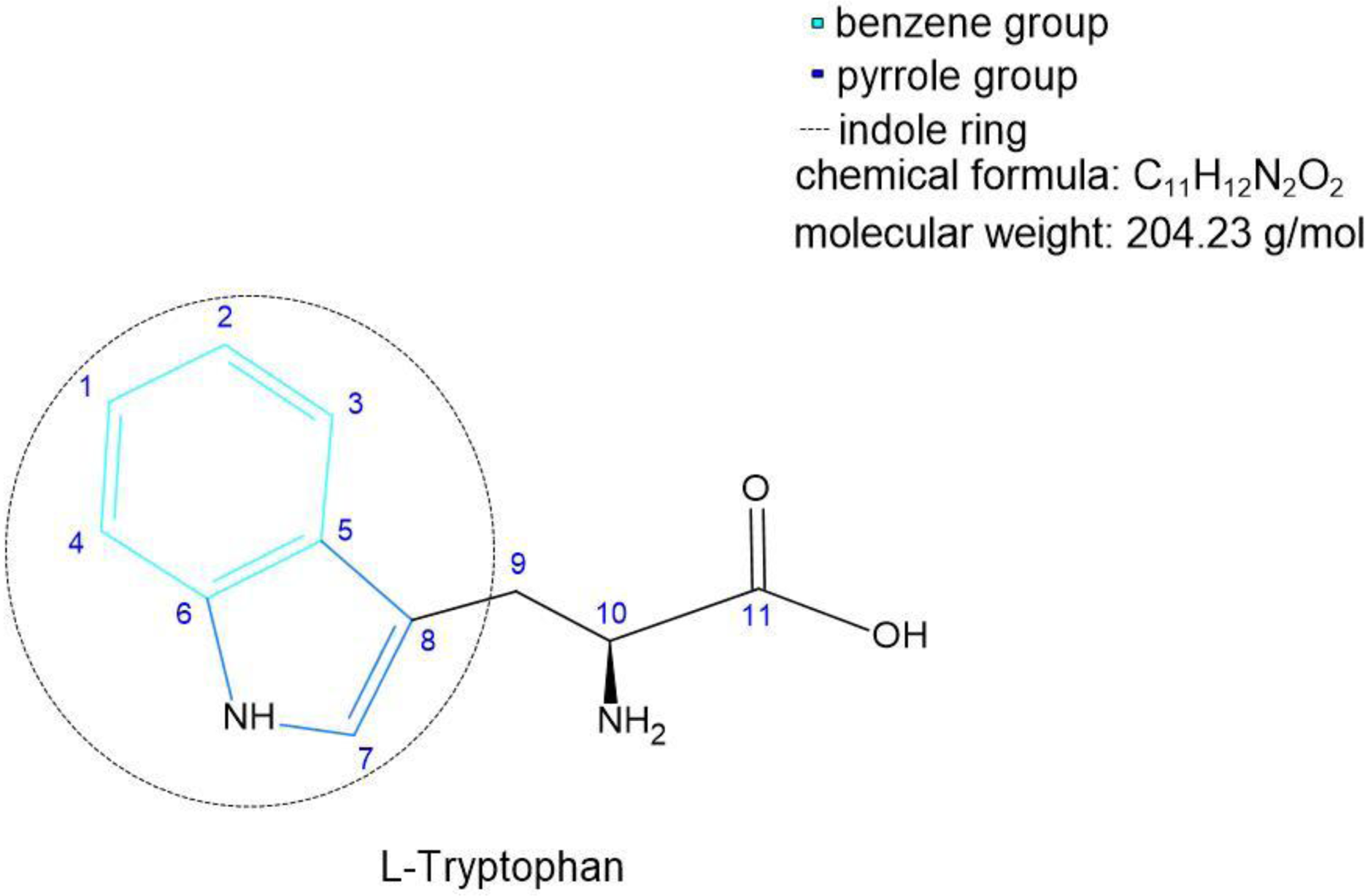
Tryptophan is the only amino acid derived from indole, a bicyclic ring formed by a benzene and pyrrole group linked to an α-carbon by a CH2 group. The presence of the indole ring in the chemical structure of TRP gives this molecule high hydrophobicity among all of the protein amino acids. TRP is the amino acid with the highest number of C atoms (C11). The TRP indole ring in residues (-R) gives unique properties to proteins and peptides, promoting structural hydrophobic protein–protein, protein–peptide, and protein–biomolecule interactions (Figure 1).
Chemical structure of tryptophan: C atoms are numbered in blue; the benzene ring is marked in turquoise; the pyrrole ring is dark blue; dashed lines represent the indole ring.

The TRP indole ring is able to stabilise structures, domains, and interactions through van der Waals forces(Reference De Jesus and Allen11). At the same time, indole-N shows a propensity to act as a hydrogen bond donor, highlighting the role of this amino acid in protein binding and recognition(Reference De Jesus and Allen11). The presence of TRP-R groups in precise domains – for instance, membrane-bound in transmembrane domains of membrane-bound proteins – is fundamental for the protein stability/assemblage to the phospholipid bilayer(Reference De Jesus and Allen11). Hydrophobic interactions between proteins and peptides or between these and other biologically active molecules are essential in cell physiology(Reference Palego, Betti, Rossi and Giannaccini12).
TRP synthesis in human and animals
TRP is essential for humans and animals. Bacteria and other eukaryotes such as fungi and plants can synthesise it from chorismic acid and anthranilate (ATA). In bacteria, fungi, and plants, the biosynthesis of TRP, tyrosine (TYR), and phenylalanine (PHE) is linked by the shikimate pathway(Reference Palego, Betti, Rossi and Giannaccini12). Phosphoenolpyruvate and erythrose 4-phosphate, derived from glycolysis and the pentose phosphate pathway, respectively, enter into a series of reactions involving the activity of seven enzymes, whose final product is chorismate, the common precursor for the synthesis of the other two primary metabolites, prephenate and ATA, the first generating tyrosine and phenylalanine, the last producing TRP(Reference Palego, Betti, Rossi and Giannaccini12).
The TRP digestive pathway of the host cell differs from that of bacteria(Reference Palego, Betti, Rossi and Giannaccini12). Many bacteria have enzymes that break down TRP to indole and indole-3-carboxaldehyde (IAld) in the indole pathway(Reference Miyamoto, Sujino and Kanai13).
The TRP pathway involves complex interactions between host cellular and bacterial processes, which lead to the production of compounds such as SER, also known as 5-hydroxytryptamine (5-HT), and KYN derivatives(Reference Miyamoto, Sujino and Kanai13).
On the other hand, host cells have enzymes involved in the KYN pathway with continuous expression of indoleamine 2,3-dioxygenase (IDO)(Reference Abd El-Fattah14–Reference Lashgari, Roudsari, Shayan, Niazi Shahraki, Hosseini and Momtaz16).
The TRP metabolism in the intestine follows the following pathways: (1) the KYN pathway in both immune cells (mainly macrophages) and intestinal epithelial cells (IECs) via IDO1, which is the primary pathway (representing 95% of ingested TRP)(Reference Chojnacki, Gąsiorowska, Popławski, Konrad, Chojnacki and Fila17,Reference Thursby and Juge18) ; (2) the direct transformation of TRP (4–6%) by the gut microbiota into tryptamine and indole metabolites via the action of the enzyme tryptophanase, expressed in many Gram–negative and Gram-positive bacteria(Reference Thursby and Juge18,Reference Taleb19) .
TRP metabolisation by human intestinal bacteria
The human intestinal tract harbours a complex and dynamic population of microorganisms, the gut microbiota, which exerts a marked influence on the host during homeostasis and disease(Reference Thursby and Juge20). Two major groups of bacteria dominate the intestinal tract: Bacillota (for example, Lactobacillus and Streptococcus species), which comprise around 65% of the total; and Bacteroidota (for example, Bacteroides intestinalis), which comprise around 30%. The remaining 5% is composed of other bacterial groups, including Pseudomonadota (for example, Escherichia coli) and Actinomycetota, such as Bifidobacteria species(Reference Yang, Xie, Li and Wei21,Reference Knight22) .
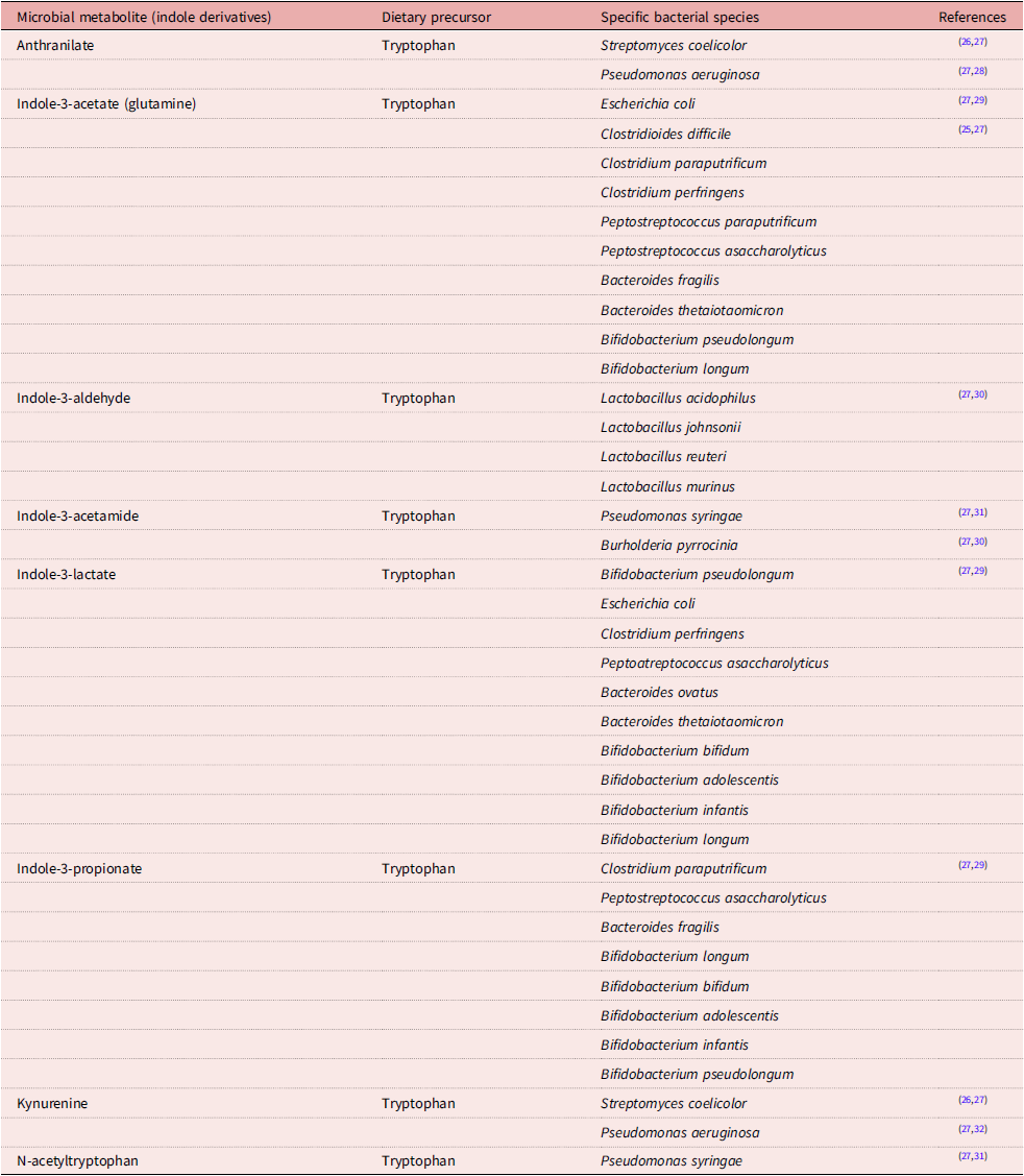
Various gut bacteria species can break down TRP as a precursor into indole catabolites, which are considered reliable sources of AhR ligands. AhR is expressed in multiple immune cells, such as myeloid cells, innate lymphoid cells, B lymphocytes, and specific subtypes of T cells(Reference Thursby and Juge18,Reference Taleb19,Reference De Juan and Segura23) . AhR activation has an overall anti-inflammatory and immunoregulatory role in innate and adaptive immunity, both in a steady state or in inflammatory scenarios such as autoimmunity or infection(Reference Thursby and Juge18,Reference Taleb19,Reference Rothhammer and Quintana24) . Many bacterial species have been demonstrated to metabolise tryptophan into indole and indolic acid derivatives, as can be seen in Table 1(Reference Sunil, François-Pierre, Kochhar and Martin25).
Tryptophan in aspects of the role of a precursor for indole metabolites and a gut bacterial producer of indole tryptophan metabolites

Table 1. Long description
A table comparing microbial metabolites derived from tryptophan, their dietary precursors, and the specific bacterial species involved in their production. The table has four columns: Microbial metabolite (indole derivatives), Dietary precursor, Specific bacterial species, and References. It contains 11 rows, each detailing a different microbial metabolite, its dietary precursor, and the associated bacterial species. The references column lists citations for each row. Row 1: Anthranilate, Tryptophan, Streptomyces coelicolor, Pseudomonas aeruginosa. Row 2: Indole-3-acetate (glutamine), Tryptophan, Escherichia coli, Clostridioides difficile, Clostridium paraputrificum, Clostridium perfringens, Peptostreptococcus paraputrificum, Peptostreptococcus asaccharolyticus, Bacteroides fragilis, Bacteroides thetaiotaomicron, Bifidobacterium pseudolongum, Bifidobacterium longum. Row 3: Indole-3-aldehyde, Tryptophan, Lactobacillus acidophilus, Lactobacillus johnsonii, Lactobacillus reuteri, Lactobacillus murinus. Row 4: Indole-3-acetamide, Tryptophan, Pseudomonas syringae, Burholderia pyrocinia. Row 5: Indole-3-lactate, Tryptophan, Bifidobacterium pseudolongum, Escherichia coli, Clostridium perfringens, Peptostreptococcus asaccharolyticus, Bacteroides ovatus, Bacteroides thetaiotaomicron, Bifidobacterium bifidum, Bifidobacterium adolescentis, Bifidobacterium infantis, Bifidobacterium longum. Row 6: Indole-3-propionate, Tryptophan, Clostridium paraputrificum, Peptostreptococcus asaccharolyticus, Bacteroides fragilis, Bifidobacterium longum, Bifidobacterium bifidum, Bifidobacterium adolescentis, Bifidobacterium infantis, Bifidobacterium pseudolongum. Row 7: Kynurenine, Tryptophan, Streptomyces coelicolor, Pseudomonas aeruginosa. Row 8: N-acetyltryptophan, Tryptophan, Pseudomonas syringae.
Dietary TRP and its role in inflammatory diseases
Dietary sources of tryptophan for adult humans are dairy products, meat, fish, eggs, bananas, oats, pumpkin and sesame seeds, chocolate, dried dates, soy, tofu, tree nuts, and peanuts. The estimated dietary need of tryptophan in adult humans ranges from 280–355 mg per day(Reference Nayak, Singh and Buttar33). The recommended daily allowance in the United States of TRP is 5 mg kg−1 d−1, or 0.35 g for a 70kg individual(Reference Fernstrom34,35) . When the supply of essential amino acids, such as methionine, cysteine, threonine, and TRP, is limited, there is greater competition associated with the immune system for the amino acids(Reference Kampman-Van de Hoek, Jansman, van den Borne, van der Peet-Schwering, van Beers-Schreurs and Gerrits36,Reference Gazi, Das, Siddique, Alam, Fahim and Hasan37) .
TRP deficiency symptoms occur at intakes as little as 25% below the requirement, affecting growth rate, skeletal development, and behaviour(Reference Moehn, Pencharz and Ball38). Vitamin B6 deficiency impairs TRP metabolism due to the coenzyme role of pyridoxal 5’-phosphate in the key enzymes kynureninase and kynurenine aminotransferase(Reference Rios-Avila, Nijhout, Reed, Sitren and Gregory39). Rios-Avila and colleagues found that moderate deficiency yielded increased 3-hydroxykynurenine and a decrease in kynurenic acid (KYNA) and anthranilic acid (ATA). More severe deficiency also yielded an increase in kynurenine and xanthurenic acid(Reference Rios-Avila, Nijhout, Reed, Sitren and Gregory39).
TRP is transported via the bloodstream(Reference Palego, Betti, Rossi and Giannaccini40). When TRP and its metabolites were quantified systemically in dried blood spots from children born vaginally or via caesarean delivery, the results showed that metabolites and microbiota have a protective effect on developing and maintaining immune system homeostasis(Reference Aust, Benesova, Vidova, Coufalikova, Smetanova and Borek41).
Gazi et al. found that low plasma TRP and an increased KYN and KYN–TRP ratio were associated with infections and chronic immune activation(Reference Gazi, Das, Siddique, Alam, Fahim and Hasan37). Elevated KYN and KYN–TRP ratios are markers of increased IDO activity(Reference Gazi, Das, Siddique, Alam, Fahim and Hasan37,Reference Pett, Kunisaki, Wentworth, Griffin, Kalomenidis and Nahra42) . A recent study carried out by Harris et al. supports the hypothesis that an elevated KYN–TRP ratio is associated with chronic inflammatory diseases (CID) and that KYN metabolism is the major route for CID-related TRP degradation(Reference Harris, Szymczak, Schuchardt, Labrenz, Tran and Welz43).
Recent ‘Westernisation’ of many countries coincides with the rise in the prevalence of asthma and allergies(Reference Eder, Ege and von Mutius44,Reference Thorburn, Macia and Mackay45) , as well as certain autoimmune diseases. Changes in dietary food and the correlation with diseases are particularly evident in epidemiological studies linking fibre and fat intake(Reference Thorburn, Macia and Mackay45). The consumption of high fibre led to a reduced risk of suffering from allergies, asthma, or colon cancer and a range of other conditions, like cardiovascular diseases and cancers, as well as infections and respiratory diseases(Reference Thorburn, Macia and Mackay45,Reference Park, Subar, Hollenbeck and Schatzkin46) .
Sinha et al. recently found that gut microorganisms catabolise TRP from dietary fibre into a wide range of metabolites, especially IPA and ILA, that are involved in both beneficial and adverse host effects such as, for example, associated chronic kidney disease(Reference Sinha, Laursen, Brinck, Rybtke, Hjørne and Procházková47), which was experimentally confirmed by using human faecal cultures and faecal–microbiota-transplanted gnotobiotic mice(Reference Sinha, Laursen, Brinck, Rybtke, Hjørne and Procházková47).
Rankin et al. investigated how perturbations in diet influence the immune–microbiome interface. They developed a mouse model of acute dietary TRP deficiency to study metabolite-based communication between the diet, commensal microbial community, and intestinal regulatory T cells (Tregs). They reported that the loss of TRP reshaped intestinal microbial communities, which included the depletion of probiotic Lactobacillus reuteri; it further drove transcriptional changes to immune response genes in the intestinal ileum and reshaped the Treg compartment. Dietary TRP deficiency also promoted the expansion of retinoic acid receptor-related orphan nuclear receptor gamma Treg (RORγt+) cells and the loss of GATA-binding protein 3 Tregs (Gata3+) in a microbiota-dependent manner. They further showed that in the absence of dietary TRP, the supplementation of the AhR ligand indole-3-carbinol (I3C) is sufficient to restore the Treg compartment. Their data showed that dietary TRP deficiency destroys the interaction between the host and its bacterial symbionts, regulating Treg homeostasis via the deprivation of bacterially derived TRP metabolites(Reference Rankin, Kaiser, de los Santos-Alexis, Park, Uhlemann and Gray48).
Immune cells, including macrophages and dendritic cells (DCs), in which the KYN pathway mainly dominates, upregulate IDO in response to stimuli(Reference Krupa and Kowalska49,Reference Krupa, Krupa and Pawlak50) .
TRP metabolites and brain health
The KYN pathway is dominated by astrocytes and microglia cells in the brain, whereas the 5-HT pathway is dominated by serotonergic neurons in the central nervous system (CNS)(Reference Haroon, Raison and Miller51–Reference Imamdin and van der Vorst53). Other cells in the peripheral tissues, such as hepatocytes, express tryptophan 2,3-dioxygenase (TDO)(Reference Miyamoto, Sujino and Kanai13,Reference Xu, Ling, Su, Su, Xu and Jiang54) . KYN, which is initially released by IDO1-expressing type 1 conventional dendritic cells (cDC1s), recruits AhR-expressing type 2 conventional DCs (cDC2s). cDC2s produce transforming growth factor-β (TGF-β), which induces anti-inflammatory forkhead box protein 3+ (Foxp3+) Treg cells. The TGF-β–IDO1–AhR loop is crucial for the generation of tolerogenic DCs, resulting in self-tolerance and lipopolysaccharide (LPS) tolerance(Reference Miyamoto, Sujino and Kanai13). Antigen-presenting cells and DCs play essential roles in the initiation and maintenance of immune responses. IDO1 expression in DCs is increased by LPS, extracellular and intracellular deoxyribonucleic acid (DNA), and type I and type II interferons(Reference Gargaro, Scalisi, Manni, Briseño, Bagadia and Durai55).
KYNA affects brain functions as it is an antagonist for the α7-nicotinic acetylcholine receptors (7nAchRs) and the N-methyl-d-aspartic acid (NMDA) receptor(Reference Chojnacki, Gąsiorowska, Popławski, Konrad, Chojnacki and Fila17,Reference Hilmas, Pereira, Alkondon, Rassoulpour, Schwarcz and Albuquerque56) . It is also an agonist for G protein-coupled receptor 35 (GPCR35)(Reference Chojnacki, Gąsiorowska, Popławski, Konrad, Chojnacki and Fila17,Reference Wang, Simonavicius, Wu, Swaminath, Reagan and Tian57) . In addition, 3-hydroxykynurenine (3-HK) is generally neurotoxic as it can support oxidative stress induction, and quinolinic acid (QUIN), as an NMDA receptor agonist, displays excitotoxic properties(Reference Chojnacki, Gąsiorowska, Popławski, Konrad, Chojnacki and Fila17,Reference Okuda, Nishiyama, Saito and Katsuki58,Reference Schwarcz and Pellicciari59) . The neurotoxic effect of kynurenines is fundamental for the SER–KYN hypothesis of depression, meaning that depression can be caused by a deficiency of serotonin or an excess of kynurenine metabolites(Reference Chojnacki, Gąsiorowska, Popławski, Konrad, Chojnacki and Fila17,Reference Oxenkrug60–Reference Miura, Ozaki, Sawada, Isobe, Ohta and Nagatsu62) . Therefore, TRP supplementation has been used in the treatment of depression(Reference Chojnacki, Gąsiorowska, Popławski, Konrad, Chojnacki and Fila17,Reference Young63) .
TRP and its metabolites can cross the blood–brain-barrier (BBB), through entering inside the substantia nigra pars compacta (SNC)(Reference Palego, Betti, Rossi and Giannaccini40), and can further regulate the immune activity of microglia and astrocytes through AhR-driven mechanisms(Reference Zhang, Tang, Chen, Xie and Tao5,Reference Rothhammer, Borucki, Tjon, Takenaka, Chao and Ardura-Fabregat64,Reference Wekerle65) . TRP metabolites such as IPA and IAld signal through AhR in astrocytes to negatively modulate NF-κB activation through the suppressor of cytokine signalling 2 (SOCS2), thereby mitigating CNS autoimmunity(Reference Zhang, Tang, Chen, Xie and Tao5,Reference Rothhammer, Mascanfroni, Bunse, Takenaka, Kenison and Mayo66,Reference Marsland67) . Moreover, AhR interacts with genes encoding vascular endothelial growth factor B (VEGF-B) and transforming growth factor α (TGF-α), suppressing the transcription of VEGF-B while potentiating that of TGF-α. In this manner, AhR activation in microglia dampens the responsiveness of neighbouring astrocytes to CNS inflammation(Reference Zhang, Tang, Chen, Xie and Tao5,Reference Rothhammer, Borucki, Tjon, Takenaka, Chao and Ardura-Fabregat64,Reference Wekerle65) .
TRP and its microbial metabolites have the potential to regulate the interaction between microglia and astrocytes, which is a key mechanism in CNS inflammation. The dysregulation of the TRP pathway plays a role in Alzheimer’s and Parkinson’s diseases, mood disorders, neuronal diseases, autoimmune diseases such as multiple sclerosis, and cancer(Reference Miyamoto, Sujino and Kanai13). One of the targeted factors of AhR in the CNS is nuclear factor-κB (NF-κB), an essential transcriptional activator in inducing glial-mediated inflammation in neurodegenerative diseases(Reference Harari and Liao68,Reference Yang, Zeng, Barlş and Tezel69) . The AhR activation significantly inhibited NF-κB signalling, reduced levels of downstream inflammatory cytokines such as tumour necrosis factor alpha (TNF-α), IL-6, and C–C motif chemokine ligand 2 (CCL2), and relieved CNS inflammation in multiple sclerosis models. At the same time, AhR deletion increased the nuclear levels of NF-κB in microglia(Reference Rothhammer, Borucki, Tjon, Takenaka, Chao and Ardura-Fabregat64,Reference Rothhammer, Mascanfroni, Bunse, Takenaka, Kenison and Mayo66) . Zelante et al. recently found that serum levels of microbial postbiotic tryptophan metabolite IAld correlated inversely with the disease duration in multiple sclerosis patients. Through AhR binding, IAld, in peripheral lymph nodes, affected the mast-cell TRP metabolism and forced the mast cells to produce SER from TRP via the enzyme tryptophan-hydroxylase 1 (Tph1)(Reference Zelante, Paolicelli, Fallarino, Gargaro, Vascelli and De Zuani70).
Moreover, in vitro, IPA protects microglia from inflammation. Kim et al. showed that IPA-treated microglial BV-2 cells exhibit a significant reduction in the concentrations of proinflammatory cytokines, interleukin 1β (IL-1β), and TNF-α following LPS stimulation compared to the LPS-alone treatment group(Reference Kim, Jung, Hwang and Shin71,Reference Kim72) .
TRP metabolites were further shown to have AhR-dependent anti-inflammatory and neuroprotective effects on retinal ischaemia/reperfusion injury(Reference Yang, Wang, Xu, Liu, Huang and Gu73). The retina is an integral part of the nervous system and considered a part of the brain. That TRP metabolism plays a role in retinal neurodegeneration in glaucoma was shown by Yang and colleagues. They detected a significant decrease of serum tryptophan metabolites and reduced retinal AhR expression in glaucoma patients. The supplementation of the TRP metabolite 2-(1’H-indole-3’-carbonyl)-thiazole-4-carboxylic acid methyl ester (ITE)(Reference Song, Clagett-Dame, Peterson, Hahn, Westler and Sicinski74,Reference Roager and Licht75) could inhibit retinal inflammation, alleviate retinal injury in retinal ischaemia/reperfusion (IR) in mice, and limit inflammation in BV2 microglia cells via AhR activation. Moreover, a cytokine antibody array revealed that ITE could significantly decrease the pro-inflammatory cytokines induced by lipopolysaccharide. At the same time, there was an increase in the anti-inflammatory cytokine interleukin-10 (IL-10), thus reducing the neurotoxicity of BV2-conditioned medium to retinal ganglion cells under IR. The authors reported that AhR activation by ITE could downregulate NF-κB and extracellular signal-regulated kinase (ERK) pathways in microglia, leading to an anti-inflammatory effect(Reference Yang, Wang, Xu, Liu, Huang and Gu73).
TRP metabolites and intestinal health
Recently, it has been shown that microbial diversity and concentrations of tryptophan metabolites were significantly higher in stools than in meconium. Further, it was highlighted that the treatment of mothers with antibiotics before or during delivery affects the metabolite composition and microbial diversity in the stool of vaginal- and caesarean-delivered newborns. The correlation between microbial and metabolite composition showed significant positive correlations between ILA, N-acetyltryptophan, and indol-3-acetic acid (IAA) and Bifidobacterium, Bacteroides, and Peptoclostridium. The positive effect of vaginal delivery on newborns’ microbiome development was further degraded when the mother was treated with antibiotics before or during delivery(Reference Aust, Vidova, Coufalikova, Smetanova, Kozeluhova and Micenkova3).
Tryptophan metabolites affect various aspects of the intestinal barrier, such as immune balance, mucus and microbial barrier, tight junction complexes, and the differentiation and proliferation of epithelial cells(Reference Shinde and McGaha76,Reference Liikonen, Gomez-Gallego and Kolehmainen77) . An intact fldC gene was shown to be essential for Clostridium sporogenes-mediated IPA generation. Mice colonised with fldC mutant C. sporogenes failed to generate IPA, exhibited enhanced intestinal permeability, and had an increased number of circulating myeloid and lymphoid cells(Reference Dodd, Spitzer, Van Treuren, Merril, Hryckowian and Higginbottom8).
Indole derivatives binding and activating AhR were shown to induce the expression of downstream cytokines as interleukin-22 (IL-22)(Reference Lee, Cella, McDonald, Garlanda, Kennedy and Nukaya78) by type 3 innate lymphoid cells (ILC3)(Reference Qiu, Heller, Guo, Chen, Fish and Fu79), thereby regulating epithelial integrity and immunity(Reference Lamas, Michel, Waldschmitt, Pham, Zacharioudaki and Louise Dupraz80). Mice deficient in AhR or IL-22 expression have been shown to succumb to Citrobacter rodentium infection(Reference Qiu, Heller, Guo, Chen, Fish and Fu79,Reference Zheng, Valdez, Danilenko, Hu, Sa and Gong81) . Remarkably, gut microbiota composition can be altered by an AhR ligand-free diet or in il-22 knock-out mice(Reference Zenewicz, Yin, Wang, Elinav, Hao and Zhao82,Reference Brawner, Yeramilli, Duck, Van Der Pol, Smythies and Morrow83) . Recently, microbiota-induced expression of AhR in colonic neurons was shown to be essential in neuronal programming by microbiota to maintain gut motility and thus to regulate intestinal physiology(Reference Obata, Castaño, Boeing, Bon-Frauches, Fung and Fallesen84). Lactobacillus reuteri metabolises TRP into IAId that activates IL-22-producing ILC3s in an AhR-dependent manner, thereby exerting colonisation resistance against mucosal Candida (Reference Zelante, Iannitti, Cunha, De Luca, Giovannini and Pieraccini85). Mdo-miRNA7267-3p, a newly identified plant-derived exosomal micro ribonucleic acid (microRNA), boosts AhR-dependent production of IL-22 by ILC3s by accelerating IAld production, ultimately strengthening barrier function(Reference Teng, Ren, Sayed, Hu, Lei and Kumar86).
Some indole metabolites, such as IAA, IAld, and tryptamine, have been shown to maintain intestinal barrier integrity and immune cell homeostasis through activation of the AhR(Reference Zelante, Iannitti, Cunha, De Luca, Giovannini and Pieraccini85,Reference Lamas, Richard, Leducq, Pham, Michel and Da Costa87) . Other studies demonstrate that IPA can improve intestinal barrier function through the activation of the PXR(Reference Taleb19). IPA diminishes intestinal mucosal permeability and suppresses inflammation in a PXR-dependent and toll-like receptor 4 (TLR4)-dependent manner(Reference Venkatesh, Mukherjee, Wang, Li, Sun and Benechet88).
The influence of TRP metabolism alterations to the outcome of inflammatory bowel disease (IBD) is still unclear. Several TRP metabolism end-products, produced by gut microbiota, were shown to be essential to intestinal homeostasis(Reference Michaudel, Danne, Agus, Magniez, Aucouturier and Spatz89). Michaudel et al. investigated the role of metabolites from the kynurenine pathway. They used targeted quantitative metabolomics in two large human IBD cohorts and experiments with dextran sodium sulphate-induced colitis experiments in mice to evaluate the effects of identified metabolites. In vitro, ex vivo, and in vivo experiments were used to decipher the mechanisms involved. The results showed that intestinal inflammation severity negatively correlates with the amount of xanthurenic acid (XANA) and KYNA acids in mice and humans. Supplementation with XANA or KYNA decreased colitis severity by modulating intestinal epithelial cells and T cells, involving AhR activation and the rewiring of cellular energy metabolism. Furthermore, direct modulation of the endogenous tryptophan metabolism, using the recombinant enzyme aminoadipate aminotransferase (AADAT), responsible for the generation of XANA and KYNA, was protective in rodent colitis models (Reference Michaudel, Danne, Agus, Magniez, Aucouturier and Spatz89).
It was further shown that the AhR/IL-22 pathway has potential as a candidate in treating post-infectious irritable bowel syndrome (IBS)(Reference Meynier, Baudu, Rolhion, Defaye, Straube and Daugey90). Meynier et al. investigated the mechanisms involved in post-infectious irritable bowel syndrome (PI-IBS) using a mouse model of C. rodentium infection to mimic bacterial gastroenteritis in humans. The study unravelled the influence of the tryptophan metabolism, particularly the microbiota-induced AhR expression, on intestinal homeostasis disturbance following gastroenteritis resolution, and evaluated the efficacy of IL-22 cytokine vectorisation on PI-IBS symptoms. In the study, the C. rodentium infection model in C57BL6/J mice was used to mimic Enterobacteria gastroenteritis (Reference Meynier, Baudu, Rolhion, Defaye, Straube and Daugey90). The authors used a Lactococcus lactis strain, which carried a eukaryotic expression plasmid for murine IL-22 (L. lactis IL-22) to induce IL-22 production in mouse colonic mucosa. C. rodentium-infected mice exhibited persistent colonic hypersensitivity and cognitive impairments and anxiety-like behaviours after pathogen clearance. Notably, post-infectious disorders were associated with low-grade inflammation, increased intestinal permeability, decreased Lactobacillaceae abundance associated with the colonic layer, and increased short-chain fatty acids (SCFAs). During the post-infection period, the indole pathway and AhR activity were decreased due to a reduction of tryptophol production. They found that the treatment with L. lactis IL-22 restored gut permeability, normalised colonic sensitivity, restored cognitive performances, and decreased anxiety-like behaviours(Reference Meynier, Baudu, Rolhion, Defaye, Straube and Daugey90). Their microbiome analysis revealed that four bacterial genera were significantly modified in infected animals, including a decrease in relative abundance of Bacteroidota and an increase in Bacillota, Pseudomonadota, and Actinomycota. At the family level, they observed a significant decreased level in the relative abundance of Moraxellaceae, Lactobacillaceae, and Rikenellaceae, whereas Desulfovibrionaceae, Oxalobacteracea, and Lachnospiraceae were increased(Reference Meynier, Baudu, Rolhion, Defaye, Straube and Daugey90).
Van et al. found in their study that the amount of consumed TRP reduced colitis and determined the transcription level of the AhR-mediated colonic T cell homing receptor, G-protein coupled receptor 15 (GPCR15), in mice, thus affecting the number of colonic Foxp3+ Treg cells and local immune homeostasis. TRP supplementation nearly doubled the colonic GPCR15+ Treg cells and substantially reduced the future risk of colitis(Reference Van, Zhang, Wigmore, Kennedy, DaSilva and Huang91).
TRP metabolites in genetic engineering
TRP metabolites have been further shown by genetic engineering to increase the expression of ILC3 in C57BL/6 mice and through microbial mimicry to repress proinflammatory cytokine production in mice.
Kouno et al. found that in C57BL/6 mice, which were further supplemented with control Escherichia coli Nissle 1917 (EcN) or engineered EcN-AhR, that the EcN-AhR ameliorated the ethanol-induced liver disease in C57BL/6 mice. EcN-AhR were engineered to overproduce TRP, and additional engineering enabled the conversion of this TRP to indoles, including IAA and ILA. EcN-Ahr was found to upregulate the intestinal gene expression of Cyp1a1, Nrf2, Il22, Reg3b, and Reg3g, and increased IL-22-expressing ILC3. Furthermore, EcN-Ahr reduced bacterial translocation to the liver. The beneficial effect of EcN-AhR was abrogated in mice lacking AhR expression in IL-22-producing immune cells(Reference Kouno, Zeng, Wang, Duan, Lang and Gao92).
Dvořák et al. showed that microbial indole metabolites have been shown to serve as a novel strategy for drug discovery through microbial metabolite mimicry. They found non-cytotoxic PXR agonists that repress proinflammatory cytokine production in cells and abrogate inflammation in mice which expressed the human PXR gene(Reference Dvořák, Kopp, Costello, Kemp, Li and Vrzalová93). The lead compound, FKK6 (Felix Kopp Kortagere 6), has been demonstrated by Dvořák et al. as a PXR ligand and agonist that induced PXR-specific target genes in vitro and in vivo in mice. FKK6 further showed a PXR-dependent protective effect in Dextran sulfate sodium (DSS)-induced colitis in mice and human intestinal organoids stimulated with pro-inflammatory cytokines(Reference Dvořák, Kopp, Costello, Kemp, Li and Vrzalová93–Reference Dvořák, Sokol and Mani95). The described examples of the anti-inflammatory effect of TRP and its indole metabolites show their potential as promising alternatives in the treatment of various inflammatory diseases.
Discussion and conclusion
TRP is an essential amino acid which needs to be incorporated into the human body with food. This amino acid can be digested by human gut microbiota into TRP catabolites. These TRP catabolites act as ligands for AhR and PXR, thereby modulating the human immune system; for example, TRP has been found to regulate the Treg homing receptor and the TRP metabolite IAld through the binding of AhR to negatively correlate with multiple sclerosis, and IPA was shown to positively regulate CNS inflammation. Peripheral targets, such as the brain, may be reached. Here, TRP supplementation was suggested for the treatment of depression. Another peripheral target, the mucosal barrier, may be reached and modulated by IPA. IPA was shown to diminish intestinal mucosal permeability and to suppress inflammation through the PXR pathway. The gut microbiome was shown to be altered by supplementation with bacterial TRP metabolites. This could be a potential treatment option and support for diseases such as inflammatory bowel disease, retinal ischaemic diseases, and neurodegenerative and autoimmune diseases, including multiple sclerosis, to positively influence the gut microbiome composition, which had been shown to be negatively altered by antibiotic use. As shown in examples given in this review, inclusive experimental models and the supplementation of TRP and its metabolites could be a new and alternative approach for treating patients. This treatment support option with TRP indole metabolites could potentially lead to clinical studies in the near future, where the TRP indole metabolites would be tested in healthy controls and patients with inflammatory diseases. This approach, as a possible supportive treatment option, has not yet been investigated, and data for these studies are lacking. Here we highlight the option of these alternative treatment opportunities; however, studies in the direction of clinical trials are yet to be conducted, as only animal preclinical models are currently available.
Furthermore, this review described the new role of TRP indole metabolites as regulators of inflammatory diseases through microbial metabolite mimicry. Here, as well, only the first results in vivo and in vitro in mice were described; no further studies are available on this topic to date.
Our analysis showed that the amino acid TRP and its indole metabolites are ligands which can modulate mucosal barrier integrity and regulate disease states.
Acknowledgements
The authors thank the RECETOX Research Infrastructure (no. LM2023069) and Cetocoen CETOCOEN Plus (CZ.02.1.01/0.0/0.0/15_003/0000469), financed by the Ministry of Education, Youth and Sports, Czech Republic, for their support. This work was supported by the European Union’s Horizon 2020 research and innovation programme under grant agreement no. 857560 (CETOCOEN Excellence). This publication reflects only the authors’ views, and the European Commission is not responsible for any use that may be made of the information it contains. A-CA and FW participate in the Deutsche Forschungsgemeinschaft-funded research group BARICADE FOR 5427/1 (project no. 466687329).
Competing interests
The authors declare no conflict of interest.
Authorship
A-CA wrote the manuscript. VT and FW supervised and reviewed the manuscript for clarity. All authors contributed to the article and approved the submitted version.

 Open access

