

Introduction
The ‘dundasite group’ (syn. dresserite-group) (Table 1), now including the new mineral grguricite, seemingly comprises eight species with three structure types but is yet to be formally ratified by the Commission on New Minerals and Mineral Names of the International Mineralogical Association (IMA–CNMNC). The minerals characteristically occur as secondary, alteration phases in aluminium or chromium-rich environments. Alumohydrocalcite is the most widespread, occurring across multiple geological environments while dundasite, probably with some bias due to the interest of collectors, is also regularly reported as a part of secondary lead mineral assemblages. The other members of the group, with Ba, Sr or Cr, are much rarer and are reported from only one or two occurrences worldwide.
Comparative data for grguricite and members of the dundasite group of minerals. Note that for consistency the Pnma standard unit-cell setting is used for all of the orthorhombic structures, transformed from the published Pbnm cell where necessary.

* Transformed from published Pbnm cell
[1] This study; [2] Stachowicz et al. (Reference Stachowicz, Parafiniuk, Wilson, Coles and Woźniak2015); [3] Jambor (Reference Jambor, Fong and Sabina1969); [4] Szymański (Reference Szymański1982); [5] Cocco et al. (Reference Cocco, Fansani, Nunzi and Zanazzi1972); [6] Birch et al. (Reference Birch, Kolitsch, Witzke, Nasdala and Bottrill2000); [7] Sajó and Szakáll (Reference Sajó and Szakáll2007); and [8] Whitfield et al. (Reference Whitfield, Mitchell, Le Page, Margeson and Roberts2010).
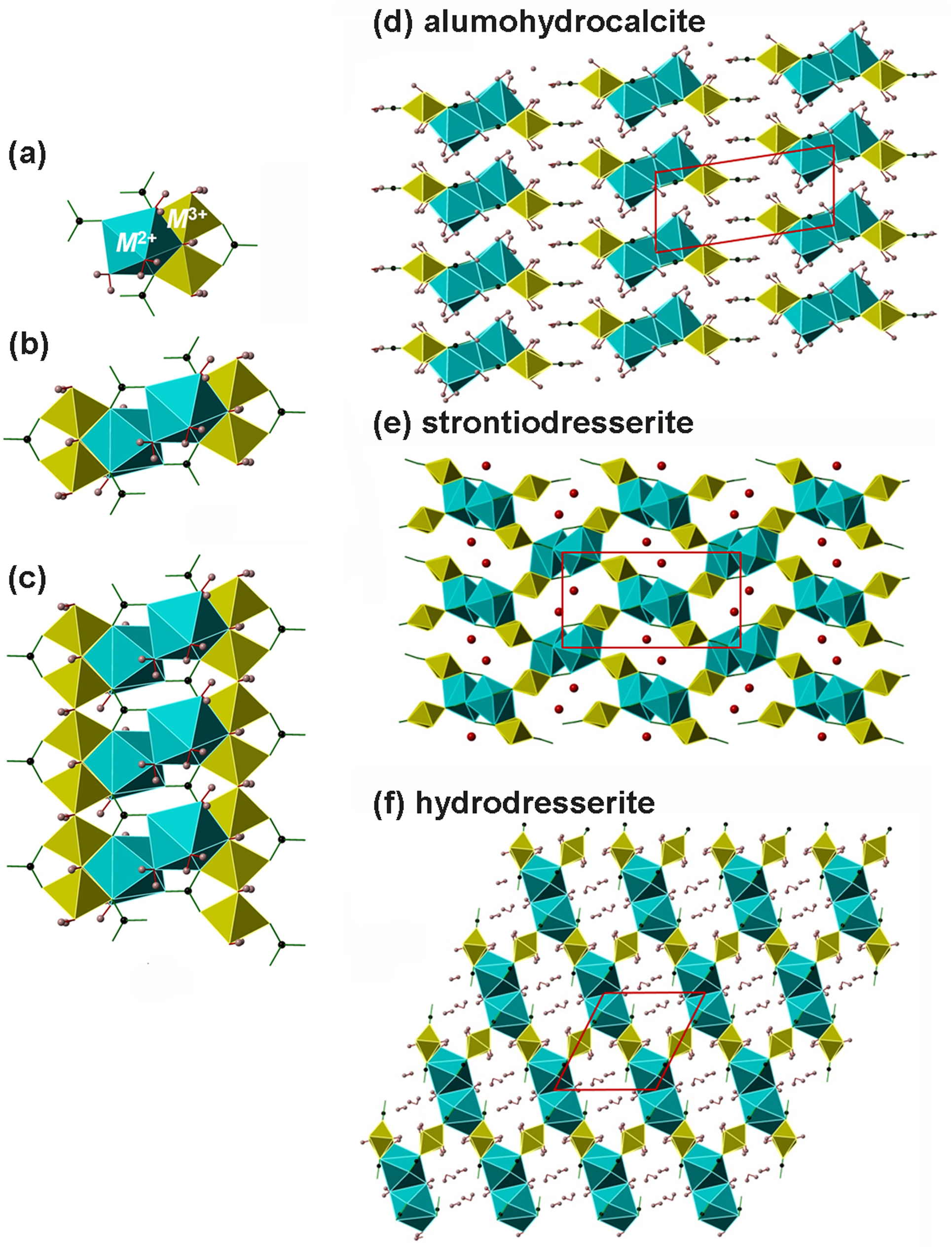
The crystal structures of alumohydrocalcite, dundasite, strontiodresserite and hydrodresserite are shown in Fig. 1. The fundamental building unit, an asymmetric polyhedral cluster (Fig. 1a) consists of two non-equivalent M 3+O2(OH)4 octahedra (M3+ = Al and Cr), one M 2+O3(OH)(H2O)4 polyhedron (M 2+ = Pb, Ca, Ba and Sr) and four CO3 groups comprising two non-equivalent pairs. A carbonate group bridges the two octahedra. A pair of these clusters, related by a centre of symmetry, produces the structural unit that is common to all dundasite-group minerals (Fig. 1b). These units are then stacked via edge-sharing of octahedra and CO3 groups to form columns (Fig. 1c). It is the way in which these columns are connected via hydrogen bonds and/or (Al/Cr)O2(OH)4 octahedra that is the variable topological feature (Figs 1d–f). Alumohydrocalcite (Stachowicz et al., Reference Stachowicz, Parafiniuk, Wilson, Coles and Woźniak2015) and now grguricite are exceptional among the group in that the columns are only connected via hydrogen bonding mediated by additional interstitial H2O molecules hydrogen-bonded to carbonate oxygens. The repeat distance of the structural unit is 5.6−5.8 Å and corresponds to one of the cell parameters of each structure.
(a) The fundamental structural unit of dundasite-group minerals that comprises two M 3+O2(OH)4 octahedra sharing edges with each other and a M 2+O3(OH)(H2O)4 polyhedron. (b) Two structural units related by a centre of symmetry to form a double-unit; (c) double-units polymerised to form a chain with connections made by CO3 groups and M 3+-centred octahedra. This chain is shared by all dundasite-group minerals. (d) Structure of alumohydrocalcite showing isolated chains viewed end-on and only connected via hydrogen bonding involving interstitial H2O groups. (e) Structure of strontiodresserite with linked chains; this polyhedral topology is shared with dundasite. (f) Structure of hydrodresserite showing different inter-chain linkages achieved by a different rotation of chain relative to their orientation in strontiodresserite. In strontiodresserite/dundasite and hydrodresserite water molecules lie in channels between polyhedral chains to which they are bound only via hydrogen bonding.

Here, the occurrence and characterisation of a new member of this group is reported. Grguricite, CaCr2(CO3)2(OH)4⋅4H2O, is the Cr analogue of alumohydrocalcite, CaAl2(CO3)2(OH)4⋅4H2O (Stachowicz et al., Reference Stachowicz, Parafiniuk, Wilson, Coles and Woźniak2015). Apart from petterdite, PbCr2(CO3)2(OH)4⋅H2O (Birch et al., Reference Birch, Kolitsch, Witzke, Nasdala and Bottrill2000), the Cr analogue of dundasite, PbAl2(CO3)2(OH)4⋅H2O (Cocco et al., Reference Cocco, Fansani, Nunzi and Zanazzi1972), no other Cr analogues are known.
Occurrence
Grguricite was identified on a single hand specimen from the Mibladen mining district of Morocco, purchased as a ‘wulfenite’ from a Moroccan vendor at a trade fair in about 2014. It is believed to be sourced specifically from the Adeghoual mine based on the distinctive associated mineralogy (Praszkier, Reference Praszkier2013). Adeghoual mine is located between the towns of Midelt and Mibladen (32°43′50″N, 4°41′48″W). All the mines and shafts local to the region work the limestone-hosted stratiform Pb–Ba (galena–baryte) deposits and are internationally famous for the occurrence of the secondary Pb–V mineral vanadinite. Other secondary minerals have been recorded from the mines, but the assemblage at Adeghoual is limited to cerussite, wulfenite and pyromorphite. Vanadinite is absent. The source of Cr for grguricite is unknown.
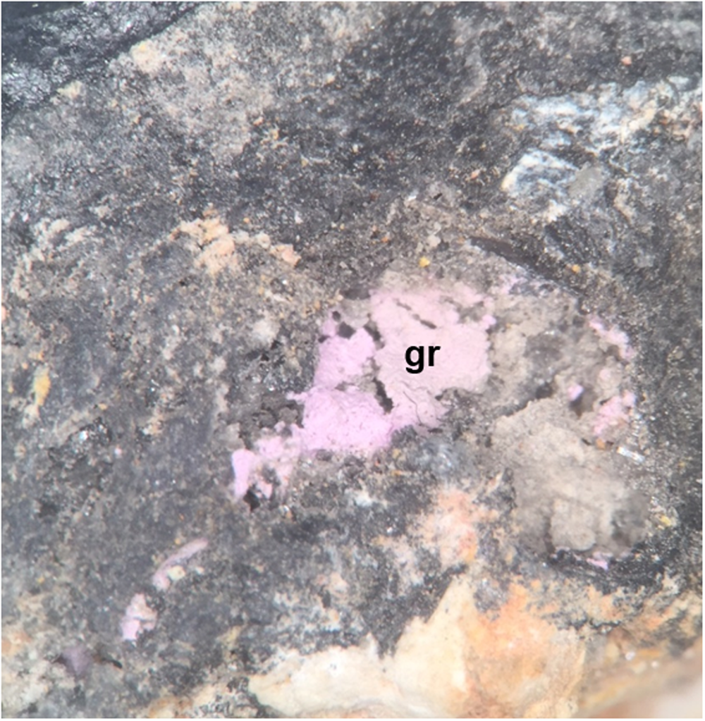
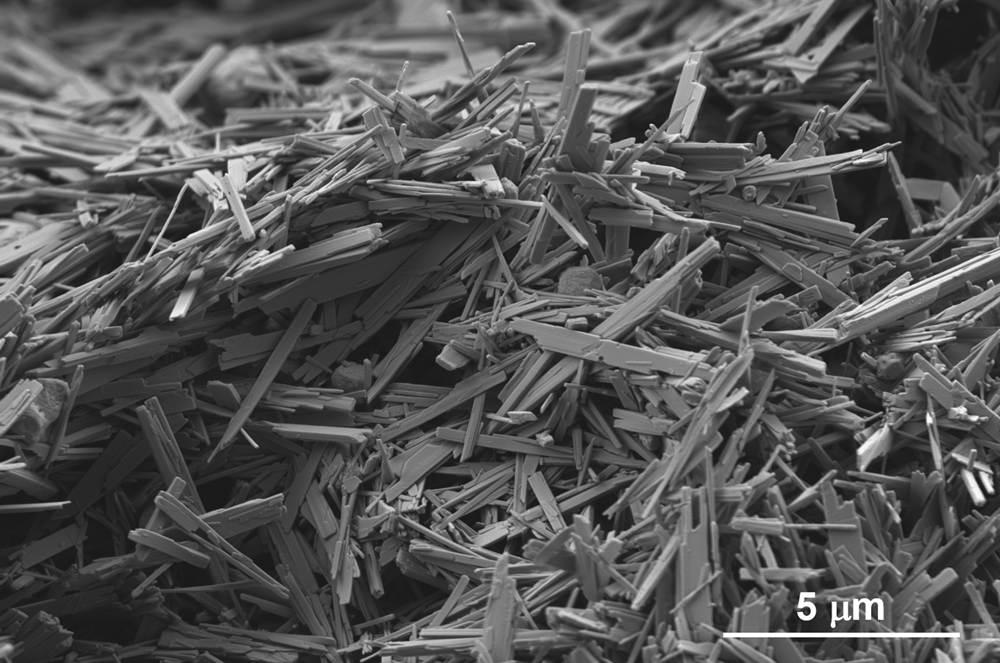
On the holotype specimen, grguricite occurs as thin, visually earthy, lilac pink, crusts, often loosely attached or completely detached in cavities, fractures and interstices in a corroded cerussite-rich alteration layer of primary galena. This layer, a few mm thick, is found underneath well-formed crystals of baryte and wulfenite, occasionally coated with a dusty-green pyromorphite (Fig. 2). On close examination the crusts comprise chaotic mats or sprays of minute (<0.01 mm) lath-like crystals, often displaying frayed terminations (Fig. 3). This mode of occurrence is characteristic of several dresserite-group minerals.
Optical image of a crust of lilac-pink grguricite (gr), formed in a cavity of corroded galena, associated with grey cerussite and unidentified grey secondary Pb carbonates, holotype specimen BM 2019,5. Field of view is 0.75 cm.

Scanning electron microscopy image of an irregularly arranged mat of grguricite crystals, showing their lath-like form and frayed terminations. Holotype specimen BM 2019,5.

Grguricite may be the same phase as that reported incompletely characterised from the Happy Jack Mine in Comet Vale, Western Australia (Grguric et al., Reference Grguric, Pring, Bevan and Downes2006) and thought at the time to be the Ca analogue of petterdite briefly reported by Birch et al. (Reference Birch, Kolitsch, Witzke, Nasdala and Bottrill2000). We have examined the sample of ‘Ca-Petterdite’ from the Happy Jack mine preserved in Museums Victoria (M35586) by quasi-Gandolfi powder XRD (see Experimental methods) and found that the small fine-grained, pale pink aggregates are composed of minerals with both the dresserite/dundasite structure type and the alumohydrocalcite structure type. However, it was not possible at this time to identify how the Cr, determined by energy-dispersive spectroscopy, was distributed between the two, so assigning a species was not possible. Another possible occurrence of grguricite from the Kempirsai Cr deposit in Kazakhstan was reported online, again as a Ca analogue of petterdite (https://www.mindat.org, accessed 17/5/2018), but was not included in this study.
Holotype grguricite consists of the specimen, several fragments thereof, a probe block and the small powder mount created for quasi-Gandolfi powder diffraction analysis, all of which are registered under catalogue number BM 2019,5 in the collections of the Natural History Museum (NHM) in London. The mineral is named grguricite (‘greg-UR-ik-ite’) for Australian geologist Ben Grguric (b. 1971), who reported the potentially equivalent ‘Ca-petterdite analogue’ from Happy Jack mine in 2006 and has published on secondary minerals and Australian topographic mineralogy (e.g. Grguric et al., Reference Grguric, Madsen and Pring2001, Reference Grguric2002, Reference Grguric, Seat, Karpuzov and Simonov2013; Clark et al., Reference Clark, Grguric and Mumm2004). The new mineral and name have been approved by the IMA-CNMNC (IMA2019-123, Rumsey et al, Reference Rumsey, Welch, Spratt and Kleppe2020).
Experimental methods
Chemical composition
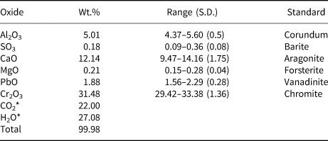
Chemical analyses of grguricite were obtained using a Cameca SX100 electron microprobe by wavelength-dispersive spectrometry (WDS) at the Natural History Museum, London. Settings were 15 kV and 10 nA with a 10 μm spot size. Only Al, Ca, Cr, Pb, Mg, S and Ca were detected. Data were corrected for elemental peak overlaps.
The loosely aggregated masses composed of very small crystals (Figs 2 and 3) did not polish well and proved to be problematic for electron-beam microanalysis. This problem was further exacerbated by the water-rich nature of the mineral.
Various combinations of counting times, element sequences and beam sizes were tested to optimise data collection. However, it was not possible to achieve optimal analysis conditions. Furthermore, as there is not enough material for CHN or TGA analysis, the sample could not be analysed directly for H or C content and significant dehydration of the sample in the beam renders totals and estimations by difference unreliable. Therefore chemical data is presented (Table 2) that is normalised relative to the calculated values of C and H taken directly from the crystallographic study that clearly shows analogy to alumohydrocalcite, which contains four H2O per formula unit (pfu) Alumohydrocalcite loses this molecular water above 150°C (Stachowicz et al., Reference Stachowicz, Parafiniuk, Wilson, Coles and Woźniak2015) and as such is very susceptible to dehydration in the electron beam of the microprobe. Petterdite was also observed to dehydrate quickly under an electron beam, becoming amorphous in a transmission electron microscope (160–200 Kv) after just 2–10 s (Birch et al., Reference Birch, Kolitsch, Witzke, Nasdala and Bottrill2000).
Electron microprobe analyses of grguricite (n = 9).

* The CO2 and H2O contents were calculated in accordance with their stoichiometry based on alumohydrocalcite.
S.D. – standard deviation
Raman spectroscopy
Unpolarised Raman spectra of holotype grguricite and an alumohydrocalcite specimen from Nowa Ruda in Poland (Stachowicz et al., Reference Stachowicz, Parafiniuk, Wilson, Coles and Woźniak2015) were collected from small crystal aggregates using a LabRAM HR800 (Horiba Jobin Yvon) at beamline I15, Diamond Light Source (UK). This apparatus was equipped with 1200 grooves/mm grating, a 20× objective (NA 0.35) and a CCD detector. The spectra were excited by the 532 nm line of a 300 mW Torus laser focused down to a 10 μm spot on the sample and collected through a 70 μm confocal aperture. Laser power at the sample position was ≤0.9 mW and exposure times were 2 × 360 s. Spectra were collected in 180° back-scattering geometry over the range 50–4250 cm–1. The intrinsic resolution of the spectrometer is <1.0 cm–1 and calibrations are accurate to ±1 cm–1. The frequency of each Raman band was obtained by fitting Voigtian line profiles using a least-squares algorithm (Peakfit software package, Systat Software Inc).
Powder X-ray diffraction
Crystals of grguricite are too small (<0.01 mm, Fig. 3) for structure determination by standard single-crystal X-ray diffraction methods. The very small quantity of grguricite available for study also precluded standard (flat-plate) powder X-ray diffraction methods. However, a quasi-randomised powder pattern of a small representative aggregate of powder (0.08 mm × 0.11 mm × 0.15 mm) was collected using a Gandolfi-like movement of ϕ and ω circles on a Rigaku Oxford Diffraction RAPID-II diffractometer equipped with an image-plate and graphite-monochromatised CuKα radiation operated at 45 kV and 40 mA.
Results
Twelve chemical analyses, each on a different area of an aggregated mass of multiple crystals from the holotype sample, were obtained by electron-microprobe (WDS). However, only nine of these points were considered of good enough quality for interpretation. The average composition (Table 2), normalised to the anion stoichiometry of grguricite, by analogy with alumohydrocalcite, is: Ca0.84Pb0.03Cr1.65Al0.39Mg0.02(CO3)2(OH)4⋅4H2O. The ideal formula is CaCr2(CO3)2(OH)4.4H2O, which requires a composition (wt.%): Cr2O3 37.60, CaO 13.87, CO2 21.78, H2O 26.74.
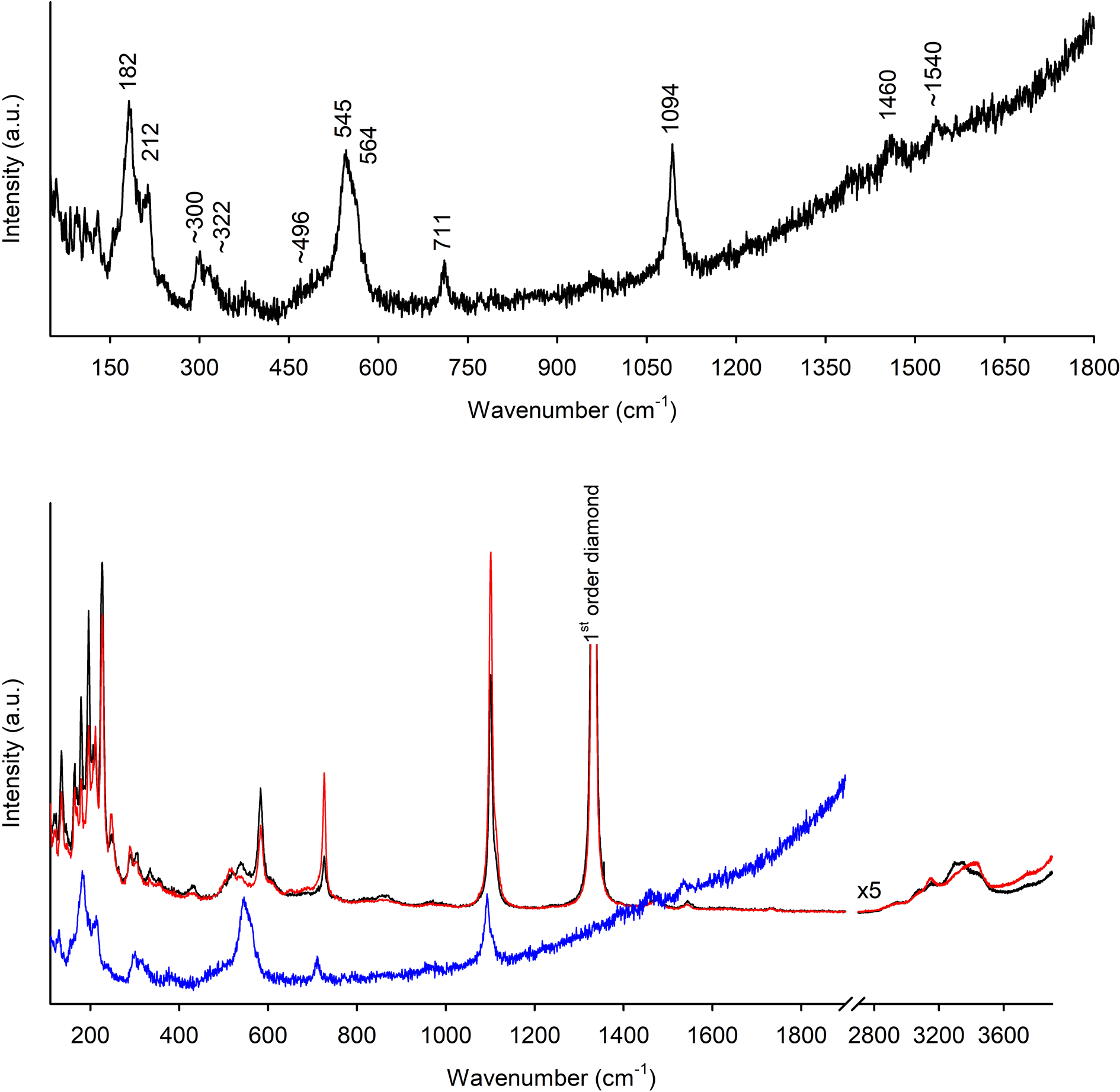
A Raman spectrum of alumohydrocalcite from Nowa Ruda in Poland, the occurrence studied by Stachowicz et al. (Reference Stachowicz, Parafiniuk, Wilson, Coles and Woźniak2015), was also collected for comparison. The spectra of both minerals are shown in Fig. 4. In the grguricite specimen, very high background fluorescence at high wavenumbers did not permit the observation of the weak Raman modes associated with OH stretching vibrations. The high Cr content of grguricite may be the cause of this high fluorescence when using a green laser. The Raman spectra of grguricite and alumohydrocalcite are otherwise similar. They comprise groups of low-wavenumber peaks at 100–250, 280–340 and 500–600 cm–1, which are interpreted as lattice modes and combination modes involving the hydrous species. The shared peaks at higher wavenumbers are assigned to internal vibrations of the CO3 structural units by comparison with mode assignments made for the free carbonate ion and carbonate minerals (e.g. Scheetz and White, Reference Scheetz and White1977). The peak at ~711 cm–1 can be attributed to the ν4 symmetric (in-plane) bending mode of the CO3 groups., the intense band at ~1094 cm–1 to the symmetric stretching vibration (ν1), and the weak peak at 1460 cm–1 to the asymmetric stretching vibration (ν3). The weak peak at 1540 cm–1 fits better with the wavenumber range of hydroxyl than the carbonate groups in the structure.
Raman spectra of grguricite and alumohydrocalcite. In the lower part of the figure the grguricite spectrum (blue) is compared with spectra of a sample of alumohydrocalcite (red and black) from the type locality (collected in two beam orientations 90° apart corresponding to the red and black spectra). The first order diamond peak visible in the alumohydrocalcite spectra is caused by the diamond-anvil that was used as a substrate. Also shown is the OH-stretching region of alumohydrocalcite (enlarged ×5 relative to the 100–1800 cm–1 spectrum). The corresponding region of the grguricite spectrum (not shown here due to the steeply rising background due to fluorescence) is featureless and it is likely that the weak OH bands are not resolved relative to the baseline, which is noisier that that of the alumohydrocalcite spectrum. The close correspondence between spectra of the two phases in the 100–1800 cm–1 region is evident.

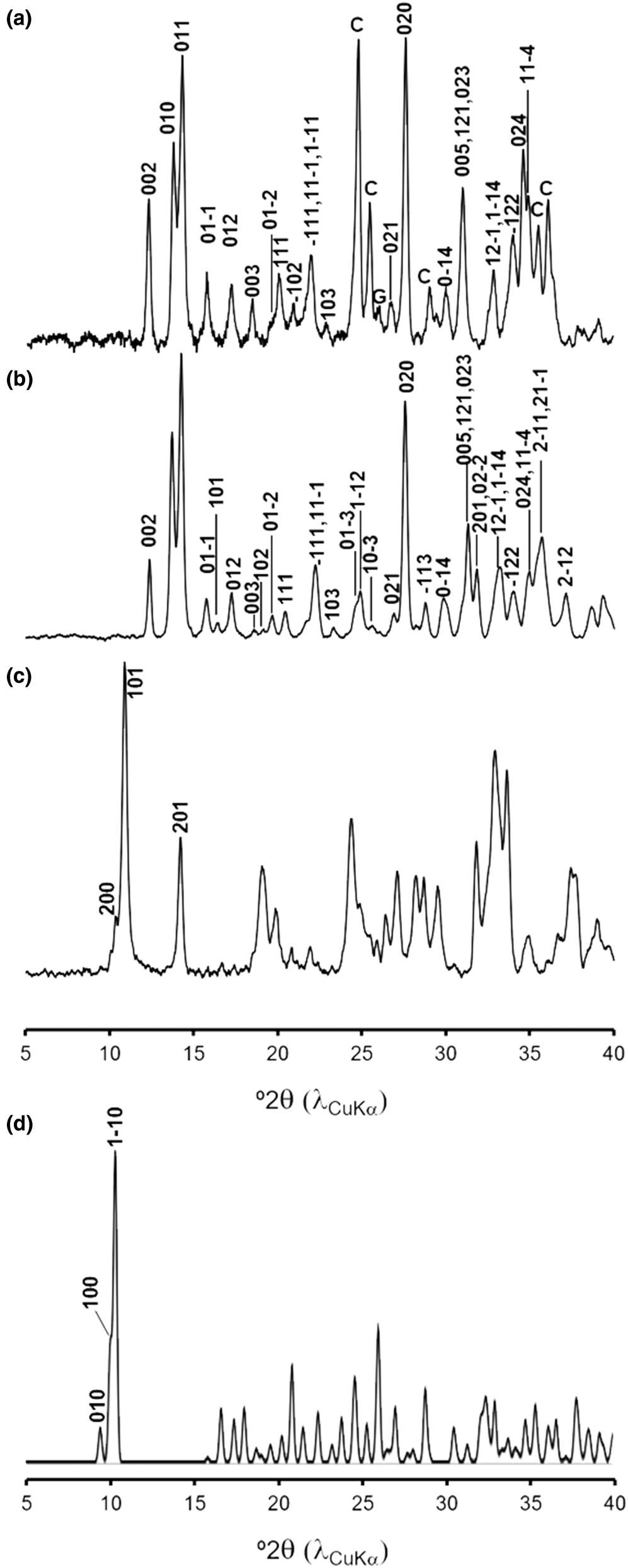
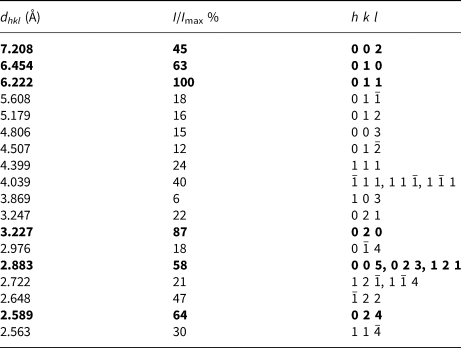
The powder diffraction patterns (Fig. 5) show that there is a very close correspondence between grguricite and alumohydrocalcite, particularly in the diagnostic region 10–15°2θ. The patterns of the other closely related topologies (dresserite, dundasite/strontiodresserite and hydrodresserite) are very different from those of grguricite and alumohydrocalcite (Fig. 5). The unit-cell parameters of grguricite were obtained by a Pawley fit of the powder diffraction pattern as implemented in the HighScore program (®PanAnalytical Industries, Degen et al., Reference Degen, Sadki, Bron, König and Néner2014), and using as a starting point for refinement the triclinic cell of alumohydrocalcite reported by Stachowicz et al. (Reference Stachowicz, Parafiniuk, Wilson, Coles and Woźniak2015): R = 0.092 and wR = 0.097, GoF = 1.120 and are (triclinic, space group P  ${\bar 1}$): a = 5.724(2), b = 6.5304(9), c = 14.646(4) Å, α = 81.682(1), β = 83.712(2), γ = 86.365(2)°, V = 537.8(2) Å3 with Z = 2. Table 3 lists the dhkl values and relative intensities of reflections of the powder diffraction patterns obtained from the Pawley fit and based upon the derived unit cell.
Quasi-random experimental powder diffraction patterns (CuKα) of (a) grguricite, (b) alumohydrocalcite and (c) dresserite. All patterns were obtained from small aggregates. In the grguricite pattern C = cerussite impurity and G = galena impurity. (d) Calculated powder diffraction pattern (CuKα) of hydrodresserite based upon the crystal structure determined by Jambor et al. (Reference Jambor, Sabina and Sturman1977). The diffraction patterns of dresserite and hydrodresserite are labelled with the hkl values of diagnostic low-angle reflections. Note that the hkl values shown for dresserite correspond to those of the Pnma setting of the unit cell.

The d spacings and relative intensities of peaks in the quasi-powder X-ray diffraction pattern of grguricite obtained by a pseudo-Gandolfi-like movement on a small fine-grained aggregate removed from the hand specimen.*

* Only peaks with I/I max >5% and with no overlap with cerussite impurity are given.
The strongest lines are given in bold
Discussion
The unit-cell parameters of grguricite and the other members of the ‘dundasite group’ are summarised in Table 1. It is evident that the unit-cell volumes of grguricite and alumohydrocalcite are distinct from other members of the group and are in this respect diagnostic. Furthermore, they are almost identical, within error, to each other. Consequently, any lilac-pink sample of a material identified as alumohydrocalcite from its powder X-ray diffraction pattern should be analysed chemically to determine its Cr content. The almost identical unit cells of grguricite and alumohydrocalcite can be rationalised by recognising that both minerals have a topology in which isolated heteropolyhedral units are only connected via hydrogen bonding involving inter-unit H2O. It might be expected, therefore, that minor structural adjustments to the heteropolyhedral unit, such as substitution of Cr for Al, will have little or no clear effect on the inter-unit configuration and, therefore, on the unit-cell parameters. This configuration contrasts with other dundasite-group structures, all of which have heteropolyhedral units that are connected directly to each other and for which substitution of Cr for Al may have a more obvious effect on unit-cell parameters.
The Raman spectra of alumohydrocalcite and grguricite may not discriminate sufficiently between these two minerals. In the present study, grguricite was first recognised qualitatively as a compositionally distinct Ca- and Cr-bearing phase by energy-dispersive spectrometry. Powder X-ray diffraction then identified the material as being closely related to alumohydrocalcite, on the basis of close agreement of their diffraction patterns and the derived unit-cell parameters. It is not a Ca analogue of petterdite, the assumption made on-line for the chemically similar incompletely characterised materials from Comet Vale or Kemparsi, which may or may not contain, or be, grguricite. The very fine-grained nature of grguricite implies that only by using non-standard X-ray methods might a full crystal structure be obtained, as was done for alumohydrocalcite using synchrotron single-crystal X-ray diffraction (Stachowicz et al., Reference Stachowicz, Parafiniuk, Wilson, Coles and Woźniak2015).
Acknowledgements
The authors would like to thank Bill Birch, Oskar Lidenmayer and Stuart Mills at Museums Victoria for information and access to the Comet Vale samples, Alex Ball of the Core Research Laboratories at the Natural History Museum, London for SEM images, and Ben Grguric for information regarding the Comet Vale occurence. Uwe Kolitsch, Martin Stachowicz and an anonymous reviewer are thanked for their comments that have improved this manuscript.