Case report
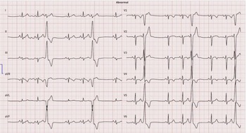
An 8-year-old boy presented with intermittent chest pain for 3 months. Physical examination was unremarkable. Electrocardiography revealed frequent premature ventricular contractions with trigeminy (Figure 1). Twenty-four-hour Holter monitoring demonstrated a premature ventricular contraction burden of 40%. Transthoracic echocardiography revealed mild systolic dysfunction (left ventricular ejection fraction: 50%). Laboratory investigations including serum troponin and metabolic screening were normal.
12-lead electrocardiogram from the patient demonstrating isolated, uniform premature ventricular contractions (PVCs) occurring in a trigeminal pattern. No signs of repolarization abnormality or ventricular hypertrophy are evident.

Initial treatment with propranolol failed to reduce the premature ventricular contraction burden. Antiarrhythmic therapy was subsequently changed to sotalol, a Class III antiarrhythmic agent, alongside an angiotensin-converting enzyme (ACE) inhibitor. This combination resulted in normalisation of systolic function (ejection fraction: 65%), and the 24-hour Holter monitoring showed a reduction in premature ventricular contraction burden from 40% to 10%.
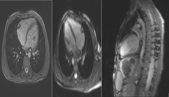
Cardiac MRI revealed no evidence of myocardial oedema on fat-suppressed T2-weighted sequences or T2 mapping. Native T1 values and extracellular volume of the left ventricular myocardium were within normal limits. First-pass perfusion imaging showed no perfusion defects in the interventricular septum or in either ventricular wall. Left ventricular systolic function was at the lower limit of normal (ejection fraction: 51%), and right ventricular systolic function was also borderline (ejection fraction: 48%). Quantitative tissue characterisation demonstrated non-ischaemic myocardial fibrosis involving more than 3% of the left ventricular myocardium. Late gadolinium enhancement imaging revealed a subtle, mid-myocardial focal enhancement in the mid-ventricular anteroseptal segment, consistent with a non-ischaemic pattern (Figure 2).
Sagittal cardiac magnetic resonance (CMR) image demonstrating normal thoracic anatomy. No myocardial oedema was observed on fat-suppressed T2-weighted sequences or T2 mapping. Quantitative tissue characterisation showed >3% non-ischaemic fibrosis of the left ventricle, with late gadolinium enhancement (LGE) in the mid-myocardial anteroseptal segment.

Genetic testing revealed variants in FLNC and CTNNA3, both implicated in arrhythmogenic cardiomyopathy.
Introduction
Arrhythmogenic cardiomyopathy is a genetically heterogeneous disorder primarily affecting the right ventricle, characterised by progressive fibrofatty myocardial replacement, ventricular arrhythmias, and an increased risk of sudden cardiac death, particularly in young individuals.Reference Mattesi, Zorzi, Corrado and Cipriani1,Reference Tadros, Miyake, Kearney, Kim and Denfield2 Although classical arrhythmogenic right ventricular dysplasia (ARVD) is predominantly linked to desmosomal gene mutations, recent studies have identified pathogenic variants in non-desmosomal genes such as FLNC (filamin C) and CTNNA3 (catenin alpha-3) that contribute to overlapping arrhythmogenic phenotypes.Reference Dong, Zhou and Zhang3,Reference Ader, De Groote and Réant4
The FLNC gene encodes filamin C, a cytoskeletal protein essential for myocardial structural integrity, and pathogenic variants have been associated with arrhythmogenic and dilated cardiomyopathies.Reference Dong, Zhou and Zhang3,Reference Alsubhi, Aldarwish and Agrawal5,Reference Baban, Alesi and Magliozzi6 CTNNA3, encoding catenin alpha-3, has also been implicated in arrhythmogenic remodelling through impaired intercellular adhesion.Reference Tadros, Miyake, Kearney, Kim and Denfield2,Reference Ader, De Groote and Réant4
While isolated variants in each gene have been described in the literature, the co-occurrence of FLNC and CTNNA3 heterozygous variants in a paediatric patient is exceedingly rare. Here, we report the case of an 8-year-old male presenting with frequent premature ventricular contractions, left ventricular dysfunction, and two concurrent heterozygous variants—FLNC c.3301G >A (p. Gly1101Ser) and CTNNA3 c.988G >A (p. Ala330Thr). This case highlights the clinical relevance of comprehensive genetic evaluation in early-onset ventricular arrhythmias and suggests potential synergistic pathogenicity of multiple non-desmosomal variants in paediatric arrhythmogenic cardiomyopathy.
Genetic analysis
Targeted single-gene mutation analysis was performed using polymerase chain reaction (PCR) and genomic DNA sequencing techniques. The test identified a heterozygous FLNC variant, c.3301G >A (p. Gly1101Ser), previously associated with cardiomyopathy phenotypes. This variant was confirmed in peripheral blood obtained from the patient.
Discussion
This case underscores the significance of evaluating genetic contributions in paediatric patients presenting with idiopathic dilated cardiomyopathy. Although truncating mutations in the FLNC gene are infrequent, they are strongly associated with life-threatening ventricular arrhythmias and sudden cardiac death.Reference Ader, De Groote and Réant4 The presence of atypical biventricular involvement may reflect overlapping cardiomyopathy phenotypes, further complicating the diagnostic approach. Timely genetic screening, multimodal imaging, and individualised management strategies are essential for improving prognosis and guiding follow-up.Reference Mattesi, Zorzi, Corrado and Cipriani1,Reference Tadros, Miyake, Kearney, Kim and Denfield2
Although FLNC variants are more commonly reported in adults, recent paediatric series have highlighted their clinical relevance. For instance, in a cohort of 500 paediatric patients, FLNC variants were reported in 14 individuals (2.8%), some of whom exhibited cardiomyopathy or arrhythmic manifestations.Reference Dong, Zhou and Zhang3,Reference Baban, Alesi and Magliozzi6 A study published in 2024 further described novel FLNC mutations in children, emphasising their potential association with dilated or mixed-type cardiomyopathy.Reference Dong, Zhou and Zhang3 Another report highlighted that FLNC-related forms of “pediatric cardiomyopathy,” while rare, represent a clinically significant subgroup.Reference Alsubhi, Aldarwish and Agrawal5,Reference Baban, Alesi and Magliozzi6
In contrast, CTNNA3 has been implicated in arrhythmogenic cardiomyopathy and ventricular arrhythmias, although existing evidence is largely derived from studies in adults or young adults.Reference Tadros, Miyake, Kearney, Kim and Denfield2 To our knowledge, there are no prior reports describing a paediatric patient presenting with this specific combination of clinical manifestations and dual FLNC–CTNNA3 heterozygous variants. Thus, this case may represent a previously unrecognised phenotype.
Conclusion
This case highlights the importance of integrating advanced cardiac imaging and genetic testing in the evaluation of paediatric patients presenting with idiopathic arrhythmias and systolic dysfunction. The identification of concurrent FLNC and CTNNA3 variants may contribute to arrhythmogenic cardiomyopathy and underscores the need for comprehensive genotype–phenotype assessment in similar clinical presentations. Further studies are needed to determine the clinical significance and long-term outcomes associated with these combined variants in paediatric populations.
Data availability statement
Data used in this study can be provided on reasonable request.
Financial support
This study did not receive financial support.
Competing interests
The authors declare that they have no conflict of interest to disclose.
Consent to participate
The informed consent form was obtained from the patient.