Introduction
Pigmented lesions of the sinonasal cavity are rare, with the most common pathology being malignant melanoma.Reference Prasad1 Other malignant possibilities include lymphoma, sinonasal undifferentiated carcinoma and olfactory neuroblastoma.Reference Prasad1 Though benign pigmented lesions of the sinonasal cavity are even rarer, an important clinical differential diagnosis to consider is the blue naevus.
Blue naevi are composed of deeply pigmented dendritic melanocytes admixed with a variable number of spindled melanocytic cells with pale cytoplasm and melanophages. This lesion is most commonly found in the skin. Rare cases have been reported in the oral cavity,Reference Ojha, Akers, Akers, Hassanein, Islam and Cohen2 cervix,Reference Craddock, Bandarchi and Khalifa3 scalp,Reference Golden, Maliawan and Mulyadi4 prostate,Reference Di Nuovo, Sironi and Spinelli5 lymph nodes,Reference Misago, Nagase, Toda, Shinoda, Koba and Narisawa6 subungual regionReference Gershtenson, Krunic, Chen, Konanahalli and Worobec7 and conjunctiva.Reference Wang, Prieto, Esmaeli, Kolda, Lazar and Ivan8 Blue naevi located in the sinonasal mucosa are exceptionally rare.
We herein report a case of sinonasal blue naevus, emphasising the patient's clinicopathological features.
Case report
A 47-year-old, Chinese man presented to our otolaryngology clinic with chronic sinusitis. His symptoms were those of intermittent nasal obstruction and hyposmia, of many years' duration. There was no associated nasal discharge or epistaxis. He had a background history of allergic rhinitis for which he had previously undergone laser inferior turbinoplasty.
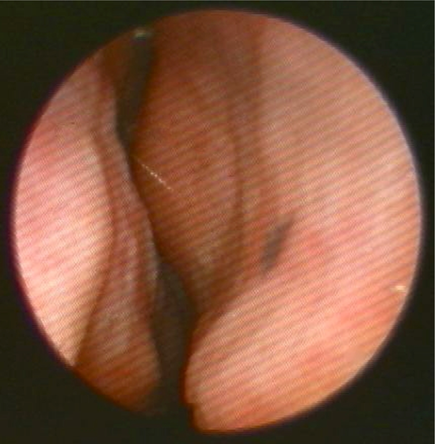
On physical examination, the patient's middle meati were oedematous and his nasal septum deviated to the left. Incidentally, a small, blackish spot, 2 mm in diameter, was noted on the right nasal septum, opposite the anterior head of the inferior turbinate (Figure 1).
Endoscopic photograph of the blue naevus.

A computed tomography scan of the sinuses showed extensive sinus opacification, but no mass lesion of the nasal septum.
Medical therapy for the patient's sinusitis was commenced.
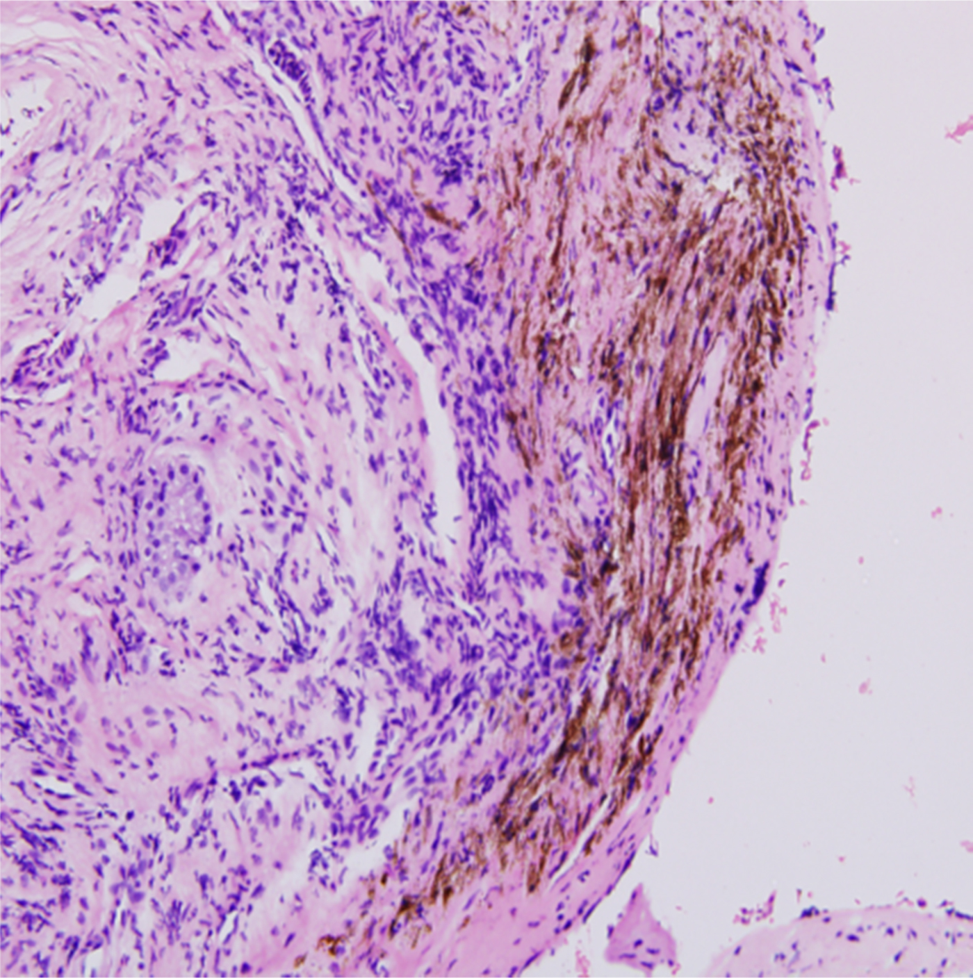
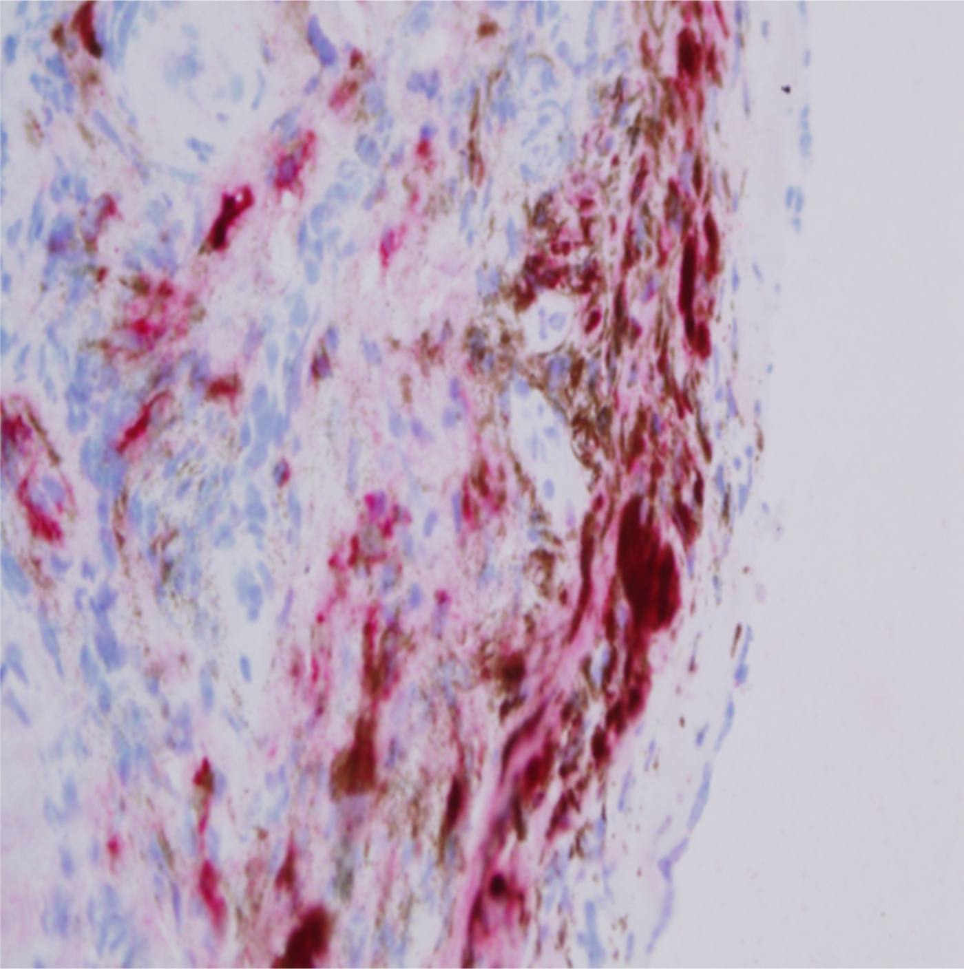
In the meantime, a complete excision biopsy of the pigmented lesion of the septum was carried out under local anaesthesia. Histological examination revealed the presence of heavily pigmented aggregates of dendritic cells within the stromal tissue, with bland nuclei and no mitotic figures (Figure 2). Upon immunohistochemical analysis, the lesional cells were reactive for S-100 and Melan-A (Figure 3). A diagnosis of conventional blue naevus (analogous to cutaneous-common type) was made.
Photomicrograph showing nasal septal tissue with stromal presence of heavily pigmented spindle cells. (H&E; original magnification ×200)

Photomicrograph showing reactivity of lesional cells to Melan-A immunohistochemical staining. (Original magnification ×400)

After the excision biopsy wound site had healed, the patient underwent endoscopic sinus surgery and septoplasty to treat his chronic sinusitis, which had proved to be refractory to maximal medical therapy.
At the most recent follow up, 13 months after blue naevus excision and 11 months after endoscopic sinus surgery and septoplasty, there had been no recurrence of the blue naevus, and the patient's sinusitis symptoms had significantly improved.
Discussion
Blue naevi are usually heavily pigmented lesions and mostly occur in the skin. Blue naevi arising in the sinonasal cavity are extremely rare. Melanocytes are derived from the neural crest and migrate to the skin and mucosa during embryogenesis.Reference Prasad1 In the sinonasal cavity, blue naevi are postulated to arise from melanocytes located in the epithelium and stroma of the respiratory mucosa.Reference Ojha, Akers, Akers, Hassanein, Islam and Cohen2
There have been only four previously reported cases of sinonasal blue naevi.Reference Chuang, Hao, Yeh and Jung9–Reference Aneiros, O'Valle, Garcia del Moral, Gomez Morales and Alvaro11 These were all small, asymptomatic and darkly pigmented lesions. Previous reported follow-up times post-excision have ranged from 13 months to 20 years, with no recurrence.
Apart from the blue naevus, the other, more sinister pigmented lesion to consider in the sinonasal cavity is the malignant melanoma, either primary or metastatic.Reference Plaza, Torres-Cabala, Evans, Diwan, Suster and Prieto12 Sinonasal melanomas are also uncommon lesions, constituting less than 1 per cent of all melanomas and less than 4 per cent of all sinonasal tumours.Reference Craddock, Bandarchi and Khalifa3 They are generally larger tumours with symptoms including epistaxis. They have a very poor prognosis, with survival rates of only 30 per cent at 60 months post-treatment.Reference Golden, Maliawan and Mulyadi4 Wide local excision with negative margins is the treatment of choice, and some patients may benefit from adjuvant post-operative radiotherapy.Reference Di Nuovo, Sironi and Spinelli5
• Although very rare, blue naevus is a differential diagnosis for a pigmented lesion of the sinonasal cavity
• It is characteristically small and asymptomatic
• Histology is required for definite diagnosis
• Treatment is complete excision for diagnostic and therapeutic purposes
• The risk of recurrence is very low
The key to the correct histological diagnosis of blue naevus of the sinonasal cavity is identification of the heavily pigmented dendritic melanocytes that are invariably present within blue naevi, and also confirmation that mitotic activity, if present, is very infrequent (i.e. less than 1 per 10 high power fields) and never abnormal in form. Immunohistochemistry provides limited additional value in the distinction of blue naevus from melanoma: blue naevi typically have uniform immunoreactivity for Human Melanoma Black-45 (‘HMB-45') while melanomas often display a patchy reaction pattern.Reference Lee, Torres, McLean, Chen and Lee13
Conclusion
Blue naevus should be included in the differential diagnosis of pigmented lesions within the sinonasal cavity, despite its rarity. It may be initially suspected by its small size and asymptomatic nature. However, histological examination is required for definitive diagnosis, with identification of the key microscopic features described above. A complete excision biopsy can usually be easily performed, for both diagnostic and therapeutic purposes. Blue naevus has an excellent prognosis with very low risk of recurrence.