





Introduction
Fungal mycelium has gained increasing attention within the context of bio-based materials; when combined with industrial waste streams, it represents a promising solution for sustainable applications in construction, design and architecture with the advantage of being a craft that can rely on local production and distribution (Elsacker et al. Reference Elsacker, Vandelook, Brancart, Peeters and De Laet2019; Rigobello Reference Rigobello2023; Vandelook et al. Reference Vandelook, Elsacker, VanWylick, Laet and Peeters2021). Within these fields, mycelium has been predominantly explored as a bulk material, often combined with organic substrates, and processed into rigid composites or structural elements that are subsequently subjected to heat treatment to inactivate the organism (Camilleri et al. Reference Camilleri, Narayan, Lingam and Blundell2025; Elsacker et al. Reference Elsacker, Vandelook, VanWylick, Ruytinx, Laet and Peeters2020). This craft is highly versatile, as variations in the type and orientation of the lignocellulosic substrate allow the material to be modulated to achieve specific mechanical performances. Such hybrid approaches have demonstrated the technical, ecological and mechanical potential of fungal-based materials (Rigobello and Ayres Reference Rigobello and Ayres2022; Rigobello et al. Reference Rigobello, Colmo and Ayres2022). However, this practice primarily frames mycelium as an inert end-product rather than as a living, adaptive system capable of regeneration and environmental interaction. As a result, mycelium-based materials are emphasised primarily through their volumetric and structural performance, limiting the expression of fungal metabolism and reducing it to a transient production phase, rather than allowing it to persist as an active material condition. Recent developments in advanced material research, particularly within the field of ELMs, foreground a “living material” paradigm by proposing materials that retain biological activity and actively interact with their surroundings. The living cells of such materials are deliberately genetically tuned towards achieving specific design objectives and therefore exhibiting functional behaviours that extend beyond conventional material performance (Nguyen et al. Reference Nguyen, Courchesne, Duraj-Thatte, Praveschotinunt and Joshi2018; Srubar Reference Srubar2021). Within the design and architectural practice, these qualities open new perspectives, enabling materials that mediate between performance and expression and between functional requirements and formal articulation. Despite growing interest in living materials, the use of living fungal mycelium in the built environment remains underexplored, particularly in applications that rely solely on living cells rather than hybrid or inactivated composites (Elsacker et al. Reference Elsacker, Zhang and Dade-Robertson2023). From a material perspective, the transition from mycelium-based bulk materials to pure biological fungal films and membranes opens new design opportunities. Operating at the scale of surfaces and interfaces, living fungal membranes introduce a material regime within architecture that is expressive, legible, communicative and responsive. These configurations reposition the material as an active mediator between environments, where interaction and metabolic exchange are foregrounded. The regulation of gas diffusion, moisture dynamics and biochemical activity becomes central to the material’s adaptive and regenerative behaviour. Living fungal materials in the form of membranes invite a rethinking of mycelium not as an inert matrix embedded within a composite, but as the primary constituent and facilitator between environments, while defining novel material behaviour and functionality through its metabolic activity.
Membranes and patterns in architectural design
Inspiration of methods to unfold membrane-like materials can be drawn by textile and tapestry craft tradition and development within the architectural design context. The ambition towards achieving architectural scales leads to the need for assemble discrete pieces of membranes through junction, as the manufacturing of a continuous and monolithic piece is hardly feasible from an operational and logistical standpoint. Moreover, patterning constitutes a fundamental dimension of the material, as it operates simultaneously as a mode of design, a vehicle for material expression and communication. Patterning operates not only as an ornamental motif, but it represents an organisational and performative strategy. In textile design, patterns can be leveraged as a cultural means in form of embroideries for storytelling as in the traditional tapestry craft, with large scale of images to express significant events. It is a way of responding to the need of preserving memories, marking time as in the case of the famous Bayeux Tapestry. Patterns can also be of structural importance, dictating material behaviour such as folding or a way to encode information across scales. In textile tradition patterns have embodied performances, representing interfaces; weaving densities can regulate light penetration (Cremers Reference Cremers2010), embroidered knitted pattern can allow for distribution of stress and also directional elasticity (Šinke et al. Reference Šinke, Tamke, Manian, Troi and Thomsen2025). In architectural scales and contexts membranes acquire an important role as screens and partition as mediators between environments, modulating and revealing visions of external details, affecting the material behaviour and offering lightweight construction alternatives (Paech Reference Paech2016). Morphological variation within a membrane functions as a mediator between geometry, aesthetic expression and performative behaviour, elevating plain surfaces into potential active interfaces (Šinke et al. Reference Šinke, Tamke, Manian, Troi and Thomsen2025). This approach resonates and can be implemented into living material research. In living fungal membranes, morphological variation is not imposed but orchestrated through morphogenesis. Form emerges as a developmental process guided by the designer, who choreographs conditions rather than applies additional material. Patterning is therefore cultivated, not constructed. Instead of being achieved through layering, printing, adding or subtracting material, it arises from the metabolic vitality of the membrane itself. Through exposure, modulation and environmental control, the organism generates surface differentiation intrinsically, transforming pattern into an expression of biological aliveness. Masking techniques and microclimate manipulation, explained in this paper, allow to induce stimulation of the growth of aerial hyphae on the living membrane with formation of hydrophobic mats organised in pattern geometries. Such bio-fabricated patterning operates in a micro and macro scales: microscopic hyphal networks develop into macroscopic variations in texture, permeability, aesthetics and morphogenesis, representing the surface extension of the membrane towards the environment. This characteristic can be leveraged to frame patterning both as functional and ornamental. The generation of living pattern opens new perspectives towards the use of this design and biological aspect, still grounded from the traditional and cultural heritage but elevated by the metabolic activity of the living fungal materiality.
Living fungal membranes
Membranes occupy a critical role in both biological and architectural systems, functioning as mediators between internal and external conditions while simultaneously supporting mechanical, chemical and spatial differentiation, acting as an interface apparatus designed for element selection between environments. Living fungal membranes can exist as hybrid composites, for commercial use is common to include a structural fabric-like material to enhance mechanical proprieties, on which the mycelium is grown (Vandelook et al. Reference Vandelook, Elsacker, VanWylick, Laet and Peeters2021); the membranes investigated in this paper are composed exclusively of living fungal cells that retain biological activity. This allows the material to express dynamic functions including; sensing, growth, adaptation and self-repair throughout its lifespan. The activation of this “fourth dimension” of time, through which material behaviour becomes animated, significantly extends the scope of potential applications especially towards advanced fungal living membranes for ELMs (Elsacker et al. Reference Elsacker, Martin, Sangosanya, Verstuyft, VanWylick and Peeters2025). Elevating living fungal membranes into ELMs expands opportunities across both the biotechnological field and the built environment, where they may operate as responsive building components, self-healing systems, environmental remediation agents and biological sensors. Living fungal membranes constitute a distinct subclass of mycelium-based materials and can be fabricated either through Solid-State Fermentation (SSF), where mycelium colonises a lignocellulosic substrate and a surface pellicle is subsequently extracted, or through Liquid-quid-state submerged (LSF) and liquid-state surface fermentation (LSSF), in which a continuous mycelial skin self-assembles at the liquid–air interface under static conditions (Elsacker et al. Reference Elsacker, Vandelook and Peeters2023; Peeters et al. Reference Peeters, Saluena and Vandelook2023). Conventionally, these approaches have been employed to produce flexible fungal tissues that are subsequently dried or heat-treated to obtain inert materials for consumer goods applications such as leather-like material (Elsacker et al. Reference Elsacker, Vandelook and Peeters2023). Elsacker et al. propose an alternative paradigm in which the membrane is maintained in a living state to exploit the intrinsic properties of fungal cells focusing on self-healing, environmental sensing and adaptation. In the study, the self-healing capacity of Ganoderma lucidum is attributed to the presence of chlamydospores, stress-induced survival cells capable of withstanding harsh conditions and initiating new hyphal growth upon reactivation. The work demonstrates that a dry, flexible mycelium membrane can remain biologically active without continuous growth and can be selectively reactivated to repair damages (Elsacker et al. Reference Elsacker, Zhang and Dade-Robertson2023). Building upon this conceptual and methodological framework, the present work investigates Ganoderma lucidum cultivated and processed as a living membrane, with a particular focus on agitated LSF as the primary material production protocol. The study extends existing research by examining the material’s inherent self-assembly and regenerative behaviours as emergent properties of this growth condition; by using tensile tests we assess the bonding strength between assemblies of living membrane elements and by applying designed masking we instigate material growth of aerial hyphae for surface patterning. Ganoderma lucidum species is selected due to its dense hyphal network, robustness during cultivation, rapid growth rates and established relevance within mycelium-based material research. Rather than relying exclusively on static growth at the liquid-air interface – which, although convenient, produced inconsistent results in preparatory growth tests, including spatial variations in membrane thickness – this study adopts agitated LSF. While this approach requires additional production steps, the separation of biomass from the liquid phase through vacuum filtration enables significantly greater control over membrane thickness and surface homogeneity. This paper adopts a design-driven material exploration that integrates both quantitative and qualitative experimentation to interrogate the intrinsic properties of living fungal membranes. Two complementary lines of inquiry structure the investigation, focusing on methods for joining multiple membrane components and for patterning their surfaces. First, tensile testing is employed to characterise the mechanical behaviour of thin living mycelium membranes, with thicknesses below 1 mm. In this approach, multiple membranes are layered one atop another, allowing the dynamics of self-assembly to be expressed and evaluated through quantifiable material performance. Second, membrane-masking experiments are conducted to investigate regenerative capacity, selective regrowth and surface differentiation. By controlling air exposure by masking selected regions, these experiments explore how timing and spatial manipulation influence aerial hyphae formation producing hydrophobic patterning while limiting risk of contamination. By integrating mechanical characterisation with growth-based surface experimentation, this paper contributes to expand the knowledge and understanding of the potentials and limitations of mycelium as a living membrane material and as a functional interface. By defining mechanical performance, functionalities and aesthetic properties, it becomes possible to assess the material’s applicability within architectural scenarios, while simultaneously outlining its limitations. These constraints, rather than restricting the use, invite design participation to negotiate, adapt and strategically respond to the material’s specific properties and boundaries. The findings position thin mycelium membranes as performative interfaces in which functional behaviour and aesthetic expression emerge simultaneously from biological activity, offering new directions for the design and application within architectural and biodesign research.
Materials and methods
Liquid culture
Ganoderma lucidum (M9726) spawns are plated in agar-agar Petri dishes, prepared using a powder mixture of malt extract purchased from Mushroom production centre GmbH with a ratio of 141g for 3L of water. A 3*3 mm piece is cut and placed in the centre of each prepared Petri dish, closed and stored in an incubator at 25 degrees for 10 days. Liquid culture media (17 g
 ${{\rm{L}}^{ - 1}}$
malt extract, 3 g
 ${{\rm{L}}^{ - 1}}$
peptone, supplemented with thiamine (0.12 mg m
 ${{\rm{L}}^{ - 1}}$
) and 10 mL of trace elements comprising the following (in mg
 ${{\rm{L}}^{ - 1}}$
): FeSO47H2O (12), MnSO47H2O (3), ZnSO47H2O (3), CoSO47H2O (1) and (NH4)6Mo7O244H2O (1)(Hadibarata and Ayu Reference Hadibarata and Ayu2012), is prepared and autoclaved for 1 h. The addition of metals is found to central to fungal metabolism and enzymatic expression (Karaffa et al. Reference Karaffa, Fekete and Kubicek2021). After cooling, for each litter of liquid culture Ganoderma lucidum biomass is scraped from the Petri dishes, added into the liquid with 1000
 ${\rm \mu {L}}$
kanamicyn (concentration of 50 mg/mL) and poured in Erlenmeyer flasks. The flasks are incubated on a rotatory shaking bed for two weeks at 150 mpr at 29 degrees.
Membrane manufacturing
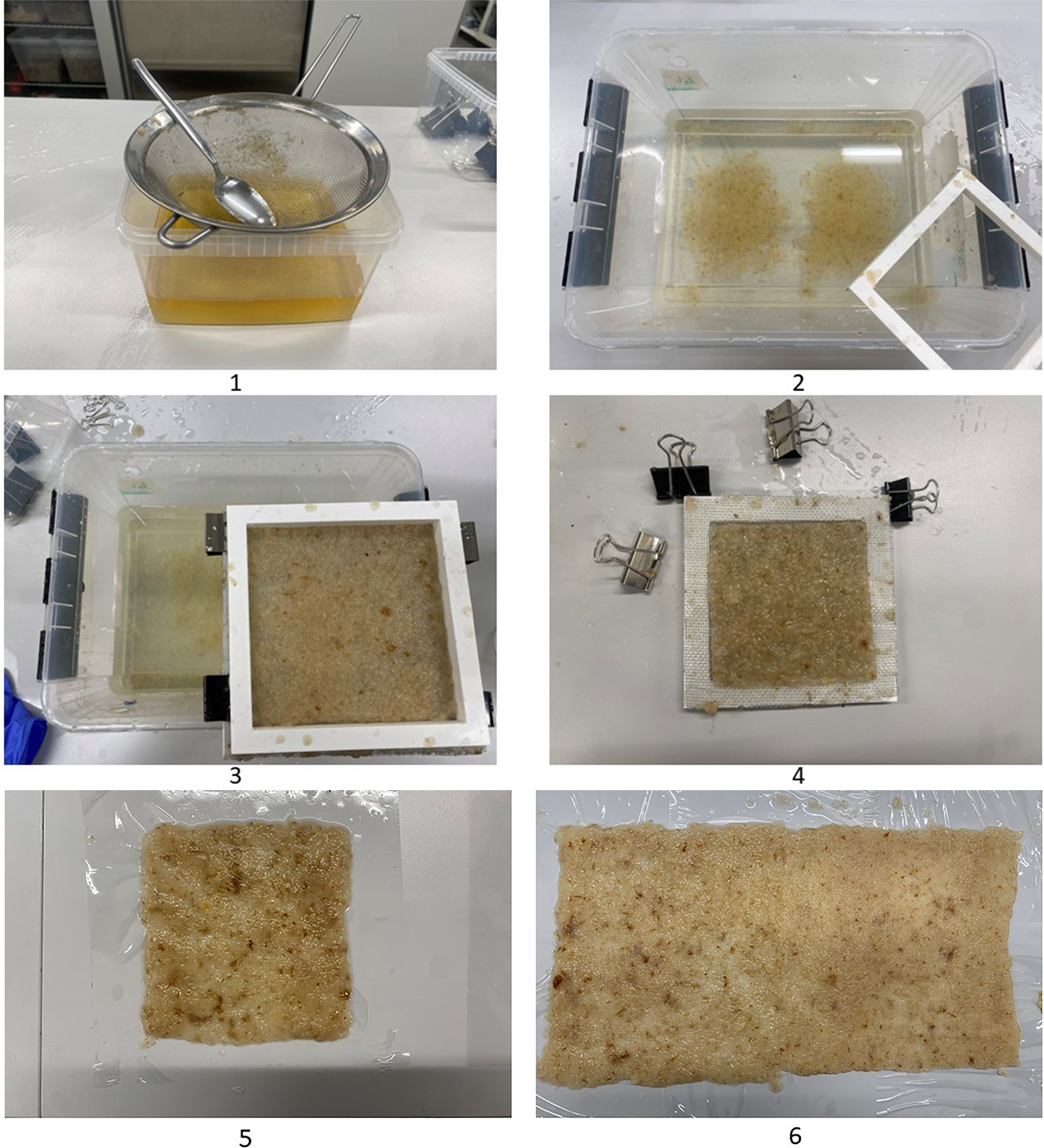
The manufacturing process of the fungal membrane is inspired by traditional paper-making techniques (Figure 1). The process begins with harvesting the fungal pellets formed in liquid culture using a strainer. The biomass is gently pressed to remove excess liquid and then transferred into a plastic container filled with demineralised water. A mould composed by of two 3d printed deckle where a metal mesh is placed in between is subsequently used to extract the biomass from the suspension, allowing it to collect as a uniform layer of
 $150 \times 150{\rm{\;mm}}$
dimensions on the mesh. The formed layer is then pressed and compacted between two sheets of cellophane, which permits gas exchange while preventing external contamination. This stage is critical as it enables the biomass comprising individual colonies to self-assemble into coherent sheets through ongoing mycelial activity. The paper-making-inspired technique allows a single frame to be used to fabricate larger membranes, as individual sheets can be formed separately and then layered and dried, enabling a sequential stacking or adjacent layering in order to achieve architectural scale up. In this paper we apply 48 h of curing, the sheet develops into a uniformly and can be further assembled by stacking multiple layers to produce thicker, multilayered membranes. These layers bond together through continued mycelial growth, effectively welding the membranes into a single, cohesive material.
Fungal membrane fabrication: 1. biomass harvesting and straining; 2.Biomass submersion into water; 3.Biomass extraction through a deckle and mesh forming a layers of
 $150 \times 150{\rm{\;mm}}$
dimension; 4. Biomass deposition on the mesh mould; 5. Biomass transfer on chellophane; 6. Sequential adjacent biomass transfer for large membranes fabrication.

Figure 1. Long description
Fungal membrane fabrication: 1. biomass harvesting and straining; 2.Biomass submersion into water; 3.Biomass extraction through a deckle and mesh forming a layers of 150 × 150mm dimension; 4. Biomass deposition on the mesh mould; 5. Biomass transfer on chellophane; 6. Sequential adjacent biomass transfer for large membranes fabrication.
Specimen design and standard reference
Using the 150
 $ \times $
150 mm frame and deckle, biomass is collected and after 48 h of curing, the resulting sheets were layered to create membranes composed of two overlapped layers. The material cross section of two layers membrane ranges between 0.15–0.20 mm thickness and four layers doubles between 0.28 and 0.40 mm thickness. Tensile testing of the living sheets is conducted to evaluate their mechanical performance, specifically strength, elongation at break and elastic modulus. While the manufacturing process is advantageous in terms of accessibility and minimal tooling requirements, it does not allow for the production of monolithic sheets. Consequently, to achieve architectural scale, larger membranes or designed geometries might be required, inviting the exploration of design assemble strategies. We investigate the potential of harnessing the living material’s regenerative properties to fuse discrete membrane elements into a continuous, larger surface, using biological regrowth as a method of connection. For this reason, the tensile tests presented in this study include investigations of overlapping seems in both shear (horizontal) and attachment (vertical) patched bio-welded seams configurations of discretised membrane elements. ASTM D882 is selected as the reference standard for tensile testing because it is specifically designed for thin plastic sheeting and membrane-like materials with thicknesses below 1 mm, which corresponds to the physical characteristics of the membranes developed in this research. ASTM D882 allows flexibility in grip separation and strain measurement, making it well suited to soft and extensible materials that may undergo large deformations prior to failure. The standard explicitly accommodates adaptations required for delicate biomaterials. This adaptability ensures that mechanical characterisation can be performed without compromising specimen integrity while remaining within a recognised testing framework. Its adoption ensures reproducibility, while aligning the mechanical testing approach with the actual geometry, thickness and mechanical behaviour of the membranes investigated in this study.
Results
Tensile result
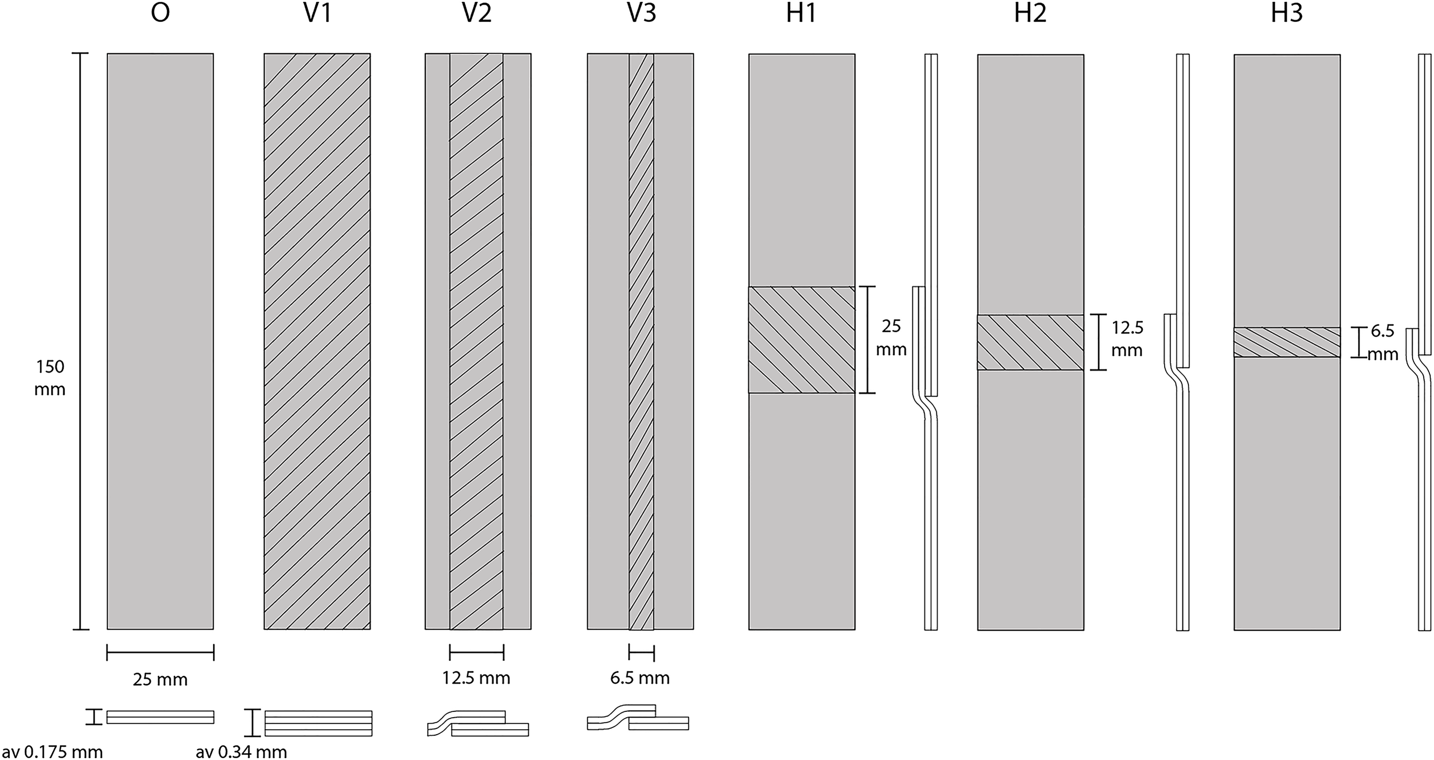
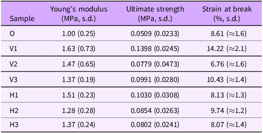
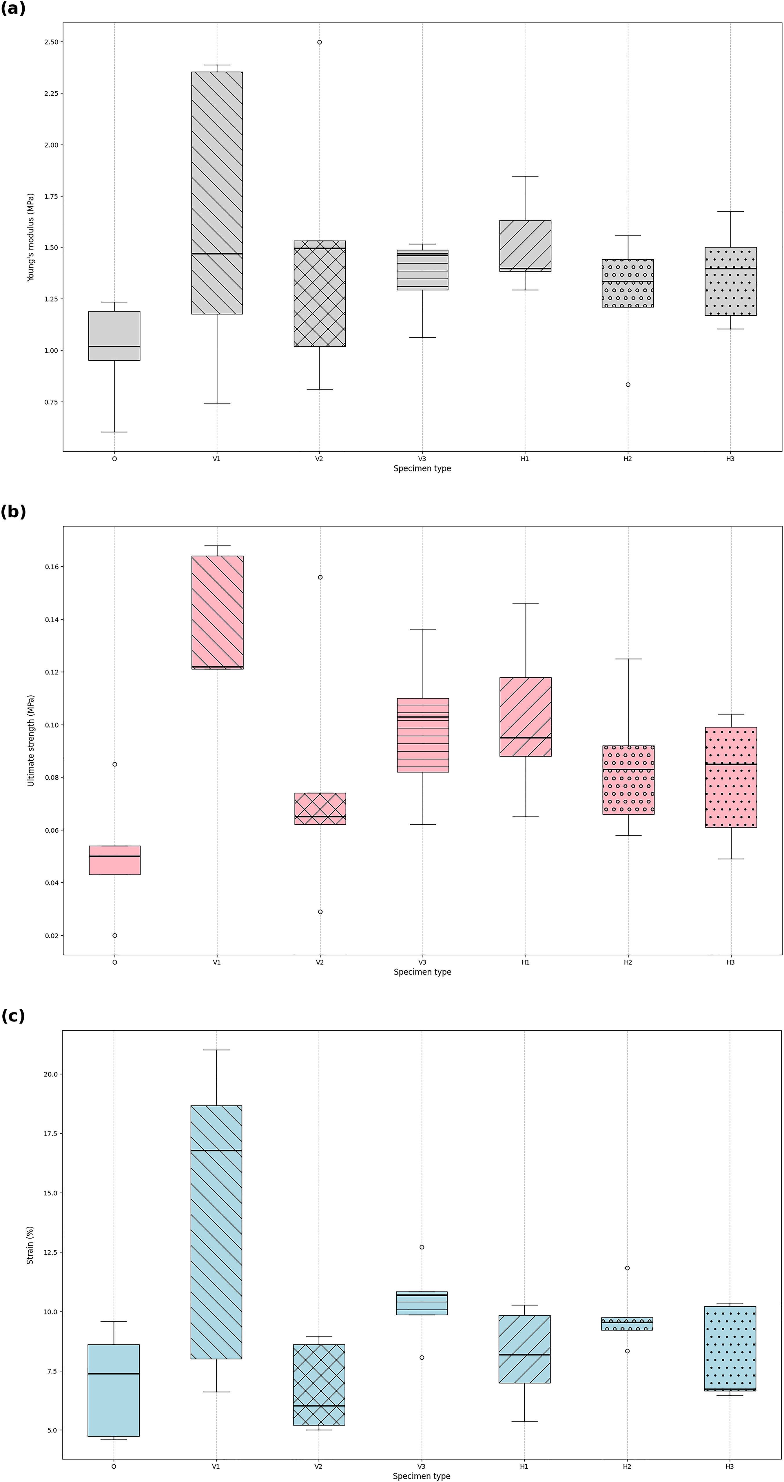
In this investigation material strips with dimension of 25.00 mm wide and 150 mm long are tested. Following the initial curing phase, the sheets are cut and reassembled to produce different overlapped dimensions; two configurations of overlapping are analysed: horizontal (H)and vertical (V), measuring (1)25.0 mm, (2)12.5 mm and (3)6.5 mm. For each configuration, five replicates were tested against a control sample (O) made of two overlapping membrane sheets (Figure 2). Tensile strength tested with a crosshead speed of 12.5 mm/min and conducted by Universal Testing Machine (UTM) machine, load cell of 100 N. with a set of customised grips. Table 1 shows the specimen type, the resulting mean Young’s modulus and ultimate strength. The tensile tests reveal a clear stiffness-deformability trade-off across the tested configurations, highlighting the strong influence of the material composition on mechanical performance. Among all samples, the V1 configuration exhibited the highest deformation capacity, reaching an elongation of approximately 8.1 mm, while simultaneously showing the highest Young’s modulus (
 $ \approx $
1.6–2.3 MPa), thereby representing the most mechanically performative system tested. In contrast, the V2 configuration showed the lowest elongation (
 $ \approx $
3.9 mm) together with high variability in stiffness, indicating a brittle response probably associated with a heterogeneous and weakly connected mycelial network. A systematic comparison between V and H configurations further demonstrates that H samples consistently sustain higher strain at break, suggesting improved load redistribution, enhanced ductility of the fungal network and a reduced tendency toward premature fracture. This difference reflects a structural effect, emphasising the role of internal organisation in governing failure behaviour. Within the O and V1 series, where O is the control sample made by 2 layers of mycelium and V1 made by 4 layers, a pronounced increase in deformation capacity is observed, with V1 exhibiting nearly double the elongation of O. Overall, deformation capacity increases progressively from V to H and O to V1 configurations, ranking from most brittle to most ductile as follows: V2, 3H
 $ \approx $
1H, O, 2H, 2V and V1. Notably V1 combines high stiffness with high elongation, identifying it overall as the most mechanically performative among the tested samples (Figure 3).
Specimen O consists of two bio-welded layers. V1 comprises four fully overlapping layers, while V2 and V3 feature a localised four-layer overlap along the central vertical axis with decreasing width. H1–H3 present horizontal four-layer overlaps positioned with progressively reduced thickness.

Figure 2. Long description
Specimen O consists of two bio-welded layers. V1 comprises four fully overlapping layers, while V2 and V3 feature a localised four-layer overlap along the central vertical axis with decreasing width. H1–H3 present horizontal four-layer overlaps positioned with progressively reduced thickness.
Specimen groups, Young’s modulus (Mpa), ultimate strength (Mpa), strain at break (%)

Table 1 Long description
A table comparing mechanical properties of different material samples. The table has 8 rows and 4 columns. The columns are labeled Sample, Young’s modulus (MPa, s.d.), Ultimate strength (MPa, s.d.), and Strain at break (%, s.d.). The row labels are O, V1, V2, V3, H1, H2, and H3. Each row provides the mean values and standard deviations for Young’s modulus, ultimate strength, and strain at break for each sample type. Notable trends include V1 having the highest Young’s modulus and ultimate strength, while O has the lowest strain at break.
Box plots for Young’s modulus results (a) and ultimate strength results (b) and Strain (c).

Statistical analysis
The result distributions are two-tailed. The distributions’ normality was verified for ultimate strength, Young’s modulus and strain at break results with the Shapiro-Wilk test (
 $\alpha $
= 0.05); the V1 group’s ultimate strength and the H3 group’s strain at break are not considered normally distributed based on this test (respectively p = 0.019733, p = 0.020632). Fisher’s defined kurtosis for these groups is respectively −1.800879 and −1.825887. Fisher-Pearson’s skewness coefficient for these groups is respectively 0.420769 and 0.401463. The distributions are considered normal as
 $\left| {kurtosis} \right|$
 $ \le $
2 and
 $\left| {skewness} \right|$
 $ \le $
1 (Hair et al. Reference Hair, Black, Babin and Anderson2013). The equality of variances was confirmed for ultimate strength, Young’s modulus and strain at break results with the Levene test (respectively p = 0.9834, p = 0.09186, p = 0.05135;
 $\alpha $
= 0.05). One-way ANOVA, Welch ANOVA and Kruskal-Wallis tests were conducted and found results not significantly different for Young’s modulus (respectively p = 0.3668, p = 0.1872, p = 0.3295;
 $\alpha $
= 0.05), yet significantly different for ultimate strength (respectively p = 0.004356, p = 0.008725, p = 0.01339;
 $\alpha $
= 0.05), and the one-way ANOVA indicated significant difference for strain at break (respectively p = 0.007823, p = 0.06133, p = 0.06636;
 $\alpha $
= 0.05). The mean values of specimen groups were found not to be significantly different with the Bonferroni corrected pairwise Welch t-test, besides ultimate strength’s groups O vs V1 (p = 0.007929,
 $\alpha $
= 0.05). Using the pairwise Games-Howell test, the most significant differences in ultimate strength are reported between the groups H3 vs V1 and O vs V1 (respectively p = 0.04649, p = 0.004298;
 $\alpha $
= 0.05). Using the Tukey’s HSD test we report significant differences in ultimate strength between the groups O vs V1 and V1 vs V2 (respectively p-adj = 0.0013, p-adj = 0.0431; FWER = 0.05). We identify pairwise difference with the Tukey’s HSD test for strain at break, between groups H1 vs V1, H3 vs V1, O vs V1 and V1 vs V2 (respectively p-adj = 0.0471, p-adj = 0.0442, p-adj = 0.0113, p-adj = 0.0084; FWER = 0.05). In conclusion, we show that Young’s modulus results are not statistically different between groups, which confirms the predictable qualities of the material. Variance in these results can be explained by methodological imprecision, prototypal production control explainable by the early nature of this research, and the heterogeneity of local properties of the cultivated material linked to the second growth process – see end of this section. We observe that groups V1, V2 and V3 have the same cross section, only the overlap and bonding strength will differ between these groups. The maximal bonding in V1 shows a contribution to ultimate strength and strain at brake compared to V2 and potentially V3 despite a lack of significant difference. This confirms that the mycelial bonding increases significantly the strength in the material, which we hypothesise is due to the homogenisation of stress across the two layers thanks to relatively long fibres mycelial connections occurrence. We observe that the failure of groups H1, H2 and H3 all happened at the extremities, not at the place of layers overlap. This suggests the shear strength resulting from the bonding is significantly higher than the tensile strength. The shear to tensile strength ratio in materials reflects its internal structure – here, that of the bonding. This can be explained by uniaxial tensile stress resulting in polymer chain alignment and stretching, cavitation and craze initiation which can localise early. Tensile strength may thus be capped by early damage mechanisms. Under shear stress, polymeric chains are sliding, rotating and disentangling, cavitation and crazing are suppressed; the hydrostatic tension is very low. Because of the friction between chains and the density of their entanglement, shearing involves more of the polymer’s specific resistance than tension. The present material looks to be therefore pressure sensitive, and we hypothesise its strength can increase with compressive normal stress. This is consistent with the methods of making of the material: the local environmental conditions will favour mycelial growth between the overlapping layers, leading to relatively long fibres linking, whereas the other parts of the material lacking oxygen access see no inner mycelial growth during the second growth process, and are made up of shorter fibres (hyphae) initiated by agitated liquid-state fermentation in the form of pellets, with a cross-pellet chemical binding driven by hydroxyl linking. We also note that the failure mode of the H1, H2 and H3 groups might be influenced by grip damage, stress concentration at grip transition and drying gradients, which are conditions which can be revised in a next iteration of experiment design to confirm the mechanical phenomena driving the material behaviour.
Masking tests
This series of experiments investigate the qualitative aspect of the living membrane, focusing on its aesthetic expression by using lasercuted cellophane sheets as templates to instigate hyphal formation on the uncovered area, while simultaneously speculating on the functional potentials these qualities offer. An 80
 $ \times $
80 mm mould is used to collect the biomass, which is then transferred between two sheets of cellophane and cured for 24 h. Following curing, the upper cellophane sheet is peeled off and replaced with a laser-cut, engraved sheet bearing the desired pattern. To reduce the risk of contamination and to maintain a localised microclimate that preserves moisture and minimises light exposure, an aluminium-foil-covered frame is placed over the sample. The sample is subsequently incubated under ambient conditions for approximately 48 to 72 hours. In areas where the cellophane is absent, active hyphal growth occurs, forming a white, hydrophobic mycelial mat that follows the negative space of the engraved pattern. In order to understand the dynamics of mycelia regeneration through this second growth and to understand the possibility to engage different masking techniques, this experimentation comprises the following investigation approaches: Pattern resolution test, pattern test with multiple masks, gradient effect with multiple patterns, complex pattern with multiple complementary masks.
Pattern resolution test
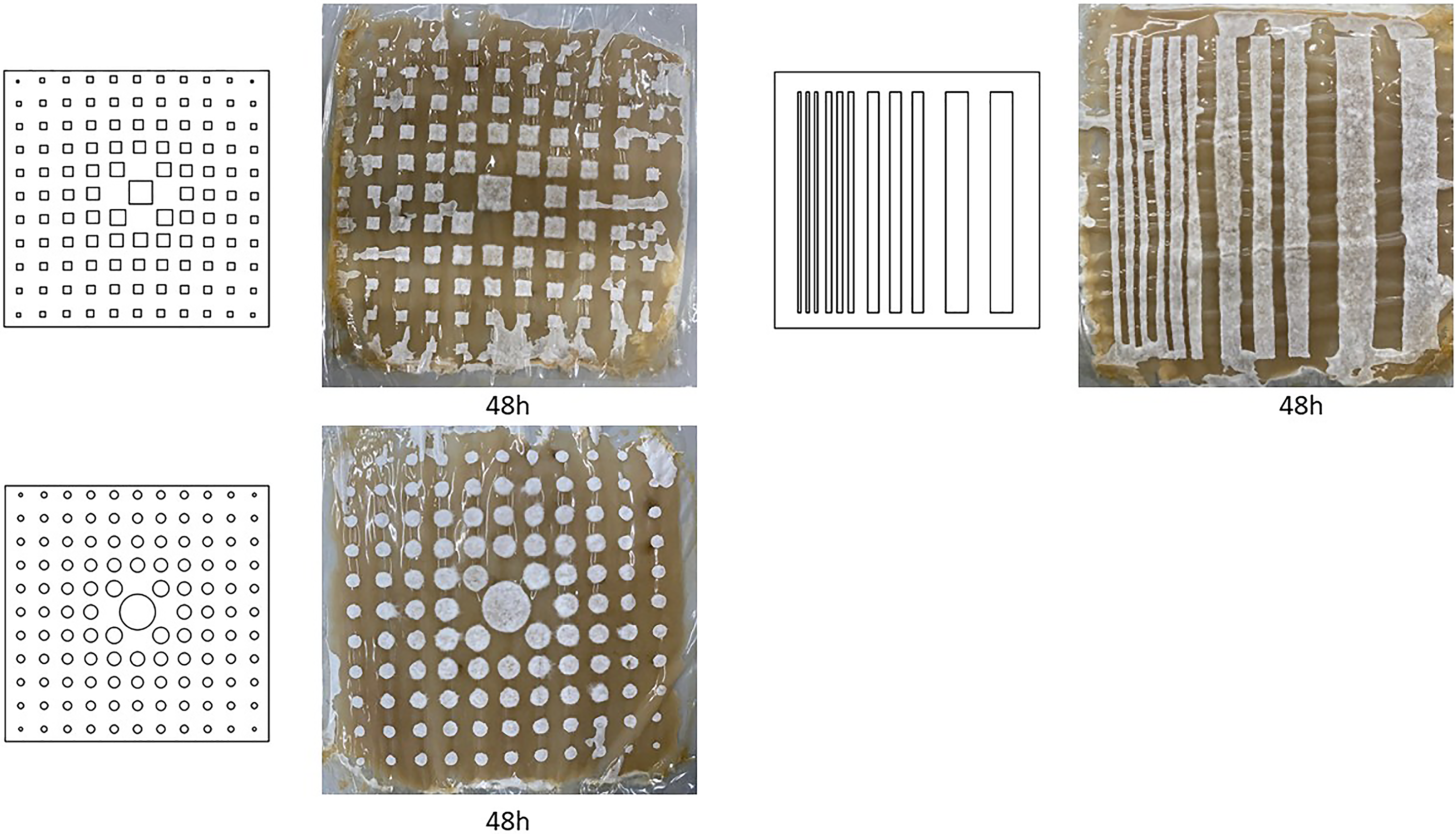
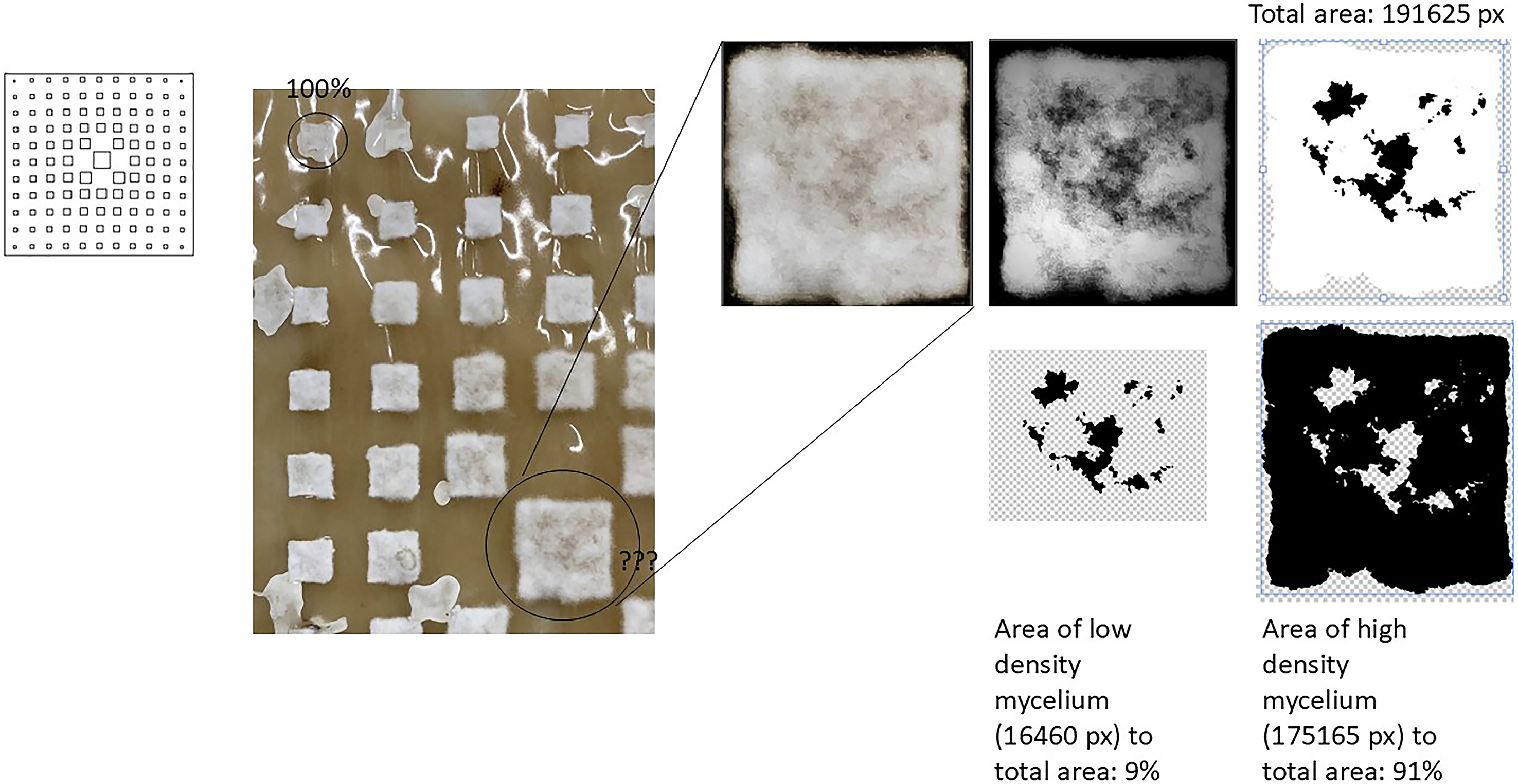
This series of experiments investigates the relationship between predefined masking patterns and the resulting resolution and density of hyphal growth. The primary objective is to examine how the extent of unmasked area correlates with hyphal densification, and how the geometric precision of the masking pattern influences the spatial resolution of the resulting mycelial mat. Three patterned samples are presented, each exploring a geometric gradient through squares, circles and linear strips (Figure 4). In the square-based pattern, particular attention is given to the resolution of sharp corners in comparison to the rounded edges of circular geometries. Initial observations indicate that while the mycelium accurately follows the imposed geometry, hyphal density decreases in larger unmasked areas relative to smaller ones. To quantify this effect, a photographic analysis is conducted (Figure 5). A small square exhibiting visually uniform, high-density white hyphal coverage is selected as a reference and defined as 100% densification. This reference is then used to assess densification across the larger square. The analysis reveals that as the unmasked area increases, hyphal density correspondingly decreases. Within the larger square, the region matching 100% densification is first isolated and subsequently subtracted from the total area, resulting in an overall densification loss of approximately 9%. These findings indicate that although hyphal growth maintains high geometric fidelity to the predefined pattern, its density diminishes at larger spatial scales. This behaviour is consistently observed across all three samples, suggesting an intrinsic relationship between growth resolution, area size and mycelial densification. The fact that by increasing the dimensions of the rectangle by 1600 % there is a consequent loss of hyphal density of 9%, strengthen the argument for this technique to be applied in small resolution on a large membrane.
Squared-based pattern, circle-based pattern and strip-based pattern testing resolution of hyphal growth against surface area with 48 h growth each.

Hyphal density resolution to surface area test using image analysis. The left top small square is taken as a reference of 100% hyphal densification and compared to the larger square, resulting loss of 9% densification in the larger square.

Geometric pattern test with multiple masks
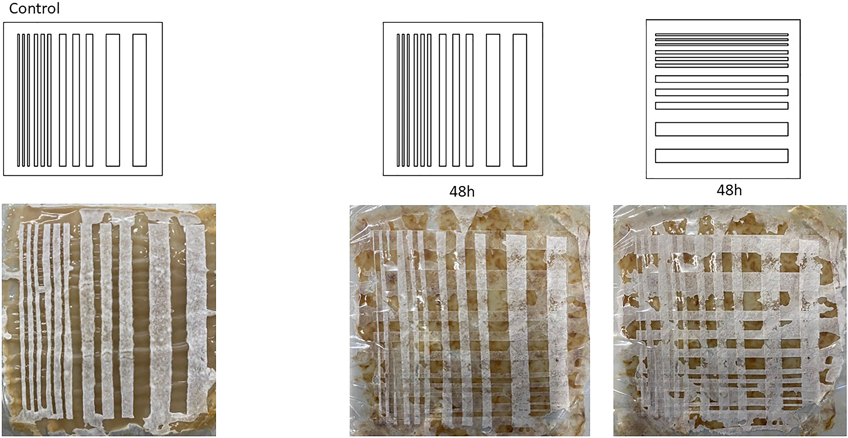
This experiment explores the potential of extending the design possibilities of hydrophobic patterning using masking technique through a two-step temporal application. Following the initial curing phase, a linear striped pattern is applied to the living membrane for 48 hours, allowing the mycelium to densify within the negative spaces of the mask. After this period, the cellophane mask is removed, rotated by 90 degrees and reapplied, introducing a second temporal and spatial constraint using the same geometric pattern. This approach investigates how geometric complexity can be expanded through sequential masking, enabling material efficiency from a manufacturing standpoint as the mask is reused, generating a composite pattern outcome that is derived from a single predefined geometry. Twenty-four hours after the application of the second mask, the newly formed negative spaces created by the juxtaposition of the two patterns begin to be colonised by white hyphae. After 48 hours, all exposed areas are visibly densified (Figure 6). However, the experiment also reveals that successful pattern resolution is highly dependent on the precision with which continuous contact between the living membrane and the mask is maintained. In this case, the decision to reuse the same mask, although material conscious, does compromise the outcome: residual moisture and material deformation introduced ripples that prevents consistent adhesion, resulting in reduced geometric clarity. This observation underscores that material behaviour in living systems is not solely determined by predefined design intent but emerges through an ongoing negotiation between designer and organism. Co-creation thus operates in the space between imposed geometric constraints and the autonomous growth tendencies of the living material.
Two-step of the same mask application. The mask is applied for the first 48 h and then turned by 90 degrees and reapplied for other 48 h.

Gradient test with multiple masks – regular and complex patterns
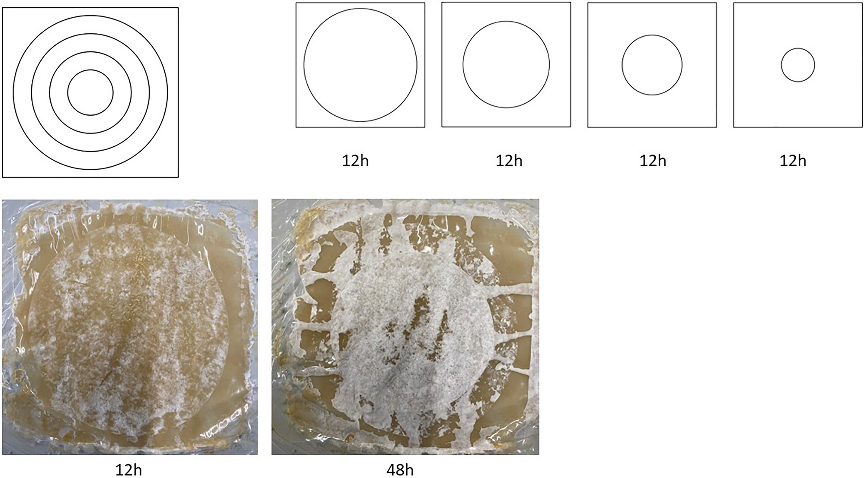
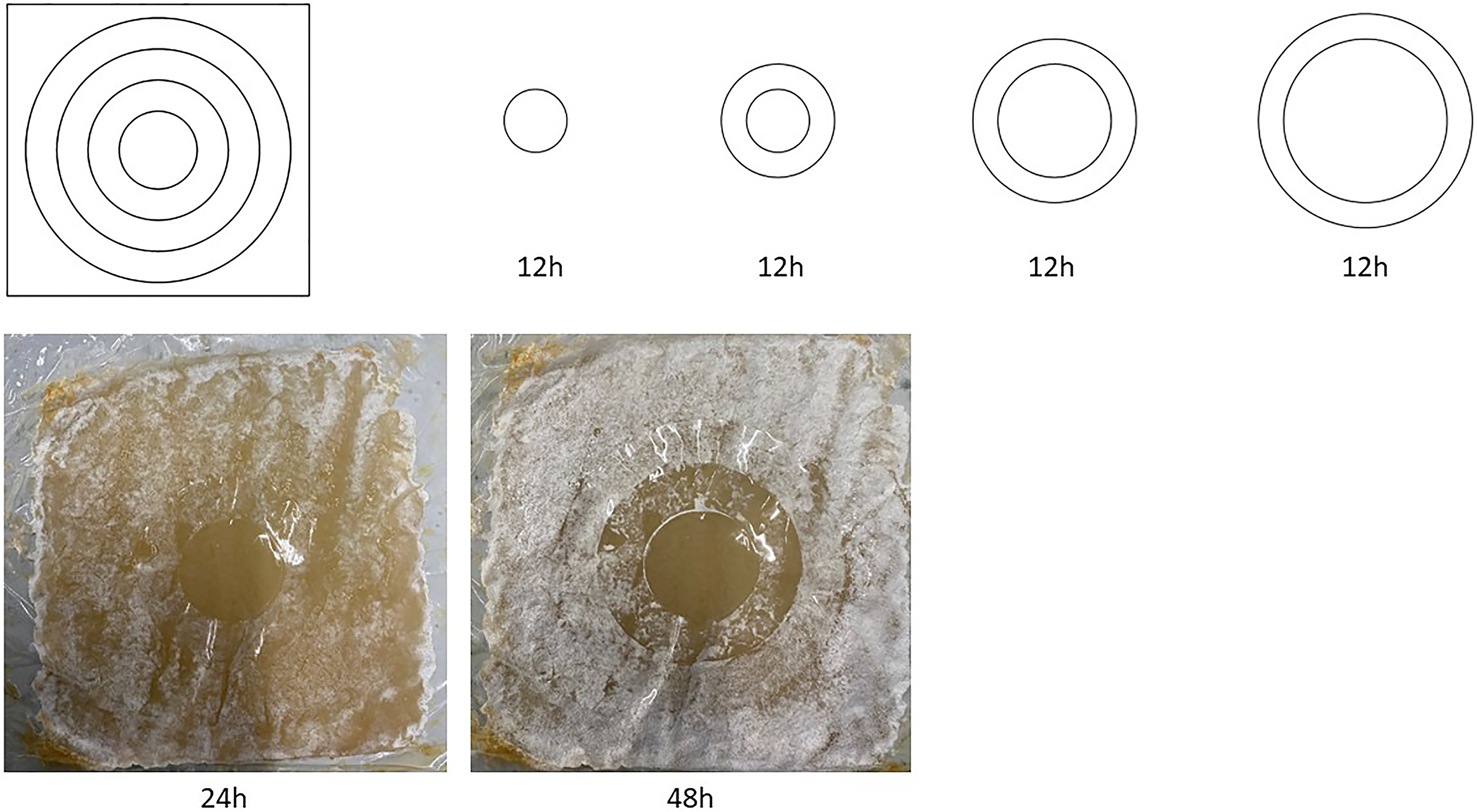
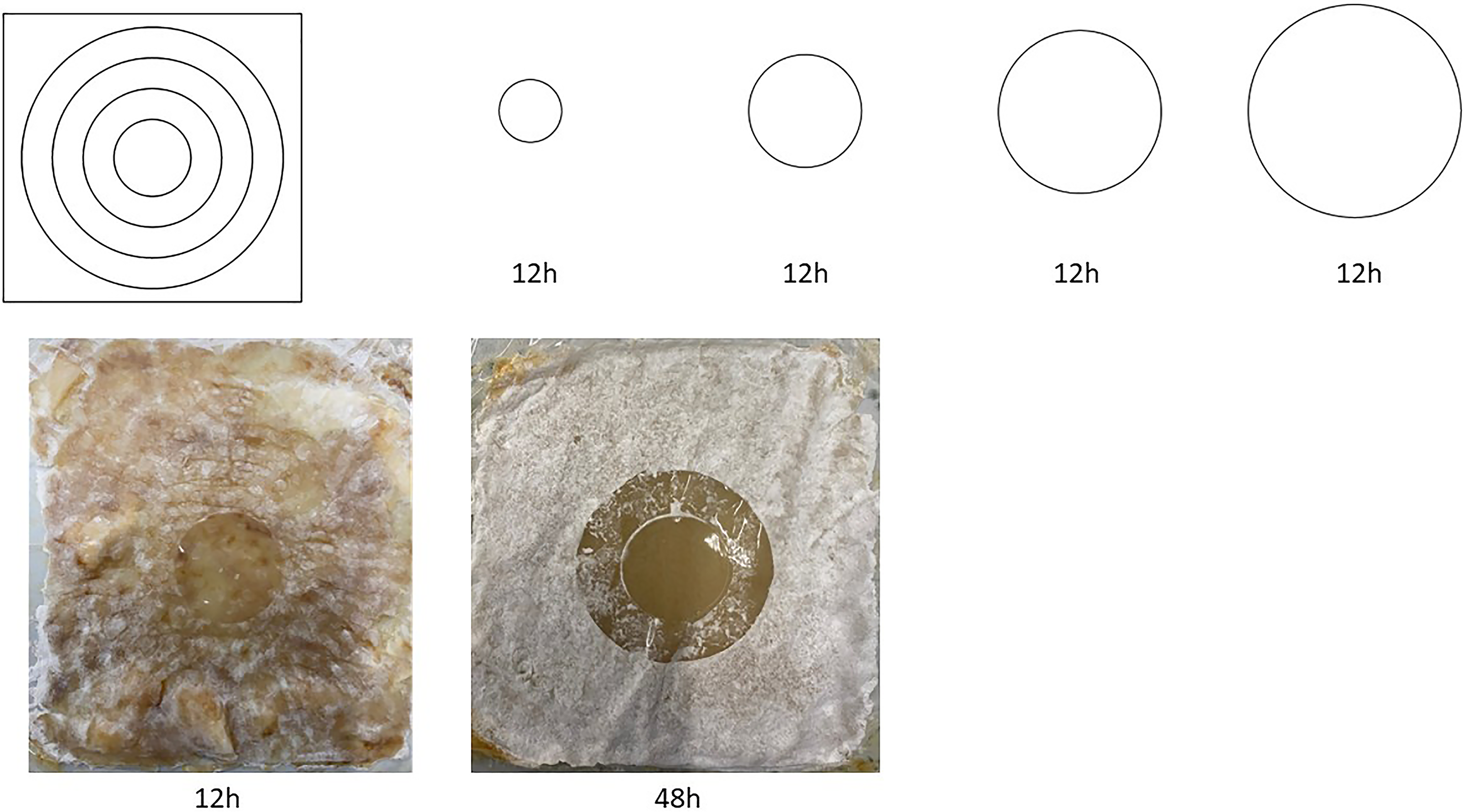
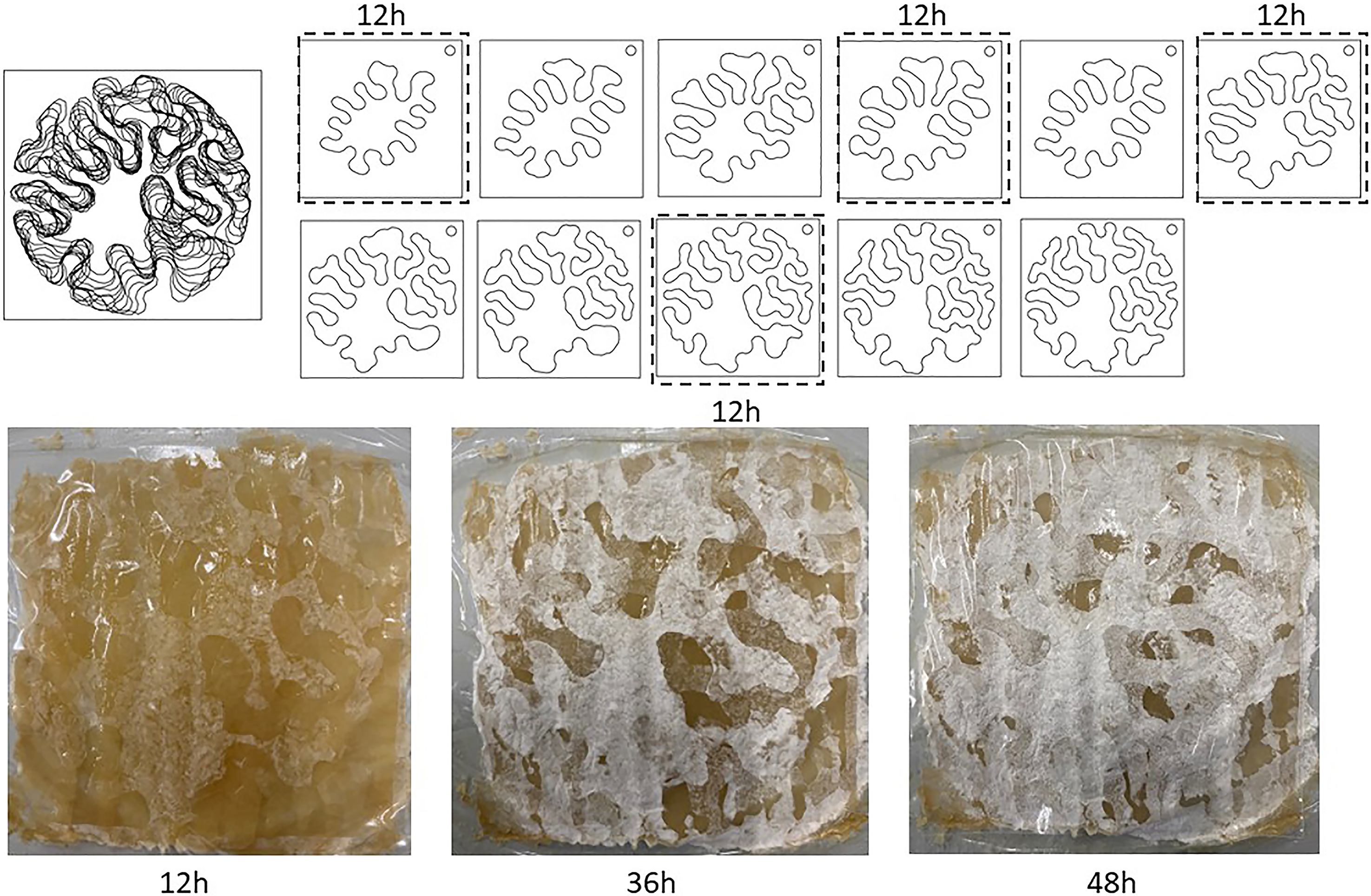
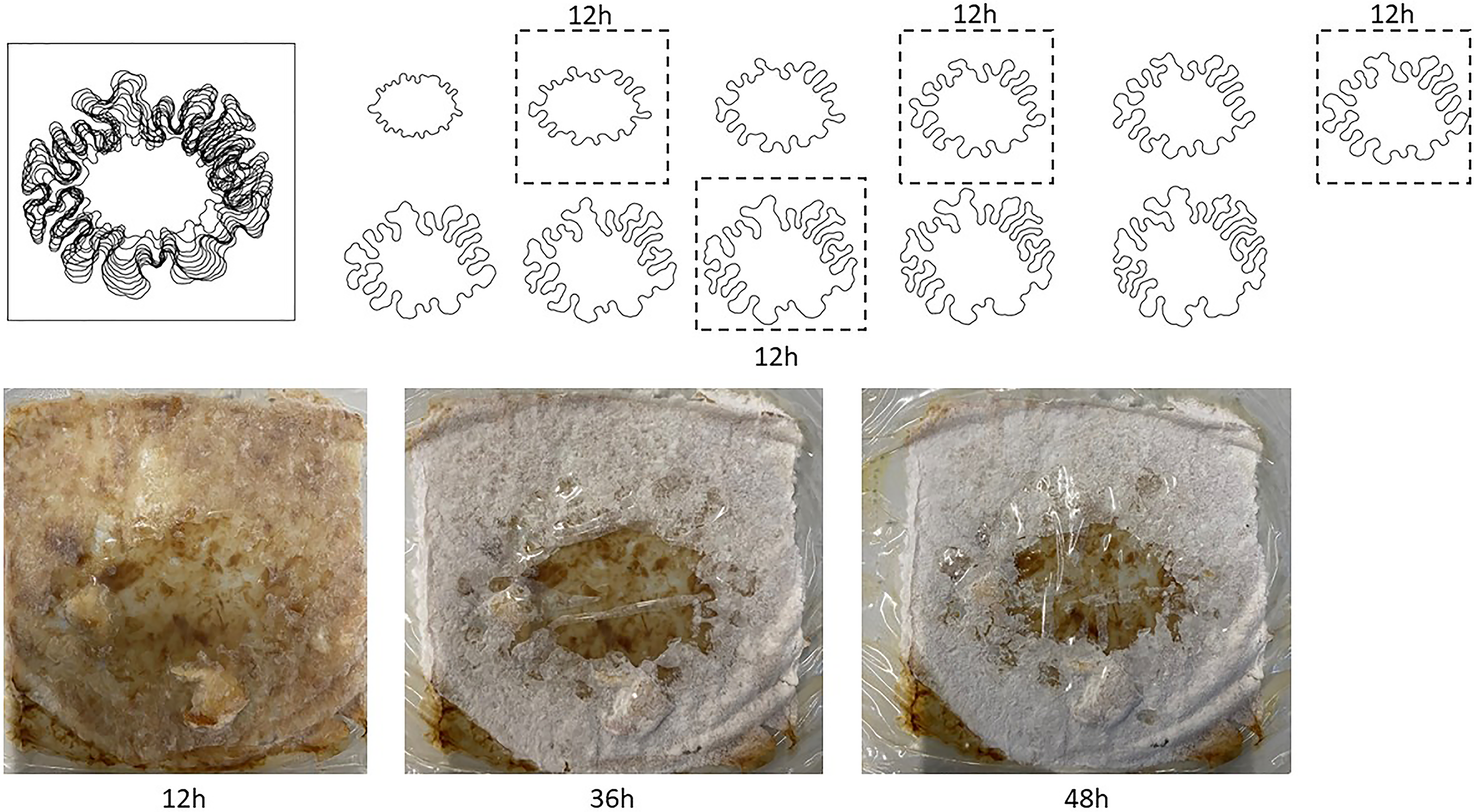
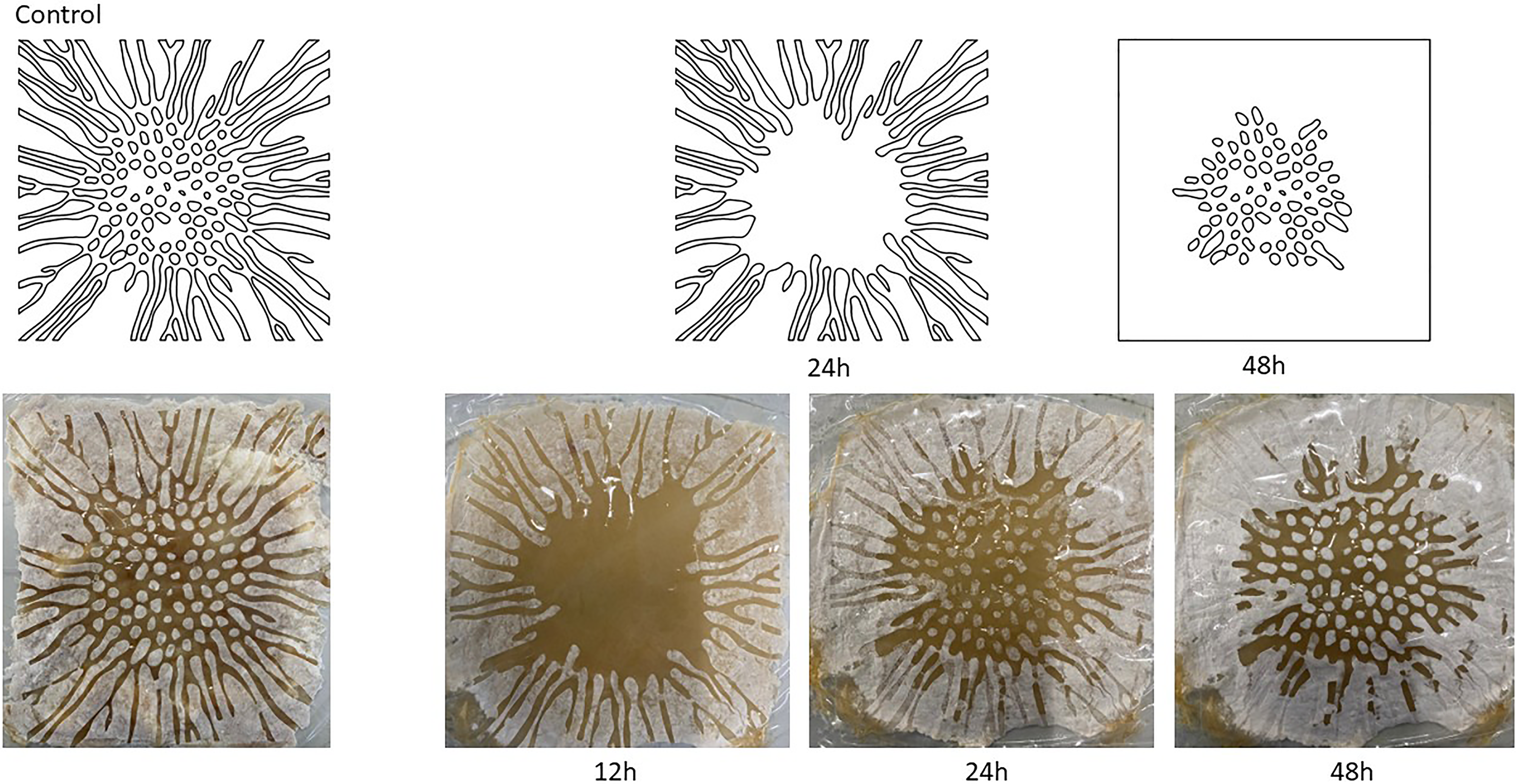
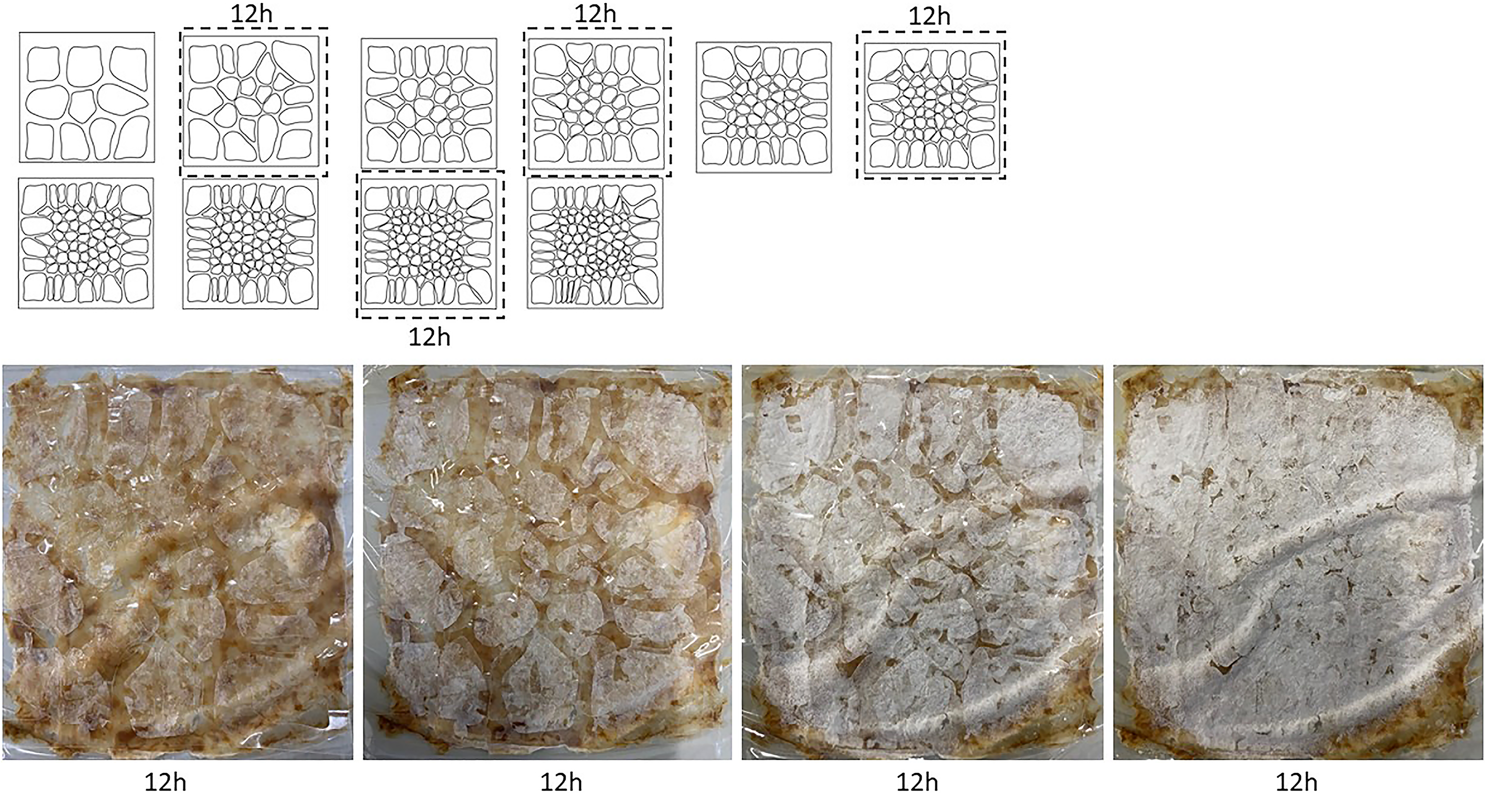
This experiment tests the idea of having the possibility of creating a density gradient growth of hyphae by masking patterns. Through the application of concentric masks within smaller time steps of 12 hours, the objective is to achieve a gradient densification of the hydrophobic mat. This set of experiments examines different strategies for generating hyphal density gradients through patterned masking. Two primary approaches are tested: a series based on regular geometric patterns of concentric circles and a second series generated through an incremental growth algorithm, producing more organic and complex geometries. Within the concentric circle series, three techniques are employed to induce gradients: (1) inward progression (Figure 7), (2) outward progression (Figure 8) and (3) overlaying of multiple patterns (Figure 9). For the complex geometry series, two methods are tested: (4) inward progression (Figure 10) and (5) outward progression (Figure 11). Overall, gradient effects are more clearly legible in the regular concentric circle patterns. However, across all samples, hyphal growth does not exhibit fully consistent gradient formation. This inconsistency is likely related to the duration of each temporal step; reducing the incubation intervals may enable greater control and yield more sharply defined gradient outcomes.
Inward hydrophobic hyphal growth progression of 4 circled masks with different dimension radius every 12 h.

Outward hydrophobic hyphal growth progression of concentric circled masks every 12 h.

Overlaying of multiple patterns for outward growth progression of 4 circled masks with different dimension radius every 12 h.

Inward progression of hyphal growth of 4 patterned using differential growth algorithm every 12 h.

Onward progression of hyphal growth of 4 patterned using differential growth algorithm every 12 h.

Complex patterns with multiple complementary designs
This experiment explores an additional strategy for generating complex patterns through sequential masking. A control sample, produced using a single mask, is compared to a second sample in which selected elements of the original geometry are extracted and reapplied through a secondary masking step after a 24-h interval. The aim is to evaluate whether this staged approach produces discernible differences in growth outcomes when compared to a single-mask application. This method is motivated by potential large-scale design scenarios, in which inaccuracies or unforeseen conditions may arise. Introducing a secondary temporal masking step offers an opportunity to adjust, correct, or refine patterning either prior to deployment or as part of an ongoing maintenance strategy. After 24 h, the initial mask, defining only the outer geometry of the original design, is removed and replaced with a second mask encoding the inner dot pattern. While the technique demonstrates potential for introducing additional pattern layers, its primary limitation lies in maintaining consistent adhesion between the mask and the living membrane. Due to the aggressive nature of mycelial growth, pattern definition begins to deteriorate after 48 hours, leading to a partial loss of resolution (Figure 12). Similar approach is applied on a second sample where multiple masks are layered in order to achieve a 3d effect from complex pattern juxtaposition (Figure 13). Here four patterns are chosen in a sequence of 10. The 3d effect partially takes form on the second and third application of the masks and fails as the mycelium covers the negative spaces created on the membrane. As a result, although sequential masking enables post hoc geometric intervention, it simultaneously compromises overall pattern clarity, indicating a trade-off between adaptability and resolution in living material systems.
Two complementary masks application applied after 12 h growth to test hyphal pattern resolution.

Complex juxtaposition of 4 patterns each applied after 12 h growth to achieve 3d effect.

Discussion
This paper contributes knowledge across the conceptualisation, scaling and patterning of living fungal membranes from an architectural standpoint. The work examines how discrete small-scale membranes can be combined to produce larger assemblies and explores methods for inducing the growth of defined patterns. Through tensile mechanical testing and masking experiments, both qualitative and quantitative properties of living fungal membranes were investigated, clarifying key limitations and intrinsic characteristics of mycelium-based living materials. These experiments establish a baseline for a design-oriented approach while revealing material behaviours that can be further developed within the framework of ELMs.
The tensile tests demonstrate that mechanical performance in mycelium-based membranes is primarily governed by internal organisation and layering. While Young’s modulus showed no statistically significant differences between groups across ANOVA, Welch and Kruskal–Wallis tests (p > 0.05), indicating consistent elastic behaviour, ultimate strength varied significantly (ANOVA p = 0.004356; Welch p = 0.008725; Kruskal–Wallis p = 0.01339). Post hoc analyses identified V1 as the primary source of variation (e.g., O vs V1, Tukey’s p-adj = 0.0013). These findings suggest that mycelial bonding enhances tensile performance, while observed failure patterns indicate that shear strength exceeds tensile strength, reflecting both internal organisation and fabrication processes. A stiffness–deformability trade-off emerges, where increasing the number of mycelial layers nearly doubles deformation capacity, highlighting the importance of network continuity in enhancing mechanical resilience. Although layering improves stiffness, it may still be insufficient to prevent breakage depending on application requirements.
The qualitative masking experiments investigate regenerative patterning strategies by selectively exposing membrane regions to air, thereby triggering localised hyphal regrowth and the formation of hydrophobic surface domains. While intricate patterns can be achieved with high fidelity, a reduction in hyphal density of approximately 9% with increased exposed surface area suggests that this method is most effective at smaller resolutions. Sequential masking enables gradient effects and post-growth geometric intervention; however, insufficient adhesion between mask and membrane compromises pattern clarity. These findings underscore the need to negotiate between predefined design intent and autonomous biological growth.
Importantly, both experimental approaches were conducted in a semi-wet state, highlighting humidity as a critical parameter influencing both tensile strength and hyphal patterning. This indicates that any practical application of living fungal membranes must incorporate sustained water availability as an operational condition.
From an architectural perspective, the integration of pattern as an expression of biological vitality introduces new functional possibilities. Unlike inert materials, living membranes exhibit adaptive behaviours such as self-healing and controlled regeneration, enabling morphological transformation in response to environmental stimuli. Patterning operates not only as a geometric or visual device but also as a mediator of structure, communication and sensory experience. Spatial differentiation achieved through masking or selective exposure produces zones with distinct material states – ranging from dense, water-saturated regions to aerial, hydrophobic hyphal formations – resulting in varied optical and tactile qualities.
Furthermore, patterned hyphal growth can function as a sensing interface. Due to the sensitivity of fungal systems to environmental factors such as humidity, temperature, nutrient gradients and pollutants, localised growth patterns can act as bio-indicators of environmental change. This extends the role of pattern beyond aesthetics, embedding environmental feedback directly into material morphology. In parallel, the metabolic activity of fungal networks – well established in bioremediation research – enables these membranes to participate in the degradation of organic contaminants (Colmo et al. Reference Colmo, Tegelaar and Ayres2025). In aqueous environments, this potential becomes particularly relevant (Dinakarkumar et al. Reference Dinakarkumar, Ramakrishnan, Gujjula, Vasu, Balamurugan and Murali2024), where patterning can influence nutrient distribution, oxygen diffusion and regeneration dynamics. In this sense, pattern becomes not only geometric but metabolic, structuring conditions for growth, repair and functional differentiation.
Conclusion
This study demonstrates that the design of living fungal membranes requires an integrated understanding of both mechanical behaviour and biological dynamics. Layered and structurally coherent mycelial assemblies emerge as the most promising strategy for enhancing mechanical performance, although trade-offs between stiffness, deformability and durability remain. At the same time, masking-based patterning offers a viable method for directing growth and introducing functional differentiation, albeit with limitations related to scale, adhesion and environmental dependency.
Together, the findings establish a foundation for a design-oriented approach to mycelium-based materials, where structural organisation, environmental conditions and biological processes are considered as interdependent parameters. By positioning pattern as both a spatial and metabolic driver, this work highlights the potential of living fungal membranes to operate as adaptive, responsive and functionally active systems. Applications can range from interior and textile design but also architectural scale can be potentially achieved by assembly through joining strategies. These insights contribute to the ongoing development of advanced ELMs, supporting future applications that integrate growth, sensing and remediation within material design.
Data availability statement
Replication data and code can be found in Zenodo Dataverse: https://doi.org/10.5281/zenodo.18715400.
Acknowledgements
We are grateful for the statistical assistance of A.R. Author.
Author contributions
Conceptualisation: C.C. Methodology: C.C Data curation: C.C.;A.R. Data visualisation: C.C. Writing original draft: C.C; A.R. Writing review and editing: C.C;A.R.;P.A Supervision: P.A. All authors approved the final submitted draft.
Financial support
This research was funded by the European Union’s HORIZON-EIC-2021-PATHFINDER CHALLENGES programme under the project FUNGATERIA, grant agreement No. 101071145.
Competing interests
None.
Ethical standards
The research meets all ethical guidelines, including adherence to the legal requirements of the study country.
 150×150mm

 Open access

