The prevalence of primary cardiac tumours in childhood is low. Histologically, most of them are rhabdomyomas. Myofibroblastic inflammatory tumours are rare entities in infancy. Initially, they were recognized in the lung, but are now known to occur in virtually every major organ of the body. The precise biological nature and the clinical characteristics of these kinds of lesions are still uncertain. They can occur at any age, but are referred to as soft tissue tumours of childhood.Reference Sebire, Ramsay, Sheppard, Malone, Harding and Risdon1
The rarity of myofibroblastic inflammatory tumours in the heart poses a diagnostic difficulty for both paediatricians and pathologists because of the clinical and morphological overlap with other intracardiac masses.Reference Li, Cerilli and Wick2
Case report
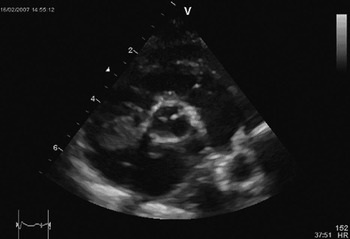
A neonate was originally admitted to our hospital at the age of 6 weeks because of haemodynamically relevant ventricular and atrial septal defects. His mother had had a history of 3 stillbirths, as well as 1 missed abortion. The current pregnancy had been the product of in-vitro fertilization, complicated by premature labour, and a ventricular septal defect that had already been suspected on fetal scans. The family history did not show any birth defects, especially no history of malignant tumours. Coagulation testing in the mother, as well as in the patient, failed to show any abnormalities. Because of progressive congestive cardiac failure, despite anticongestive treatment, surgery was performed when the baby was aged 8 weeks. After an uneventful postoperative course, the patient developed a mass in the right atrium, measuring 2 by 1.4 centimetres (Fig. 1), which was detected echocardiographically 8 weeks later. Until then the patient had been asymptomatic.
The transthoracic short-axis view shows an inhomogenous echogenic mass adherent to the right atrial wall 8 weeks after surgical closure of ventricular and atrial septal defects.

Because of the closeness to the superior caval vein, and a relatively broad base on the atrial septum, we initially suspected formation of thrombus. On this account, the patient was put on unfractioned heparin, with a target partial thromboplastin time of 60 to 70 seconds. Lysis with recombinant tissue plasminogen activator was also initiated. As there was no change of the echocardiographic findings within 7 days, we decided to remove the mass surgically. Intraoperatively, the formation was still suggestive of thrombus. It could be excised almost in its entirety with a sharp spoon. A few days later, the surprising diagnosis of a myofibroblastic inflammatory tumour was established on the basis of the histopathological findings (Fig. 2).
Microscopic sectioning of the tumour shows its inhomogenous, fissured, surface (a); and the spindle shaped tumour and muscle cells with inflammatory reaction (b).

As in this low grade lesion metastatic invasion is very rare, we decided to dispense with staging, although a complete resection due to the broad infiltration had not been possible. Additional chemotherapy or radiotherapy seemed not to be indicated because of the known low malignancy of the tumour. In order to avoid a thrombotic aggregation at the site of the excision, we put the patient on coumadin for the first 3 month after the operation. Detailed echocardiographic follow-up for 18 months has revealed no signs of a local relapse of the tumour. Clinically, the patient has been stable since the operation.
Discussion
Newly recognised echogenic masses within the heart after a cardiac operation are most often related to formation of thrombus.Reference Li, Cerilli and Wick2 Cardiac tumours in general are rare in children, with the majority of cases detected in infancy being rhabdomyomas. The inflammatory myofibroblastic tumour was initially described in the lung in 1939. Biologically, it is classified as a low grade spindle cell lesion presenting with only mild dysplasia. Since its first description, there have been reports of such tumours in almost all organ systems, including the heart.Reference Murdison, Septimus, Garola and Pizarro3 In virtually all of them, the tumour originated from the right atrium. Only a minority of the patients have been asymptomatic. The majority showed signs of cardiac disease, such as coronary ischaemia due to involvement of the coronary arteries, as well as dyspnoea, cyanosis, or pericardial effusion.
Although inflammatory myofibroblastic tumours are described as being low grade lesions, with a low potential for invasive growth, a slight risk remains for penetration of neighbouring structures, such as coronary arteries or valves. In our patient, the tumour could be excised almost in total, and the patient has since been well.