





Introduction
Rheumatic heart disease remains a major cause of cardiovascular morbidity in children and adolescents worldwide, particularly in low- and middle-income countries, despite a global decline in the incidence of acute rheumatic fever. Reference Watkins, Johnson and Colquhoun1–Reference Zühlke, Engel and Lemmer3 Rheumatic heart disease represents the chronic sequela of acute rheumatic fever and is characterised by progressive valvular inflammation, fibrosis, and remodelling, most commonly affecting the mitral and aortic valves. Reference Marijon, Mirabel and Celermajer4,Reference Passos, Nunes and Aikawa5 While echocardiography plays the central role in the diagnosis and staging of rheumatic heart disease, it provides limited insight into the underlying inflammatory activity that may contribute to disease persistence and progression.
Increasing evidence indicates that chronic, low-grade inflammatory activity persists beyond the acute phase of acute rheumatic fever and contributes to progressive valvular fibrosis and remodelling in rheumatic heart disease. Reference Passos, Nunes and Aikawa5,Reference Rwebembera, Marangou and Mwita6 In this context, easily accessible haematologic parameters derived from routine complete blood counts have gained attention as potential markers of systemic inflammation and endothelial dysfunction. Chronic inflammatory processes may influence erythropoiesis and red blood cell maturation, leading to increased heterogeneity in erythrocyte size reflected by red cell distribution width. Reference Salvagno, Sanchis-Gomar and Picanza7 Similarly, systemic inflammation and endothelial activation may affect platelet production and turnover, resulting in larger and more reactive platelets, as reflected by increased mean platelet volume. Reference Bath and Butterworth8–Reference Korniluk, Koper-Lenkiewicz and Kamińska11 Various haematologic parameters have been proposed as surrogate markers of systemic inflammation and cardiovascular involvement, reflecting different components of the inflammatory response, including leukocyte activation, erythrocyte heterogeneity, and platelet reactivity. Reference Salvagno, Sanchis-Gomar and Picanza7–Reference Korniluk, Koper-Lenkiewicz and Kamińska11
Inflammatory markers derived from peripheral blood may reflect different temporal phases of the disease process. Leukocyte-based indices have been primarily associated with acute inflammatory activity, whereas erythrocyte- and platelet-related parameters have been suggested to capture more sustained or low-grade inflammatory states. Reference Salvagno, Sanchis-Gomar and Picanza7,Reference Korniluk, Koper-Lenkiewicz and Kamińska11,Reference Forget, Khalifa and Defour12 Whether these distinct haematologic profiles are associated with persistent valvular involvement and disease stage in paediatric rheumatic heart disease remains incompletely defined.
Several studies have evaluated haematologic markers in the acute phase of acute rheumatic fever, reporting associations between leukocyte-derived indices such as neutrophil-to-lymphocyte ratio and platelet-to-lymphocyte ratio and disease severity. Reference Ozdemir, Karadeniz and Doksoz13–Reference Giray and Hallioglu15 However, data regarding the role of these parameters in persistent rheumatic heart disease, particularly in relation to echocardiographic disease stage and valvular involvement, remain limited in the paediatric population.
Therefore, the present study aimed to explore the relationship between baseline haematologic parameters and derived inflammatory indices and both valvular involvement and persistent rheumatic heart disease stage in children with a history of acute rheumatic fever.
Materials and methods
Study design and population
This single-centre, retrospective observational study was conducted at a tertiary paediatric cardiology centre. Medical records of children followed for acute rheumatic fever and rheumatic heart disease between January 2015 and December 2025 were reviewed. During this period, 63 patients with a documented history of acute rheumatic fever and echocardiographic evidence of rheumatic valvular involvement, for whom complete clinical and echocardiographic data were available, were identified.
Patients were eligible for inclusion if they met all of the following criteria:
-
1. A documented diagnosis of acute rheumatic fever based on the 2015 revised Jones criteria of the American Heart Association, applicable at the time of presentation; Reference Gewitz, Baltimore and Tani16
-
2. Echocardiographic findings consistent with rheumatic valvular involvement;
-
3. Availability of at least two standardised transthoracic echocardiographic examinations, including:
-
• one performed at the time of initial presentation, and
-
• a follow-up examination performed ≥6 months after the initial evaluation to confirm persistence of valvular abnormalities;
-
-
4. Availability of baseline laboratory data obtained during the acute presentation period, defined as blood samples collected within the first 72 hours after symptom onset or, when the exact onset time was unavailable, within the first 72 hours after hospital admission.
Patients were excluded if they had CHD, non-rheumatic valvular pathology, known chronic inflammatory or haematologic disorders, known iron deficiency anaemia requiring treatment, vitamin B12 or folate deficiency, chronic liver or renal disease, or any other systemic condition that could significantly affect haematologic parameters.
After application of these criteria, 45 patients were included in the final analysis. All included patients represented their first documented episode of acute rheumatic fever. No recurrences of acute rheumatic fever were identified during the follow-up period based on available clinical records.
Echocardiographic definition and staging of persistent rheumatic heart disease
Persistent rheumatic heart disease was defined as echocardiographic evidence of pathological rheumatic valvular involvement persisting at least 6 months after the acute episode of acute rheumatic fever, in line with longitudinal echocardiographic follow-up definitions.
For disease severity analyses, patients were categorised according to the 2023 World Heart Federation echocardiographic staging framework Reference Rwebembera, Marangou and Mwita6 into Stage A, Stage B, or Stage C based on valve morphology, regurgitation severity, and hemodynamic impact.
-
• Stage A: mild valvular involvement without significant structural or haemodynamic consequences;
-
• Stage B: moderate valvular disease with more pronounced regurgitation and/or structural changes;
-
• Stage C: more advanced valvular disease characterised by more pronounced morphological abnormalities and/or at least moderate regurgitation with greater hemodynamic impact.
No patients met criteria for Stage D, and severe regurgitation was not observed in this cohort.
All echocardiographic assessments were performed by experienced paediatric cardiologists within the same institutional team using standardised protocols. Due to the retrospective design, images were not re-evaluated by a single blinded reviewer; however, all examinations were conducted in accordance with consistent institutional and guideline-based criteria.
Pathological mitral and aortic regurgitation were defined according to the 2023 World Heart Federation echocardiographic criteria, Reference Rwebembera, Marangou and Mwita6 requiring visualisation in at least two echocardiographic views, fulfilment of valve-specific jet length and velocity thresholds, and demonstration of pan-systolic or pan-diastolic flow, as appropriate.
Regurgitation severity was graded as mild, moderate, or severe using an integrated echocardiographic approach based on colour Doppler, spectral Doppler, and structural valve assessment, in accordance with established echocardiographic recommendations for valvular regurgitation assessment. Reference Zoghbi, Adams and Bonow17
Laboratory measurements
Baseline laboratory parameters included antistreptolysin O, C-reactive protein, erythrocyte sedimentation rate, serum albumin, and complete blood count parameters (white blood cell count, neutrophil count, lymphocyte count, monocyte count, platelet count, mean corpuscular volume, red cell distribution width, platelet distribution width, and mean platelet volume). As stated above, laboratory measurements were considered baseline only if obtained within 72 hours of symptom onset or hospital admission, to reflect inflammatory status at initial presentation.
Derived haematologic indices were calculated using standard formulas:
-
• Neutrophil-to-lymphocyte ratio: neutrophil count / lymphocyte count
-
• Platelet-to-lymphocyte ratio: platelet count / lymphocyte count
-
• Monocyte-to-lymphocyte ratio: monocyte count / lymphocyte count
-
• Systemic immune-inflammation index: neutrophil × platelet / lymphocyte
-
• Systemic inflammation response index: neutrophil × monocyte / lymphocyte
All haematologic indices were calculated using absolute cell counts obtained from automated haematology analysers.
Statistical analyses
Statistical analyses were performed using SPSS version 31 (IBM Corp., Armonk, NY, USA). Categorical variables were presented as frequencies (n) and percentages (%). Continuous variables were expressed as median (interquartile range) for non-normally distributed data.
Normality of distribution was assessed using the Shapiro–Wilk test, in addition to visual inspection of Q-Q plots and histograms. Comparisons between independent groups were conducted using the Mann–Whitney U test for two groups and the Kruskal–Wallis test for more than two groups. For significant Kruskal–Wallis results, Dunn’s test with Bonferroni correction was performed for post hoc pairwise comparisons.
To identify predictors of higher stage rheumatic heart disease (Stage B–C versus Stage A), binary logistic regression analyses were performed. Initially, univariable logistic regression analyses were conducted for each independent variable. Variables with a p-value < 0.25 in univariable analyses, along with those considered clinically relevant, were included in the multivariable logistic regression model.
All selected variables were entered simultaneously using the Enter method. Adjusted odds ratios with 95% confidence intervals and p-values were calculated. Model fit was evaluated using the Hosmer–Lemeshow goodness-of-fit test. A two-sided p-value < 0.05 was considered statistically significant.
Results
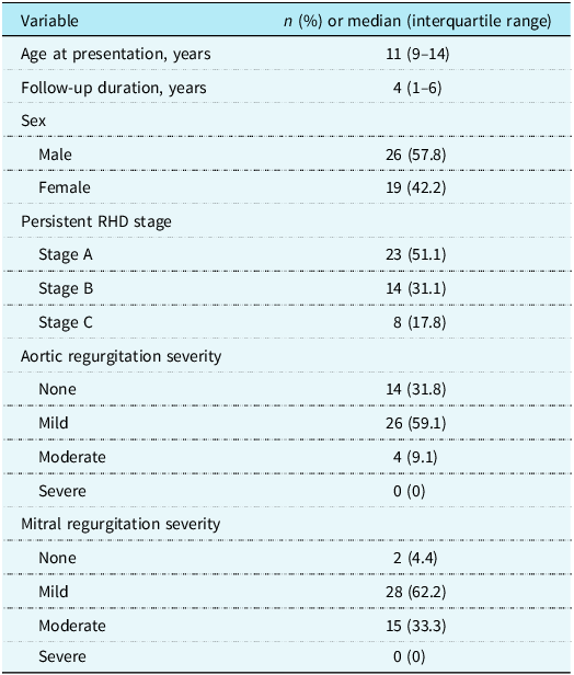
A total of 45 children with persistent rheumatic heart disease were included in the final analysis. The median age at presentation was 11 years (9–14), and the median follow-up duration was 4 years (1–6). Twenty-six patients (57.8%) were male.
According to persistent rheumatic heart disease staging, 23 patients (51.1%) were classified as Stage A, 14 (31.1%) as Stage B, and 8 (17.8%) as Stage C. Regarding valvular involvement, mild mitral regurgitation was the most common finding (62.2%), followed by mild aortic regurgitation (59.1%). Moderate aortic regurgitation was observed in 4 patients (9.1%), and moderate mitral regurgitation in 15 patients (33.3%). No patient demonstrated severe (grade 3) mitral or aortic regurgitation during the study period (Table 1).
Baseline clinical characteristics of the study population (n = 45)

Table 1. Long description
A table summarizing clinical characteristics of a study population of 45 individuals. The table has 10 rows and 2 columns. The columns are labeled ‘Variable’ and ‘n (%) or median (interquartile range)’. The rows detail various clinical characteristics. Row 1: Age at presentation, years; 11 (9-14). Row 2: Follow-up duration, years; 4 (1-6). Row 3: Sex; Male; 26 (57.8). Row 4: Sex; Female; 19 (42.2). Row 5: Persistent RHD stage; Stage A; 23 (51.1). Row 6: Persistent RHD stage; Stage B; 14 (31.1). Row 7: Persistent RHD stage; Stage C; 8 (17.8). Row 8: Aortic regurgitation severity; None; 14 (31.8). Row 9: Aortic regurgitation severity; Mild; 26 (59.1). Row 10: Aortic regurgitation severity; Moderate; 4 (9.1). Row 11: Aortic regurgitation severity; Severe; 0 (0). Row 12: Mitral regurgitation severity; None; 2 (4.4). Row 13: Mitral regurgitation severity; Mild; 28 (62.2). Row 14: Mitral regurgitation severity; Moderate; 15 (33.3). Row 15: Mitral regurgitation severity; Severe; 0 (0).
RHD = rheumatic heart disease; AR = aortic regurgitation; MR = mitral regurgitation.
Stage A–C definitions are provided in the Methods section.
When valve phenotypes were examined according to disease stage, Stage A patients predominantly exhibited isolated mild MR (82.6%), whereas Stage B was mainly characterised by combined mild aortic regurgitation and mild mitral regurgitation (71.4%). Stage C patients exhibited more advanced and heterogeneous valve patterns, characterised by moderate regurgitation and frequent combinations of moderate mitral and/or aortic involvement (Supplementary Table 1).
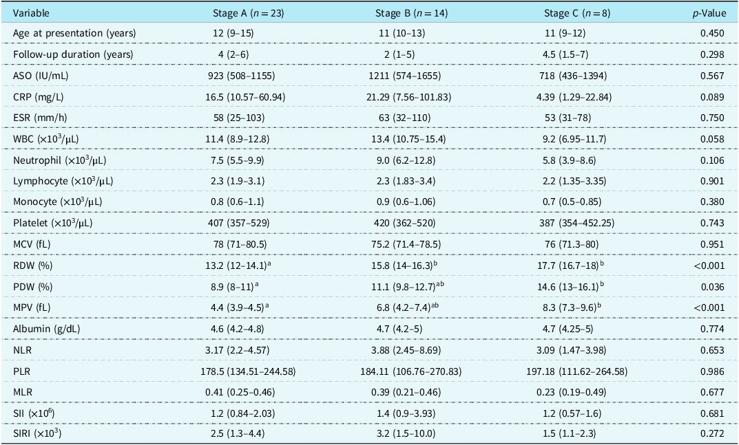
Clinical characteristics, acute-phase reactants, and haematologic parameters were compared across persistent rheumatic heart disease stages (Stages A, B, and C) (Table 2). There were no statistically significant differences among groups in terms of age at presentation or follow-up duration (p = 0.450 and p = 0.298, respectively).
Comparison of clinical and laboratory parameters according to persistent RHD stage

Table 2. Long description
A table comparing clinical and laboratory parameters across persistent rheumatic heart disease stages A, B, and C. The table has 18 rows and 6 columns. Column headers are Variable, Stage A (n = 23), Stage B (n = 14), Stage C (n = 8), and p-Value. Row labels include Age at presentation (years), Follow-up duration (years), ASO (IU/mL), CRP (mg/L), ESR (mm/h), WBC (x10^3/µL), Neutrophil (x10^3/µL), Lymphocyte (x10^3/µL), Monocyte (x10^3/µL), Platelet (x10^3/µL), MCV (fL), RDW (%), PDW (%), MPV (fL), Albumin (g/dL), NLR, PLR, MLR, SII (x10^3), and SIRI (x10^3). Each row provides specific values for each stage and a p-value indicating statistical significance. Notable trends include variations in CRP, RDW, PDW, and MPV across stages, with some parameters showing significant differences.
Data are presented as median (interquartile range). Comparisons were performed using the Kruskal–Wallis test.
a,bGroups not sharing a common superscript letter differ significantly in post-hoc pairwise comparisons.
ASO = antistreptolysin O; CRP = C-reactive protein; ESR = erythrocyte sedimentation rate; RDW = red cell distribution width; PDW = platelet distribution width; MPV = mean platelet volume; MCV = mean corpuscular volume; NLR = neutrophil-to-lymphocyte ratio; PLR = platelet-to-lymphocyte ratio; MLR = monocyte-to-lymphocyte ratio; SII = systemic immune-inflammation index; SIRI = systemic inflammation response index; WBC = white blood cell count.
Markers reflecting acute inflammatory activity, including antistreptolysin O, C-reactive protein, erythrocyte sedimentation rate, leukocyte count, neutrophil count, and derived leukocyte-based indices (neutrophil-to-lymphocyte ratio, platelet-to-lymphocyte ratio, monocyte-to-lymphocyte ratio, systemic immune-inflammation index, and systemic inflammation response index), did not differ significantly across stages (all p > 0.05).
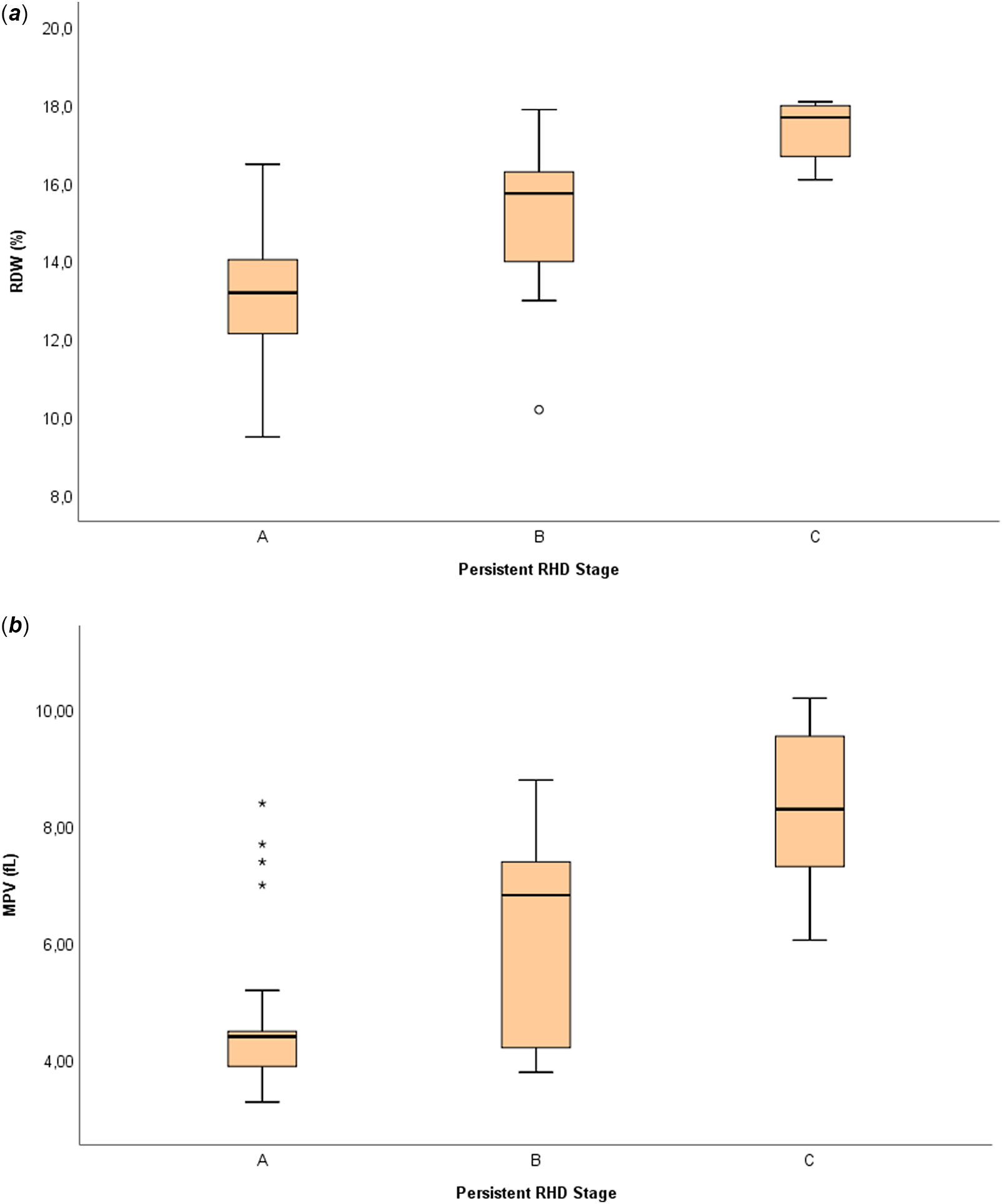
In contrast, erythrocyte and platelet-related parameters demonstrated significant stage-dependent differences. Red cell distribution width values increased progressively across stages (p < 0.001) (Figure 1a), with post-hoc analysis showing significantly higher red cell distribution width levels in both Stage B and Stage C compared with Stage A. No significant difference was observed between Stage B and Stage C.
Distribution of RDW and MPV according to persistent RHD stage. ( a ) Red cell distribution width (RDW) and ( b ) mean platelet volume (MPV) across persistent rheumatic heart disease stages (Stage A, Stage B, Stage C). The central line represents the median, boxes indicate the interquartile range, and whiskers represent the data range. Both RDW and MPV increased significantly across stages (Kruskal–Wallis test, p < 0.001).

Similarly, mean platelet volume differed significantly among groups (p < 0.001) (Figure 1b), with higher values observed in Stage C compared with Stage A. platelet distribution width also showed a significant overall difference (p = 0.036), driven primarily by higher values in Stage C compared with Stage A. However, no statistically significant differences were detected between adjacent stages for platelet distribution width or mean platelet volume. Serum albumin levels were comparable across all stages (p = 0.774).
When patients were stratified according to the presence or absence of aortic regurgitation (mild/moderate versus none), no significant differences were observed in clinical or laboratory parameters (Supplementary Table 2). Likewise, comparison of patients with mild versus moderate mitral regurgitation revealed no statistically significant differences in haematologic indices (Supplementary Table 3).
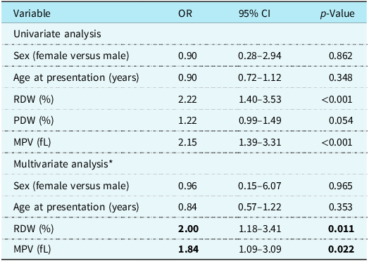
To explore factors associated with advanced persistent rheumatic heart disease stage, binary logistic regression analysis was performed. To account for small sample sizes and ensure adequate statistical power, Stages B and C were combined into a single group for comparison with Stage A (Table 3). Variables with p < 0.25 in univariate analysis (red cell distribution width, platelet distribution width, and mean platelet volume), together with clinically relevant variables (age and sex), were selected for multivariable modelling.
Binary logistic regression analysis for factors associated with advanced persistent RHD stage (Stage B–C versus stage A)

Table 3. Long description
A table comparing factors associated with advanced persistent rheumatic heart disease stage through univariate and multivariate analysis. The table has 8 rows and 4 columns. Column headers are Variable, OR, 95% CI, and p-Value. Row labels include Sex (female versus male), Age at presentation (years), RDW (%), PDW (%), and MPV (fL) for both univariate and multivariate analysis. Notable trends include significant p-values for RDW (%) and MPV (fL) in both analyses, indicating their strong association with the disease stage. The bold values in the table signify statistical significance.
Variables with p < 0.25 in univariate analysis and clinically relevant variables were included in the multivariate model. Due to collinearity between PDW and MPV, only MPV was entered into the final model. The multivariate model was constructed using the enter method. Model fit was assessed using the Hosmer–Lemeshow test (p = 0.421). Nagelkerke R2 = 0.607. Bold values indicate statistically significant p-values (p < 0.05).
OR = odds ratio; CI = confidence interval; RDW = red cell distribution width; PDW = platelet distribution width; MPV = mean platelet volume.
Before constructing the multivariate model, multicollinearity among independent variables was assessed. Because platelet distribution width and mean platelet volume showed strong correlation, only mean platelet volume was retained in the final model to avoid collinearity effects.
In univariate analysis, higher red cell distribution widthand mean platelet volume values were significantly associated with advanced-stage disease, while platelet distribution width showed a borderline association. Age at presentation and sex were not related to rheumatic heart disease stage.
In multivariate analysis, both red cell distribution width and mean platelet volume remained significantly associated with advanced persistent rheumatic heart disease stage after adjustment for age and sex. Each 1 unit increase in red cell distribution width was associated with a twofold increase in the odds of Stage B–C disease (odds ratio 2.00; 95% confidence interval 1.18–3.41; p = 0.011). Similarly, each 1 unit increase in mean platelet volume was associated with a 1.84 fold increase in the odds of advanced stage (odds ratio 1.84; 95% confidence interval 1.09–3.09; p = 0.022).
The model demonstrated acceptable calibration (Hosmer–Lemeshow p = 0.421) and moderate explanatory power (Nagelkerke R 2 = 0.607).
Discussion
In this pilot study, we evaluated the relationship between baseline haematologic parameters and persistent rheumatic heart disease stage in children with a history of acute rheumatic fever. Leukocyte-based inflammatory indices were not associated with disease stage, whereas erythrocyte and platelet-related parameters showed significant differences across stages. Red cell distribution width and mean platelet volume emerged as key markers associated with more advanced persistent rheumatic heart disease after adjustment for age and sex.
Rheumatic heart disease is the chronic outcome of acute rheumatic fever and is characterised by progressive valvular fibrosis. Despite resolution of the acute phase, structural valve abnormalities may persist and evolve, indicating that immunologically mediated remodelling processes may continue beyond the initial episode. Reference Passos, Nunes and Aikawa5,Reference Carapetis, Beaton and Cunningham18
In the present study, leukocyte-based indices such as neutrophil-to-lymphocyte ratio, platelet-to-lymphocyte ratio, monocyte-to-lymphocyte ratio, systemic immune-inflammation index, and systemic inflammation response index were not associated with rheumatic heart disease stage. This finding contrasts with previous studies reporting associations between leukocyte-derived inflammatory indices and disease severity during the acute phase of acute rheumatic fever. Reference Giray and Hallioglu15 Several factors may explain this difference. First, our study focused on persistent echocardiographic rheumatic heart disease stage during follow-up, whereas prior studies primarily evaluated acute-phase disease activity. Second, all laboratory parameters were obtained at initial presentation and were not reassessed during follow-up, limiting their ability to reflect ongoing inflammatory activity. Third, the relatively small sample size and absence of severe rheumatic heart disease may have reduced the ability to detect such associations.
Red cell distribution width showed a clear stage dependent increase and remained independently associated with advanced disease in multivariable analysis. Red cell distribution width has been associated with systemic inflammation and adverse cardiovascular outcomes in different populations. Reference Felker, Allen and Pocock19,Reference Tonelli, Sacks and Arnold20 Previous studies have also demonstrated an association between red cell distribution width and the presence and severity of rheumatic mitral valve disease. Reference Akboga, Abaci and Canpolat21 Our findings support the hypothesis that red cell distribution width reflects cumulative inflammatory and remodelling burden in paediatric rheumatic heart disease.
Similarly, mean platelet volume was independently associated with advanced stage rheumatic heart disease. Mean platelet volumes considered a marker of platelet activation and has been linked to prothrombotic and inflammatory states in cardiovascular disease. Reference Chu, Becker and Berger22,Reference Sansanayudh, Anothaisintawee and Muntham23 Beyond atherosclerotic disease, mean platelet volume reflects platelet reactivity and inflammatory activation in a variety of cardiovascular conditions. Reference Noris, Melazzini and Balduini24 In paediatric cardiac populations, increased mean platelet volume has also been associated with adverse clinical outcomes. Reference Martínez-Quintana, Rodríguez-Hernández and Riaño-Ruiz25 Increased mean platelet volume has previously been reported in patients with rheumatic mitral stenosis, supporting the concept of enhanced platelet activation in chronic rheumatic valve disease. Reference Varol, Ozaydin and Turker26 The independent association observed in our study supports the hypothesis that platelet-related mechanisms may contribute to disease progression in persistent rheumatic heart disease.
Notably, despite the relatively small sample size, the multivariable model demonstrated moderate explanatory power (Nagelkerke R2 = 0.607). This suggests that red cell distribution width and mean platelet volume capture clinically relevant aspects of disease severity in paediatric rheumatic heart disease. However, these findings should be interpreted with caution and require validation in larger prospective cohorts.
Although these markers are not intended to replace echocardiographic assessment, they may provide complementary information at the time of initial evaluation. Routine blood parameters obtained at presentation could help identify children who may be at higher risk for developing more advanced stages of persistent rheumatic heart disease. In such patients, closer clinical and echocardiographic follow-up may be considered, particularly during the early post-acute period. However, given the exploratory nature of our findings, these markers should not yet be used to guide management decisions, and their role in guiding therapeutic decisions remains to be established.
We did not observe significant associations between haematologic indices and isolated valve severity when patients were stratified by the presence of aortic regurgitation or by mitral regurgitation grade. This may be explained by the limited number of patients with moderate lesions and the absence of severe valve disease in our cohort. The absence of severe rheumatic heart disease in our cohort may be explained by several factors, including early diagnosis, regular clinical follow-up, and the implementation of secondary prophylaxis, which may have limited disease progression. In addition, the relatively small sample size may have contributed to the underrepresentation of more advanced cases. It is also possible that these haematologic markers are more closely related to overall disease stage rather than isolated valve involvement.
Importantly, all patients in our cohort represented a first episode of acute rheumatic fever, and no recurrences were identified during the follow-up period. This reduces the potential confounding effect of repeated inflammatory insults and supports the interpretation that the observed associations between red cell distribution width, mean platelet volume, and disease stage may reflect mechanisms related to the initial disease process rather than cumulative recurrence-related damage.
In this pilot cohort of children with persistent rheumatic heart disease, red cell distribution width and mean platelet volume were independently associated with advanced disease stage, whereas leukocyte-based inflammatory indices were not. These findings suggest that erythrocyte and platelet-related parameters may be more closely associated with persistent echocardiographic disease stage than leukocyte-based acute inflammatory indices in this cohort. However, because serial inflammatory measurements were not available, these results should not be interpreted as direct evidence of ongoing chronic inflammation. Larger prospective studies are needed to confirm these observations and to clarify the potential role of these readily available markers in clinical risk stratification.
Limitations
This study has several limitations. The sample size was relatively small, particularly in Stage C, which may have reduced statistical power for subgroup comparisons. Although the study period covered 10 years, the final sample size was limited due to strict inclusion criteria requiring confirmed acute rheumatic fever diagnosis, standardised echocardiographic follow-up, and availability of baseline laboratory data within 72 hours. While these criteria may have reduced sample size, they ensured a well-defined and homogeneous cohort, which strengthens the internal validity of the study. The retrospective, single-centre design may also have introduced selection bias. Although patients with known conditions affecting haematologic parameters were excluded, subclinical or unrecognised conditions such as mild iron deficiency or nutritional deficiencies could not be completely ruled out. Although echocardiographic examinations were performed according to standardised institutional protocols, assessments were conducted by different physicians within the same team, and interobserver variability cannot be fully excluded.
In addition, laboratory parameters were obtained only at initial presentation, and no serial measurements were available during follow-up. Therefore, these values reflect acute-phase measurements and do not allow assessment of longitudinal inflammatory activity.
Furthermore, potential interactions between haematologic parameters and age or sex were not formally tested. Therefore, we cannot exclude the possibility that the strength of these associations may vary across different demographic subgroups. Finally, the absence of patients with severe valvular disease limits the generalizability of our findings to more advanced stages of rheumatic heart disease.
Supplementary material
The supplementary material for this article can be found at https://doi.org/10.1017/S1047951126124615
Data availability statement
The datasets generated and/or analysed during the current study are not publicly available due to institutional and ethical restrictions regarding patient confidentiality but are available from the corresponding author on reasonable request.
Acknowledgements
The authors thank the paediatric residents of our clinic for their assistance in medical record retrieval and patient follow-up.
Author contributions
Nida Gülderen Kalay Şentürk, Nergis Akay and Sevim Aslı Ehlil designed the study and reviewed patient records. Ali Karakaş and Samed Cihad Çelik performed data entry and database management. Büşra Albayrak and Hazal Cansu Çulpan conducted the statistical analyses and prepared the tables and figures. Nida Gülderen Kalay Şentürk drafted the manuscript. Hazal Cansu Çulpan, İlker Kemal Yücel, Özgür Kasapçopur and Reyhan Dedeoğlu critically revised the manuscript for important intellectual content. All authors reviewed and approved the final version of the manuscript.
Financial support
The authors received no financial support for the research, authorship, and/or publication of this article.
Competing interests
The authors declare no competing interests.
Ethical standard
The study was approved by the Institutional Ethics Committee of Istanbul University-Cerrahpaşa, Cerrahpaşa Faculty of Medicine (Approval No: E-24687260-604.01-1539638). Due to the retrospective design, informed consent was waived.


 Open access

