Implications
The pig is born with minimal adipose tissue energy stores. The causative factors for this limited fat storage have not been identified. α 1-Acid glycoprotein (AGP) is the most prevalent protein in late fetal and neonatal pig serum. Previous research suggests that AGP can alter adipose tissue metabolism. The present study found that addition of AGP to tissue culture medium inhibited the lipogenic activity of porcine adipose tissue explants as assessed by analysis of gene expression and metabolic activity in vitro. The data suggest AGP may act to limit accumulation of body fat in fetal and perinatal swine.
Introduction
Acute phase proteins (APP) have been used to assess swine herd health and immune status (Chen et al., Reference Chen, Lin, Fung, Ho, Yang, Lee, Lee and Chu2003; Murata et al., Reference Murata, Shimada and Yoshioka2004). Among these proteins is α1-acid glycoprotein (AGP, also known as orosomucoid), whose serum concentrations appears to parallel the extent of infection (Itoh et al., Reference Itoh, Tamura, Izumi, Motoi, Kidoguchi and Funayama1992). AGP has some anti-inflammatory and immunomodulatory properties (Williams et al., Reference Williams, Weiser, Pechet, Kobzik, Moore and Hechtman1997; Hochepied et al., Reference Hochepied, Berger, Baumann and Libert2003); however, unlike other species that have been evaluated, AGP in swine appears to be a negative APP (Heegaard et al., Reference Heegaard, Miller, Sorensen, Soerensen and Skovgaard2013).
The liver is the major site of AGP expression as determined by Northern blot with the expression rising throughout fetal development and then rapidly declining in the early postnatal period (Stone and Maurer, Reference Stone and Maurer1987). AGP is the single highest source of protein in the serum of the pig at birth (Lampreave and Piñeiro, Reference Lampreave and Piñeiro1984; Martin et al., Reference Martin, Tesouro, González-Ramón, Piñeiro and Lampreave2005) with the serum concentration rapidly declining in the first 21 days after birth (Caperna et al., Reference Caperna, Shannon, Blomberg, Stoll and Ramsay2013).
Recently, Rødgaard et al. (Reference Rødgaard, Stagsted, Christoffersen, Cirera, Moesgaard, Sturek, Alloosh and Heegaard2013) have demonstrated the expression of AGP by porcine subcutaneous and visceral adipose tissues. Much of the focus on the relationship of APP, AGP and adipose tissue is because of the association of APP with metabolic syndrome and insulin resistance in animal models of obesity (Conde et al., Reference Conde, Scotece, Gómez, López, Gómez-Reino, Lago and Gualillo2011). However, the true function of AGP in the adipose is not yet known in swine. More importantly, the domestic pig is not obese, so its role in the non-obese state is even less clear.
Lee et al. (Reference Lee, Choi, Hwang, Lee, Lee, Kim, Huh, Koh, Koh, Son, Masuzaki, Hotta, Alfadda and Kim2010) have reported that AGP can alter glucose utilization in vitro by murine 3T3-L1 adipocytes and increase glut4 messenger RNA (mRNA) abundance in vivo in adipose tissue of obese, diabetic mice. Those data suggest that AGP can alter adipose tissue metabolism to increase glucose uptake and presumably enhance lipid accretion. However, in the pig the highest serum concentrations of AGP occur at the same time as minimal adipose tissue mass, as the pig is born with <2% total body fat (Mersmann et al., Reference Mersmann, Goodman and Brown1975). The causative factors for the limited adipose tissue mass at birth in the pig have not been identified. Therefore, the present study was performed to determine if AGP can increase lipid metabolism within adipose tissue of the neonatal pig in vitro as determined using adipose tissue explants and stromal-vascular/preadipocytes cell cultures. Our hypothesis is that AGP does not enhance glucose utilization by neonatal porcine adipose tissue and thus does promote lipid synthesis and accretion in the neonatal pig.
Methodology
Isolated cell culture
Isolated cell cultures were used for the analysis of gene expression. Dorsal, subcutaneous adipose tissue was obtained from between the second and fourth thoracic vertebrae of 3- to 7-day-old male, cross-bred pigs (Yorkshire × Landrace × Poland China) following i.v. pentobarbital sodium administration (200 mg/kg BW). Primary cultures containing pig preadipocytes were prepared by methods previously published (Ramsay et al., Reference Ramsay, White and Wolverton1989). Animal procedures were approved by the Beltsville Animal Care and Use Committee. Briefly, tissue was minced into sections of ~1 mm2 with scissors and then incubated with five ml/g of tissue in a digestion buffer comprised of Dulbecco’s modified Eagle medium/F12 (DMEM/F12, Gibco, Grand Island, NY, USA), 25 mM Hepes, 1.5% bovine serum albumin (BSA, wt/vol, fatty acid free, Sigma A-6003, St. Louis, MO, USA), pH 7.4 containing 2 mg/ml collagenase (Type 1, Worthington Biomedical Corp., Lakewood, NJ, USA). A fivefold excess of digestion buffer (room temperature, excluding collagenase) was added to the digestion flask after 45 min of incubation at 37°C in a shaking water bath (90 oscillations/min). Flask contents were mixed and filtered through nylon screens with 250- and 20-micron mesh openings to remove undigested tissue and large cell aggregates. The filtered cells were centrifuged at 300×g for 10 min to separate the floating adipocytes from the pellet of stromal-vascular cells. The stromal-vascular cells were then incubated with erythrocyte lysis buffer (0.154 M NH4Cl, 10 mM KHCO3, 0.1 mM ethylenediaminetetraacetic acid (EDTA)) at room temperature (Hauner et al., Reference Hauner, Entenmann, Wabitsch, Gaillard, Ailhaud, Negrel and Pfeiffer1989) for 10 min, followed by centrifugation at 500×g for 10 min. The stromal-vascular cell pellet was washed with DMEM/F12, centrifuged and resuspended in DMEM/F12 containing 10% fetal bovine serum (FBS, vol/vol, Sigma, St. Louis, MO, USA).
Aliquots of the stromal-vascular fraction were removed, stained with Rappaport’s stain and counted on a hemocytometer. Cells were then diluted in DMEM/F12 containing 10% FBS (vol/vol), 10 000 U/l penicillin sodium, 100 mg streptomycin sulfate/l, 250 µg amphotericin B/l (plating medium). Cells were seeded on six-well tissue culture plates for gene expression analysis at a density of 1.0×10⁴ cells/cm2 in plating medium. Cells were kept at 37°C in a humidified, 5% CO2 atmosphere.
Cells were maintained in plating medium until confluency (day 4 to 6 of culture), with medium changed every 2 days. At confluency, cultures were induced to differentiate and accumulate lipid using DMEM/F12 medium containing 2.5% pig serum (Sigma, St. Louis, MO, USA), 10 nM porcine insulin and 100 ng hydrocortisone/ml medium. This medium typically produces a population of adipocytes that comprises 30% to 40% of the total cell population. After 7 days of lipid filling, cultures were washed free of this medium, incubated overnight in DMEM/F12 containing 1% pig serum and then used for the experiment. The next morning, this medium was washed from the cultures with serum-free DMEM/F12 and replaced with treatment medium for 24 h. Treatment media were comprised of serum-free DMEM/F12 with 0.5% dialyzed BSA, 10 000 U/l penicillin sodium, 100 mg streptomycin sulfate/l, 250 µg amphotericin B/l, 1.0 nM insulin and supplemented with 0, 100, 1000, or 5000 ng porcine AGP/ml medium. The concentrations of AGP used in these experiments were based upon identifying the minimal effective concentration that affects metabolism, as the extreme cost of the protein prohibits the use of physiological concentrations while maintaining species specificity. Second, these low concentrations were used to minimize non-specific binding by AGP. Porcine AGP (Life Diagnostics, Inc., West Chester, PA, USA) was prepared in phosphate-buffered saline, pH 7.4. Individual insulin aliquots were thawed for each day of use and diluted in incubation medium to the appropriate concentration. The experiment was repeated five times, with cell cultures derived from five separate pigs, using duplicate tissue culture wells/treatment for each pig.
Following 24 h of incubation, treatment medium was washed from the wells with Hanks’ balanced salt solution (HBSS), wells were blotted and then the cells were scraped and transferred in 1.0 ml Qiasol extraction solution (Qiagen, Valencia, CA, USA) to a microfuge tube for RNA extraction using Qiagen RNeasy spin columns (Qiagen, Valencia, CA, USA) according to the manufacturer’s protocol.
Tissue explant culture
Subcutaneous adipose tissue explants were used for analysis of the effect of AGP on lipogenic enzyme activity or 14C-glucose and 14C-palmitate metabolism. This was done primarily due to the much higher number of adipocytes present in intact tissue than the number of differentiated preadipocytes produced by in vitro methods. Briefly, adipose tissue was dissected using sterile technique from 14- to 16-day-old pigs (n=5), with different pigs used for each trial of the experiment. Dorsal subcutaneous adipose tissue samples from between the second and fourth thoracic vertebrae were acquired following euthanasia by pentobarbital sodium injection (200 mg/kg BW). Adipose tissue strips were placed in HBBS buffer (37°C, pH 7.4, Sigma-Aldrich, St. Louis, MO, USA) and then were dissected clean of any extraneous muscle tissue and further separated into 1 cm2 cubes. Adipose tissue explants (100±10 mg) were prepared by slicing these tissue cubes with a Stadie-Riggs microtome (Thomas Scientific, Swedesboro, NJ, USA) in a laminar flow hood by procedures previously described (Ramsay and Richards, Reference Ramsay and Richards2004). Tissue slices were rinsed twice with fresh HBSS (37°C, pH 7.4), blotted free of excess liquid and weighed. Tissue slices were then transferred to 12-well tissue culture plates containing 2.0 ml of DMEM/F12 medium with 25 mM Hepes, 1.0 nM insulin and 0.5% dialyzed BSA at pH 7.4 for enzyme analysis experiments, whereas metabolic experiments utilized a similar medium except the insulin concentration during the 24 h incubation was reduced to 1.0 ng/ml medium. These media were then supplemented with 0, 100, 1000 or 5000 ng porcine AGP/ml medium. Duplicate tissue slices were incubated with these media in a tissue culture incubator at 37°C with 95% air/5% CO2 for 24 h.
Following the incubation with AGP, tissue slices were rinsed three times with HBSS and transferred to a 2.0 ml microfuge tube containing 1.0 ml of ice cold homogenizing buffer. Homogenizing buffer was comprised of 0.25 M sucrose, 1 mM EDTA, 5 mM Tris base and 1 mM dithiothreitol at pH 7.4. Tissue slices were homogenized for 60 s at maximal setting using a Polytron homogenizer (Kinematica AG, Bohemia, NY, USA). An additional 1.0 ml of homogenizing buffer was then added to the tubes and mixed. Tubes were then centrifuged for 30 min at 24 000×g. Supernatants were transferred to fresh tubes for use in analysis for the activities of the lipogenic enzymes ATP citrate lyase (CL; EC 4.1.3.8), malic enzyme (ME; EC 1.1.1.40), fatty acid synthase (FASN; EC 2.3.1.85) and for quantification of protein content.
Enzyme analysis
ME was analyzed according to the procedures of Ochoa (Reference Ochoa1955). Analysis of CL was performed using the assay of Cottam and Srere (Reference Cottam and Srere1969). Activity of FASN was measured using the procedures of Katiyar et al. (Reference Katiyar, Cleland and Porter1975). Assays were linear for sample concentration and time. Enzymatic data were calculated as nanomoles of reducing equivalents utilized per min per µg of culture protein. Protein in cell homogenates was determined by a modified Lowry procedure following NaOH solubilization of trichloroacetic acid-precipitated material (Nerurkar et al., 1981). Bovine serum albumin was used as the protein standard.
Glucose and fatty acid metabolism
Tissue slice incubations to measure 14C-glucose and 14C-palmitic acid metabolism were performed according to procedures previously described (Wolverton et al., Reference Wolverton, Azain, Duffy, White and Ramsay1992). Incubation medium was comprised of DMEM/F12 supplemented with 25 mM Hepes, 10 mM glucose, 0.5% dialyzed BSA, 1.0 ng/ml insulin and 0.5 μ Ci D-[U-14C]-glucose/ml (Moravek Biochemicals, Brea, CA, USA), whereas palmitate incubation medium contained 1.0 mM Na palmitate (Sigma, St. Louis, MO, USA) and substituted 0.5 μ Ci 1-14C-palmitic acid (Moravek Biochemicals, Brea, CA, USA) for 14C-glucose. These media were supplemented with 0, 100, 1000 or 5000 ng porcine AGP/ml medium. A second experiment was performed to assess the effect of insulin concentration on the metabolic response to AGP; insulin was used at the concentrations of 0, 0.1, 1.0 and 10 ng/ml medium while in the presence of 1.0 µg AGP/ml medium.
Test media (2 ml) were placed in 25 ml erlenmeyer flasks. Following addition of radiolabeled medium and explants, flasks were gassed for 1 min with 95% air, 5% carbon dioxide and then capped with rubber stoppers containing a center well. Flasks were incubated at 37°C for 2 h with shaking (90 oscillations/min). Following 2 h of incubation, 0.5 ml 1 N H2SO4 was injected through the stopper and into the medium to kill the metabolic activity of the tissue. Methylbenzythonium hydroxide (Sigma-Aldrich, St. Louis, MO, USA) was injected into the center well after 10 min to permit capture of CO2 for the next 30 min. Center wells were subsequently transferred to scintillation vials containing 10 ml of scintillation cocktail (Bio-safe II, Research Products International Corp., Mount Prospect, IL, USA) for counting. Tissue explants were removed with forceps, blotted and transferred to screw-capped tubes containing 5 ml of Dole’s solution for lipid extraction by the method of DeCingolani (Reference DeCingolani1972). Incorporation of 14C-glucose into fatty acids or 14C-palmitate into total lipids (a measure of esterification) was determined following saponification according to the methods of Azain and Martin (Reference Azain and Martin1983). The experiment was repeated three times using three different animals and using duplicate explants/treatment for each pig. Data were calculated as nanomole substrate utilized/h per 100 mg tissue but are represented in the graph as relative to tissue incubated without AGP, a control whose value was arbitrarily set to 1.0.
Real-time PCR analysis of gene expression
Total RNA was isolated using Qiagen RNeasy spin columns according to the manufacturer’s protocol. Integrity of RNA was assessed via agarose gel electrophoresis and RNA concentration was determined spectrophotometrically using A260 and A280 measurements.
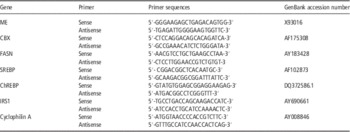
Primers used for real-time PCR analysis are displayed in Table 1. Primer sets for the mRNAs encoding the lipogenic enzymes ME, acetyl CoA carboxylase (CBX) and FASN were designed to span an intron as previously described and utilized for real-time PCR (Ramsay and Azain, Reference Ramsay and Azain2007; Ramsay et al., Reference Ramsay, Blomberg and Caperna2013). Primers for genes important for insulin responsiveness of adipose tissue were also examined and included sterol regulatory element-binding protein 1 (SREBP), carbohydrate regulatory element-binding protein (ChREBP) and insulin receptor substrate 1 (IRS1). Primers for SREBP were obtained from Gondret et al. (Reference Gondret, Perruchot, Tacher, Bérard and Bee2011), whereas primers for ChREBP were acquired from Zhang et al. (Reference Zhang, Lu, Chen, Zhao, Guo, Yang and Zang2014). IRS1 primers were derived from Jiao et al. (Reference Jiao, Lei, Wang, Mo, Lan and Xiong2011). Cyclophilin was used as a relative standard for comparisons in parallel reactions.
Primer sequences for PCR products

ME=malic enzyme; CBX=acetyl CoA carboxylase; FASN=fatty acid synthase; SREBP=sterol regulatory element-binding protein 1; ChREBP=carbohydrate regulatory element-binding protein; IRS1=insulin receptor substrate 1.
Thermal cycling and data acquisition were performed with a Bio-Rad iCycler IQ system (Bio-Rad Laboratories, Inc., Hercules, CA, USA). Reverse transcription (RT) and real-time PCR analysis were performed in a two tube assay as previously described (Ramsay and Caperna, 2009). RT was done using a Superscript First-Strand Synthesis System for RT-PCR kit (Invitrogen, Carlsbad, CA, USA). Master mix was made containing random hexamers (50 ng/µl), 10 mM dNTP mix, RNase-free H2O, and RNA (1 µg/µl). The RNA mix was annealed at 65°C for 5 min. A second master mix was prepared with 10× RT buffer, 25 mM MgCl2, 0.1 mM dithiothreitol and 1.0 µl RNaseOut (ThermoFisher Scientific, Grand Island, NY, USA). This second master mix was added to the RNA mix and incubated at 25°C for 2 min. Superscript II was then added and incubated at 25°C for 10 min, 42°C for 50 min and 70°C for 15 min. An aliquot of RNase H (1.0 µl) was then added and incubated at 37°C for 20 min.
Real-time PCR was done using the IQ Sybr green supermix kit (Bio-Rad Laboratories, Inc., Hercules, CA, USA). A 24 µl reaction mix was made containing 12.5 µl Sybr green supermix, 1.0 µl forward primer (10 µM), 1.0 µl reverse primer (10 µM) and 9.5 µl sterile water. This reaction mix was added to each well, followed by 1.0 µl RT product (25 µl total volume). Parameters for all reactions were as follows: one cycle at 95°C for 15 min (PCR activation), followed by 30 cycles, 94°C for 15 s, 58°C for 30 s, 72°C for 30 s, with a final extension at 72°C for 8 min.
Melting curve analysis was performed on all real-time PCR reactions to confirm specificity of the real-time PCR products. A non-template control was run for every assay. Specificity of real-time PCR products was further confirmed by agarose gel electrophoresis.
Quantification of gene expression
At the end of the PCR, baseline and threshold crossing values (CT) for all analyzed genes were calculated using the Bio-Rad software and the CT values were exported to Microsoft Excel for analysis. The relative expression of the genes of interest, standardized against the amount of cyclophilin mRNA, was calculated using the ∆∆CT method (Winer et al., 1999; Livak and Schmittgen, 2001). Values were calculated as the mean±SEM of duplicate determinations from duplicate tissue culture wells derived from each of five individual animals. Data are expressed relative to values determined for incubations without AGP, whose value was arbitrarily set at 1.0.
Statistical analysis
Data were analyzed by ANOVA using SigmaPlot 12 software (SPSS Science, Chicago, IL, USA) to test for the effects of treatment with AGP. Mean separation was analyzed using Student–Newman–Keuls test. Paired t-test was used to evaluate the effect of AGP on the insulin response by adipose tissue explants. Means were defined as different at P<0.05.
Results and discussion
The newborn pig is born with <2% body fat (Mersmann et al., Reference Mersmann, Goodman and Brown1975). The consumption of milk results in a rapid increase in lipid accretion by the adipose tissue, which can then serve as a critical source of energy in times of increased energy demand or an unavailability of milk from the dam. However, the causative factors for the limited lipid accretion prenatally in the pig have not been identified.
Liver is the primary source of AGP and hepatic mRNA abundance increases during gestation in the fetal pig, with very high levels of expression just before birth and a subsequent and rapid fall following parturition (Stone and Maurer, Reference Stone and Maurer1987). This glycoprotein represents the major circulating protein in the serum of newborn pigs (Lampreave and Piñeiro, Reference Lampreave and Piñeiro1984; Martin et al., Reference Martin, Tesouro, González-Ramón, Piñeiro and Lampreave2005); serum concentrations in the newborn vary from 8000 to 10 000 µg/ml serum (Caperna et al., Reference Caperna, Shannon, Blomberg, Stoll and Ramsay2013). The present data suggest that the very high concentrations of AGP in the serum may contribute to this lack of adipose tissue lipid accumulation in the fetus through an inhibition of lipogenesis, thus disproving the initial hypothesis as based upon studies with mouse cell lines (Lee et al., Reference Lee, Choi, Hwang, Lee, Lee, Kim, Huh, Koh, Koh, Son, Masuzaki, Hotta, Alfadda and Kim2010).
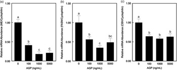
Incubation of neonatal adipose tissue cell cultures with increasing concentrations of AGP resulted in large reductions in lipogenic enzyme mRNA abundance for ME and FASN, and a more moderate decrease in CBX gene expression. Treatment with porcine AGP at 100 ng AGP/ml medium for 24 h resulted in 59% decline in ME mRNA abundance in primary cultures derived from neonatal porcine adipose tissue (P<0.01; Figure 1a). Increasing AGP concentration in the medium caused a further decline to ~20% of ME expression in cultures not treated with AGP (P<0.001). Expression of FASN was reduced to 54% by treatment with 100 ng AGP/ml medium (P<0.01; Figure 1b). Incubation for 24 h with 1000 ng AGP/ml medium resulted in a further decrease in FASN expression to 34% of control incubations without AGP (P<0.001). Acetyl coA CBX mRNA abundance was reduced to a similar extent by all three concentrations of AGP added to the medium, by approximately 36% to 40% (P<0.001; Figure 1c). The lowest concentration tested was 100 ng AGP/ml medium, which is far below the physiological range for neonatal pigs, yet was effective in inhibiting lipogenic enzyme gene expression. A concentration of 1000 ng AGP/ml medium appeared to produce the maximal response, again well below physiological concentrations in the pig. Whether physiological concentrations would produce a different response is unknown, as the testing of concentrations in the physiological range is currently not economically feasible, due to the very large quantity of purified protein that would be necessary. Second, non-specific binding by AGP at concentrations of 8 to 10 mg/ml (physiological for the newborn pig) could potentially confound the results or the data interpretation.
Relative messenger RNA (mRNA) abundance for lipogenic enzymes in primary cultures derived from neonatal subcutaneous adipose tissue in response to 24 h incubation with α1-acid glycoprotein (AGP). Cultures were incubated with 0, 100, 1000 or 5000 ng porcine AGP/ml medium. Relative malic enzyme (ME) (a), fatty acid synthase (FASN) (b) and acetyl CoA carboxylase (CBX) (c) mRNA abundance were measured by real-time PCR as described in the methodology. Data are expressed relative to cyclophilin A mRNA abundance in each cell culture. a,b,cMeans not sharing a common superscript letter are different (n=5; P<0.05).

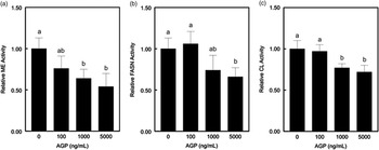
These decreases in mRNA abundance were associated with only a limited reduction in the activities of lipogenic enzymes with AGP treatment. ME activity was reduced by 1000 ng AGP/ml medium by ~40% (P<0.01; Figure 2a). This reduction in ME activity thereby suggests a decrease in reducing equivalents available for fatty acids synthesis. However, incubation with AGP had no effect on FASN activity in homogenates from adipose tissue explants of neonatal swine (P>0.05; Figure 2b). CL activity was reduced by incubation with a minimum of 1000 ng AGP/ml medium (P<0.05; Figure 2c), suggesting a reduction in the provision of acetyl CoA for lipogenesis due to the actions of AGP. The smaller changes in lipogenic enzyme activity (ME, FASN) relative to the changes in gene expression with AGP treatment are not specific to AGP. Iritani et al. (Reference Iritani, Fukuda and Tada1996) have previously demonstrated that the induction of lipogenic enzyme activity is much less than changes in mRNA abundance within adipose tissue in response to dietary manipulation of rodents.
Relative lipogenic enzyme activity in adipose tissue explants derived from neonatal pigs in response to 24 h in vitro incubation with α1-acid glycoprotein (AGP). Explants were incubated with 0, 100, 1000 or 5000 ng porcine AGP/ml medium. Relative malic enzyme (ME) (a), fatty acid synthase (FASN) (b) and ATP citrate lyase (CL) (c) activities were measured using spectrophotometric assays described in the methodology. Data are expressed relative to explant protein content. a,b,cMeans not sharing a common superscript letter are different (n=5; P<0.05).

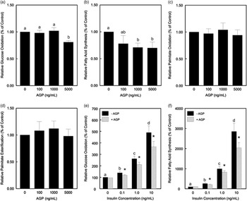
A reduction in the lipogenic potential of the adipose tissue in response to incubation with porcine AGP was confirmed by the measurement of 14C-glucose incorporation into fatty acids. Glucose oxidation as measured by 14CO2 capture was reduced only at the highest concentration of AGP tested, 5000 ng/ml medium (P<0.05; Figure 3a). However, glucose incorporation into the fatty acid fraction of the lipids was reduced by ~25% by 1000 ng AGP/ml medium (P<0.05; Figure 3b). A higher concentration of AGP had no greater effect than 1000 ng/ml medium on lipogenesis. These results confirm the observations made of decreases in mRNA abundance and enzyme activity of various lipogenic enzymes. Palmitate metabolism was unaffected by AGP treatment. Neither palmitate oxidation (Figure 3c) nor incorporation into total lipids (Figure 3d) were affected by any concentration of AGP tested (P>0.05). These results suggest that AGP appears to function through carbohydrate metabolism. As the first experiment to measure glucose metabolism included insulin in the medium, the question arose whether AGP may function through some interaction with insulin. AGP had no effect in the absence of insulin (Figures 3e; P>0.05). Incubation of tissue slices in the presence of as little as 0.1 ng porcine insulin/ml medium enhanced glucose oxidation and the rate increased with insulin concentration (Figures 3e; P<0.05). Glucose oxidation was consistently reduced by 15% to 25% in adipose tissue explants with the addition of 1000 ng AGP/ml to the medium (P<0.05). Similarly, glucose incorporation into fatty acids was elevated by increasing concentrations of insulin (Figure 3f; P<0.05), whereas addition of AGP (1000 ng/ml medium) reduced fatty acid synthesis by 16% to 28% in adipose tissue explants (P<0.05).
Relative 14C-glucose and 14C-palmitic acid metabolism in adipose tissue explants derived from neonatal pigs in response to 24 h in vitro incubation with α1-acid glycoprotein (AGP). Explants were incubated with 0, 100, 1000 or 5000 ng porcine AGP/ml medium in the presence of 1 nM insulin. Relative glucose oxidation (a) or glucose carbon incorporated into fatty acids (fatty acid synthesis) (b) were measured during a 2 h incubation period following a 24 h incubation with AGP according to procedures described in the methodology. Relative palmitic acid oxidation (c) or palmitic acid incorporated into total lipids (esterification) (d) were measured during a 2 h incubation period following a 24 h incubation with AGP according to procedures described in the methodology. Relative glucose oxidation (e) or incorporation into fatty acids (f) in response to insulin stimulation (0, 0.1, 1.0 or 10 ng/ml medium) was measured during a 2 h incubation of porcine adipose tissue explants in the absence or presence of 1000 ng AGP/ml according to procedures described in the methodology. Data are expressed relative to cultures incubated without AGP. a,b,c,dMeans not sharing a common superscript letter are different (n=3 to 4; P<0.05). *AGP effect on insulin response by tissue explants (n=3; P<0.05 according to paired t-test).

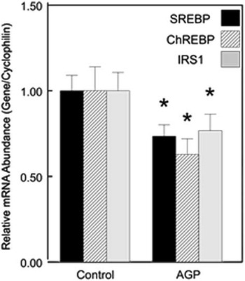
Further analysis was performed to examine potential mechanisms for this AGP-induced reduction in lipid metabolism by assessing the responses by genes for key regulatory proteins in carbohydrate metabolism; SREBP-1c and ChREBP. Studies of the promoter regions of FASN (Latasa et al., Reference Latasa, Griffin, Moon, Kang and Sul2003) and CBX (Oh et al., Reference Oh, Park, Kim, Ahn, Park and Kim2003) identified SREBP as a key transcription factor mediating insulin’s effects on lipogenesis (Foufelle and Ferre, 2002). More recently, the transcription factor ChREBP has been demonstrated to be a mediator of glucose action on lipogenic gene expression and lipid synthesis (Postic et al., Reference Postic, Dentin, Denechaud and Girard2007). The genes for both of these transcription factors are regulated by insulin (He et al., 2004; Cagen et al., 2005; Dif et al., 2006). In the present study, the mRNA abundance for both SREBP and ChREBP were reduced by incubation with 1 µg AGP/ml medium (P<0.01; Figure 4a and b). These changes in gene expression substantiate a potential role for AGP in modifying carbohydrate metabolism within porcine adipose tissue through altering insulin action as seen in the present study.
Relative messenger RNA (mRNA) abundance for insulin responsive transcription and signaling proteins in primary cultures derived from neonatal subcutaneous adipose tissue in response to 24 h incubation with α1-acid glycoprotein (AGP). Cultures were incubated with 0 or 1000 ng porcine AGP/ml medium. Relative sterol regulatory element-binding protein 1 (SREBP), carbohydrate regulatory element-binding protein (ChREBP) and insulin receptor substrate 1 (IRS1) mRNA abundance were measured by real-time PCR as described in the methodology. Data are expressed relative to cyclophilin A mRNA abundance in each cell culture. *AGP effect relative to control (n=5; P<0.05).

Lee et al. (Reference Lee, Choi, Hwang, Lee, Lee, Kim, Huh, Koh, Koh, Son, Masuzaki, Hotta, Alfadda and Kim2010) have previously reported that AGP can increase tyrosine phosphorylation and reduce serine phosphorylation at IRS1 in 3T3-L1 adipocytes, suggesting an enhancement in insulin sensitivity by AGP in the 3T3-L1 adipocyte. Analysis of IRS1 mRNA abundance in the present study demonstrated a decrease in response to the addition of 1 µg AGP/ml medium (P<0.05; Figure 4c); implying a reduction in insulin sensitivity by porcine adipose tissue, which was evident in the glucose metabolism experiments represented in Figure 2. Differences in the cellular composition of the two experimental models may contribute to the different metabolic responses and differences in IRS1 response observed between the present study using primary cultures of porcine adipose tissue and the research of Lee et al. (Reference Lee, Choi, Hwang, Lee, Lee, Kim, Huh, Koh, Koh, Son, Masuzaki, Hotta, Alfadda and Kim2010) using the 3T3-L1 cell line. If AGP alters insulin signaling, the present data imply that AGP may interact with insulin to reduce insulin binding to its receptor or alternatively reduces insulin availability by sequestering insulin. Previous research has demonstrated that AGP binds proteins such as plasminogen activator inhibitor type 1 (PAI-1) at concentrations similar to used in the present experiment (Boncela et al., Reference Boncela, Papiewska, Fijalkowska, Walkowiak and Cierniewski2001); however, in that study AGP did not interfere with PAI-1 activity.
As previously mentioned, serum AGP concentrations increase during late gestation in the pig and are highest at birth (Lampreave and Piñeiro, Reference Lampreave and Piñeiro1992), then rapidly decline postnatally (Caperna et al., Reference Caperna, Shannon, Blomberg, Stoll and Ramsay2013; Heegaard et al., Reference Heegaard, Miller, Sorensen, Soerensen and Skovgaard2013). The interaction of AGP with insulin to reduce insulin-mediated lipogenesis may be especially important in the fetal pig during late gestation when lipid accumulation within adipose tissue is restricted due to the relative unavailability of exogenous lipids and the consequent reliance on glucose for de novo lipogenesis in the fetus (Mersmann, Reference Mersmann1974). Lipid accretion in the fetal pig has been demonstrated to respond to exogenous insulin (Garssen et al., Reference Garssen, Spencer, Colenbrander, Macdonald and Hill1983); however, under physiological conditions there is minimal lipid accretion (Mersmann et al., Reference Mersmann, Goodman and Brown1975).
Differentiation of preadipocytes in tissue cultures derived from fetal pig adipose tissue has been demonstrated to be insulin sensitive (Hausman, Reference Hausman1992). Elevated serum AGP concentrations in late gestation fetal pigs may interfere with the stimulatory effect of insulin on preadipocyte proliferation and differentiation, thus reducing the pool of potential adipocytes and reducing the total lipogenic potential of the perinatal adipose tissue at that age. In a previous study (Caperna et al., Reference Caperna, Shannon, Blomberg, Stoll and Ramsay2013), serum AGP concentrations were higher in runt pigs than normal-sized littermates following birth, whereas Rehfeldt and Kuhn (Reference Rehfeldt and Kuhn2006) demonstrated that low birth weight pigs are born with less carcass lipid than high birth weight littermates. Morise et al. (Reference Morise, Sève, Macé, Magliola, Le Huërou-luron and Louveau2009) reported that low birth weight pigs at 7 days of age have reduced perirenal adipose mass but similar adipocyte volumes, potentially suggesting a reduced adipocyte number in the perirenal adipose tissue of the low birth weight pigs. However, whether AGP can affect preadipocyte proliferation or differentiation either directly or indirectly through inhibiting insulin action is currently unknown.
Immediately following birth, these limited fetal lipid stores can be an important energy source if fetal glycogen reserves become depleted. This can be due to excessive competition in large litters or anything that may limit suckling during the first 24 h after birth (Mersmann, Reference Mersmann1974). During suckling, the sow’s milk provides adequate fatty acids to be esterified and stored in the adipose tissue. The present data demonstrate that AGP does not appear to affect fatty acid esterification. Thus, despite relatively high AGP levels in the preweaning pig at 21 days of age (600 to 900 µg/ml; Caperna et al., Reference Caperna, Shannon, Blomberg, Stoll and Ramsay2013), lipid storage may not be affected by AGP in the adipose tissue of the suckling preweaning pig. Once the pig transitions through weaning and glucose becomes the major substrate for lipogenesis, the role of AGP may change in relevance but that requires further investigation.
Acknowledgments
The authors thank M. Stoll for her assistance with the RNA extractions and real-time PCR. The authors thank A. Shannon for her assistance with animal collection. The authors also thank the swine herd staff, USDA-Beltsville, for their work in animal care.