What’s already known about this topic?:
-
• Left aortic arch with aberrant right subclavian artery is not uncommon
-
• Families are sometimes incorrectly counselled to be aware of vascular ring symptoms in the setting of left aortic arch/aberrant right subclavian artery because clinicians have a poor understanding of the actual basis for a vascular ring with this anatomical variant. This article delineates the anatomical substrate for when aberrant right subclavian artery can cause a vascular ring, which is when accompanied by left aortic arch and right arterial duct.
What does this study add?
-
• Our study identifies the small subset of these patients, who have an additional abnormality of right-sided duct, therefore providing an anatomical set-up for vascular ring.
-
• Clear and detailed antenatal diagnosis allows accurate prognostication and counselling of families. In addition, it identifies the small subset of cases where clinical follow-up is advisable and cross-sectional imaging should be considered (in the context of clinical symptoms in those with this anatomical substrate for vascular ring), thereby avoiding over-investigation of children without anatomical substrate for a vascular ring.
-
• In addition, this study reports the genetic association in this rare presentation which may allow clinicians to present information to families, aiding decision-making around antenatal/postnatal genetic testing and providing information about long-term outcomes.
Key embryological concepts:
-
• The aortic and ductal arches are formed from the right and left 4th and 6th pharyngeal arch arteries between week 4 and 8 of gestation.Reference Rosen and Bordoni1
-
• Whilst both a left and right aortic arch (and ductal arch) are present early on, usually the right aortic and ductal arches regress, leaving the usual arrangement of unilateral left aortic and left ductal arches.Reference Rosen and Bordoni1
-
• The ascending aorta develops from the truncus arteriosus. The spiral conotruncal septum divides the truncus into ascending aorta and main pulmonary artery.
-
• The transverse aortic arch develops from the 4th pharyngeal artery (usually from persistence of the left 4th pharyngeal arch, resulting in left aortic arch).Reference Sadler2
-
• The right 4th pharyngeal arch also gives rise to the right subclavian artery.Reference Sadler2
-
• The ductal arch develops from either the left or right 6th pharyngeal arch (depending on which side regresses or persists).Reference Rosen and Bordoni1
-
• Aberrant right subclavian artery develops due to persistence of the distal part of the right dorsal aorta and regression of the right fourth arch artery.
Introduction
A left aortic arch with aberrant right subclavian artery can be readily identified on fetal echocardiogram. When these findings occur with a left arterial duct, there is no substrate for a vascular ring. However, on the rare occasion that aberrant right subclavian artery and left aortic arch occur with a right arterial duct, a vascular ring substrate is created. Aberrant right subclavian artery is usually a variant of the normal branching pattern of the arch, with an incidence in the general population of 1.5%.Reference Borenstein, Minekawa, Zidere, Nicolaides and Allan3,Reference Scala, Leone Roberti Maggiore and Candiani4 However, some studies suggest it is more common in fetuses with chromosomal anomalies (particularly trisomy 21). Its identification on routine scan should prompt a detailed evaluation looking for other markers of genetic abnormalities.
When any variation in aortic branching pattern is identified, the specific arrangement of vessels must be delineated (Figure 1 ( a )-( d )). In most cases of left aortic arc (± aberrant right subclavian artery), there is no substrate for a vascular ring; if not fully understood, this can result in incorrect prenatal counselling. We present the rare incidences when a left aortic arch occurs with a right-sided arterial duct and aberrant right subclavian artery, which constitutes a vascular ring and can lead to postnatal feeding and breathing difficulties (Videoclip S2).Reference Young, Hornberger and Haberer5
( a ) Left aortic arch, left arterial duct, normal right subclavian artery; ( b ) left aortic arch, left arterial duct, aberrant right subclavian artery; ( c ) left aortic arch, right arterial duct, normal right subclavian artery; ( d ) left aortic arch, right arterial duct, aberrant right subclavian artery. Ao = Aortic Arch; Duct = Arterial Duct; T = Trachea; E = Oesophagus; ARSA = Aberrant Right Subclavian Artery; RSCA = Right Subclavian Artery.

Patients and methods
We analysed all prenatally diagnosed cases of left aortic arch and right arterial duct (between 2016 and 2025) at three fetal cardiac centres (2 UK and 1 Austrian). Departmental databases and prenatal/postnatal patient records were reviewed. Over the study period >21,000 fetal echocardiograms were performed.
Cases with associated major cardiac abnormality were excluded; isolated ventricular septal defects were included.
Referral indication, gestational age at diagnosis, origin of right subclavian artery and associated abnormalities including karyotype/comparative genomic hybridisation array results were noted. Postnatal outcome, symptoms, CT findings and operative findings were included.
Results
Demographics
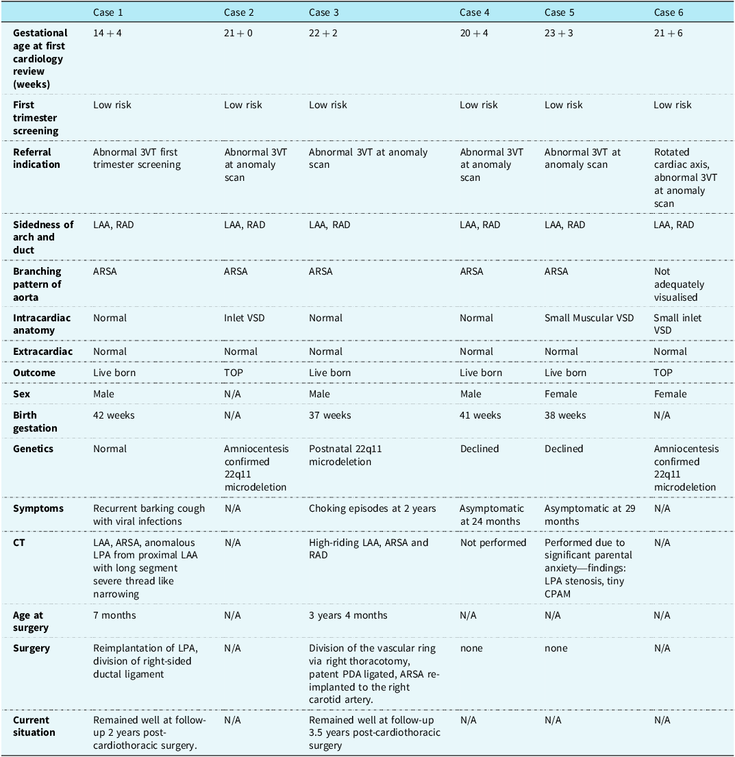
Six cases of left aortic arch and right arterial duct were identified (Table 1). All were referred due to abnormal three-vessel-trachea view and had low-risk first-trimester screening for major trisomy.
Summary of cases with left aortic arch, right arterial duct, and aberrant right subclavian artery

Table 1. Long description
6 cases along top, features detailed in each row were gestation at first cardiology review (weeks), first trimester screening, referral indication, sidedness of arch and duct, branching pattern of aorta, intracardiac anatomy, extrcardiac features, outcome, sex, birth gestation, genetics, postnatal symptoms, CT scan results (if done), age at surgery (if done), details of surgery completed and current situation.
3VT = 3 vessel trachea view; 3VV = 3 vessel view; LAA = left aortic arch; RAD = right arterial duct; ARSA = aberrant right subclavian artery; VSD = ventricular septal defect; TOP = termination of pregnancy; LPA = left pulmonary artery; PDA = persistent ductus arteriosus; CPAM = congenital pulmonary airway malformation.
Five were identified following routine 20-week anomaly scan. One case of suspected aortic arch abnormality was identified at the 12-week scan. Further details of the aortic arch pattern were not known at time of referral.
Initial fetal cardiology assessment was completed at 14–24 weeks gestation.
One patient diagnosed at 14 + 4 weeks required a second scan at 17 + 4 weeks to obtain full details of aortic branching. In two patients, a differential diagnosis of left aortic arch/right arterial duct or double aortic arch was considered (at 21 + 6 and 22 + 2, respectively). One family terminated the pregnancy due to chromosomal abnormality, before reimaging was possible; the other had a diagnosis of left aortic arch/right arterial duct/aberrant right subclavian artery confirmed at 28 + 4 weeks.
Cardiac diagnosis and counselling
In 5/6 cases of left aortic arch with right arterial duct, aberrant right subclavian artery was identified. Families were counselled regarding the potential for vascular ring symptoms and possible chromosomal abnormality. In the other case, the origin of the right subclavian artery could not be ascertained due to suboptimal imaging quality.
Three fetuses had ventricular septal defects (perimembranous inlet ventricular septal defects in two patients and small muscular ventricular septal defects in the third) and three had normal intracardiac anatomy. No fetuses had extracardiac abnormalities.
Genetics
Overall (3/6), 50% of patients had a confirmed genetic abnormality. Three had amniocentesis with polymerase chain reaction and comparative genomic hybridisation array testing. This was normal in one patient, and 22q11 microdeletion was confirmed in two (both with associated inlet ventricular septal defects). One other patient had postnatal confirmation of 22q11 microdeletion. Two families declined genetic testing.
Outcomes
There were four livebirths between 37 and 42 weeks of gestation. Two pregnancies, with 22q11 microdeletion, resulted in termination.
Two infants developed vascular ring symptoms: One had a recurrent barking cough and viral infections from 6 months of age; the other had choking episodes aged 2 years. Two patients remained asymptomatic at 24 and 29 months, respectively.
The two symptomatic patients underwent cardiac CT angiography. Anomalous origin of the left pulmonary artery from the underside of the proximal left aortic arch was identified in one case, with severe, long segment left pulmonary artery narrowing. Surgical patching and reimplantation of left pulmonary artery was completed aged 7 months, with division of the right-sided ductal ligament.
In the second patient CT confirmed a high-riding left aortic arch with descending aorta to the right of the spine, becoming midline at the level of the diaphragm. It also confirmed aberrant right subclavian artery and right arterial duct. The aorta was causing extrinsic oesophageal compression and right upper lobe collapse. Ligation of the ductal ligament and reimplantation of aberrant right subclavian artery to the right carotid artery was completed at 3 years of age.
A CT was completed on one asymptomatic patient in view of significant parental anxiety. A tiny congenital pulmonary airway malformation was noted incidentally, which had not been seen on fetal MRI. Findings were discussed at the local pulmonary multidisciplinary meeting, with an expectant approach advised. This family declined genetic testing.
Discussion
We describe six cases of left aortic arch, 5 of whom, with the combination of right arterial duct and aberrant right subclavian artery, had the substrate for a vascular ring, 50% of which were associated with DiGeorge syndrome. This combination is the only example of aberrant right subclavian artery causing vascular ring, as a left aortic arch, left arterial duct, aberrant right subclavian artery does not encircle the oesophagus and trachea (Figure 1( b )). The vascular ring set-up of left aortic arch, right arterial duct, aberrant right subclavian artery (Figure 1( d )) is comparable to the most common form of vascular ring: right aortic arch, left-sided arterial duct, and aberrant origin of the left subclavian artery.
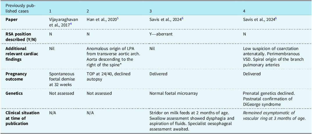
To date, only three papers (4 cases) detailing prenatal diagnosis of left aortic arch with right arterial duct (without additional major structural heart disease) have been reported, none with long-term follow-up data. These are detailed in Table 2. Reference Vijayaraghavan, Senthil and Latha6–Reference Savis, Oakley and Van Poppel8
Summary of current literature detailing prenatal diagnosis of LAA with RAD

Table 2. Long description
4 papers identified for further analysis. The details of each was assessed with relation to whether or not the RSA position was described (only completed in one of the paper), whether additional relevant cardiac findings were identified (this was the case in 2 papers), outcome of pregnancy (2 delivered, one spontaneous fetal demise at 32 weeks, one TOP at 24/40, declined autpsy), genetics were detailed in 2 cases - one with normal microarray, the other with postnatal confirmatino of DiGeorge syndrome), current situation described in the the two cases which survived to live birth - one had stridor on mild feeds at 2 months of age, the other remained asymptomatic of vascular ring symptoms at 3 months of age).
RSA = right subclavian artery; LPA = left pulmonary artery; VSD = ventricular septal defect; TOP = termination of pregnancy.
* similar to one case in our series.
Ultrasound identification of an aberrant right subclavian artery with a vascular ring substrate
Usually, both the aorta and duct pass to the left of the trachea creating a V–configuration on the three-vessel-trachea view. The normal origin of the right subclavian artery is from the first branch of the aorta (innominate artery) which crosses anteriorly to the trachea and divides into the right common carotid and right subclavian artery. When there is an aberrant right subclavian, most cases have left aortic arch and left arterial duct, therefore again creating the usual V-configuration, but with the right subclavian artery arising separately as a 4th vessel from the arch. In this situation, the aberrant right subclavian artery is seen posterior to the trachea coursing towards the right and without the tracheal encirclement which causes a vascular ring (Figure 1( b )).
In the rare circumstance described in our study, imaging of the three-vessel-trachea view demonstrates the aorta to the left of the trachea and duct to the right of the trachea. This arrangement also creates a U-configuration with the trachea in the middle (Figure 1( c ) and ( d )), rather than the usual V-shape with both the aortic and ductal arch on the left (as is seen in normal left aortic arch/left arterial duct). Extra care is required to accurately identify the vessels in cases of abnormality as one arch may lie in a plane slightly above the level of the other (Figure 2). When a left arterial duct and right arterial duct occurs in combination with aberrant right subclavian artery, a complete vascular ring is formed with a risk of symptoms.
Images obtained from transabdominal ultrasound scan of case 2 at 21 + 0 weeks of gestation. ( a ) Image showing LAA and RAD encircling trachea; ( b ) dual image: 2D and colour image shows the LAA and ARSA; ( c ) dual image showing 2D and colour images of the U-shaped configuration of the RAD coursing posteriorly around the right side of the trachea to meet the left-sided aortic arch before it descends to the left of the spine; ( d ) single colour image showing the relationship of the LAA, MPA continuing in to the RAD and the distal course of the ARSA after it has passed behind the trachea. HB = Head Below; LAA = Left Aortic Arch; ARSA = Aberrant Right Subclavian artery; RAD = Right Arterial Duct; MPA = Main Pulmonary Artery; T = Trachea.

Left arterial duct with aberrant right subclavian artery is the most common aortic arch abnormality, but reports of requirement for intervention are rare, limited to case reports or small case series.Reference Janssen, Baggen and Veen9,Reference Stone, Jn, Fowl, Garg, Bower and Money10 This observation is further supported by a recent publication showing a very low rate of intervention, 1.3% of 528 prenatally diagnosed cases.Reference Zidere and Vigneswaran11
Since inclusion of the three-vessel-trachea view into the national UK screening programme in 2015,12 a significant rise in the identification of arch differences has been appreciated. Whilst some variations carry a risk of vascular ring, most do not. An understanding that an aberrant right subclavian with left arterial duct does not create the substrate for a vascular ring is important to correctly guide patients and avoid unnecessary follow up, postnatal investigations and parental anxiety.
Genetics
Isolated aberrant right subclavian, with left aortic and ductal arches and normal intracardiac anatomy, is not thought to be associated with a significant increase in the chance of a genetic abnormality. However, our data suggest that when this vascular difference (aberrant right subclavian) co-exists with a right ductal arch, genetic testing should be offered.Reference Li, Qiu and Shi13,Reference Maya, Kahana and Yeshaya14 In our study, 50% of all patients had 22q11 deletion (origin of the right subclavian artery unknown in one case) but genetics remain unknown in two cases. Three also had a ventricular septal defect (one normal genetics, two patients 22q11 deletion), suggesting the finding of a ventricular septal defect in addition to right arterial duct and aberrant right subclavian artery may convey an increased chance of genetic abnormalities. Whilst the small number of cases of left aortic arch/right arterial duct/aberrant right subclavian artery limits the ability to draw firm conclusions, the higher rate of genetic anomaly in this cohort versus isolated aberrant right subclavian artery may be expected in the context of two variations in the vascular arrangement (i.e. aberrant right subclavian artery and right arterial duct). An appreciation of the potentially increased risk of co-existent genetic diagnosis enables more thorough antenatal counselling.
One patient (with normal genetics) had an additional vascular abnormality with the left pulmonary artery arising from the aortic arch.
Limitations
Left aortic arch, right arterial duct with aberrant right subclavian artery is an uncommon finding antenatally limiting the ability to observe the outcome in a larger series. Due to regression of the arterial duct after birth, the prevalence of right arterial duct is unknown, and it cannot definitively be known whether cases of left aortic arch and aberrant right subclavian artery requiring surgical intervention had this pattern antenatally.
Conclusion
Left aortic arch with aberrant right subclavian artery is not an uncommon prenatal ultrasound finding. The vast majority of cases have a left-sided arterial duct and therefore do not have the substrate for a vascular ring. The cases reported in this study highlight a small subgroup of patients who have a right-sided rather than left-sided arterial duct with aberrant right subclavian artery. This combination does form a vascular ring substrate with potential for vascular ring symptoms therefore requiring surveillance for symptoms of airway or oesophageal compression. Our study also indicates an increased risk of 22q11 microdeletion (Di George syndrome), particularly when seen in combination with a perimembranous ventricular septal defect.
We also found additional anatomical abnormalities in all 3 patients who had a CT scan, including one child who was asymptomatic. This suggests CT angiography should be considered in patients with this anatomical vascular ring substrate.
Supplementary material
The supplementary material for this article can be found at https://doi.org/10.1017/S1047951126113936.
Acknowledgements
None.
Financial support
This research received no specific grant from any funding agency, commercial, or not-for-profit sectors.
Competing interests
None.