Congenitally corrected transposition is a rare condition in which the cardiac ventricles are connected to morphologically inappropriate atrial chambers, and the arterial trunks also arise from their inappropriate ventricles. Thus, the morphologically left ventricle pumps blood to the pulmonary trunk, while the morphologically right ventricle acts as the systemic ventricle. We present a male patient with congenitally corrected transposition, but without associated cardiac malformations, who was diagnosed in childhood and followed up as an adult. He underwent two consecutive cardiac gated single photon emission computed tomographic images.
Case report
Because of a heart murmur, the patient had been referred to a paediatric cardiologist. The auscultatory findings at that time included a click and a scratchy systolic murmur at the lower left sternal border, and a loud second heart sound. An electrocardiogram showed a QS pattern over the right percordial leads, and no Q waves over the left-sided ventricle. At the age of 10 years, he underwent cardiac catheterization, which confirmed the clinical impression of congenitally corrected transposition. There were no intracardiac shunts were present and all intra-cardiac pressures were normal. The right-sided mitral valve showed prolapse, but no significant insufficiency. The left-sided tricuspid valve was redundant, but again without any regurgitation. He was followed regularly with frequent complaints of non-exertional chest pain and palpitations. For that reason, he was placed on Corgard, which he subsequently discontinued. He engaged in normal physical activity without any symptoms. At the age of 18, he complained of chest pain of two or three hours of duration, which continued off and on for the next week. The electrocardiogram, however, was unchanged, but an echocardiogram showed the systemic morphologically right ventricle to be slightly reduced in contractility. The left-sided tricuspid valve remained redundant, but now showed mild to moderate regurgitation.
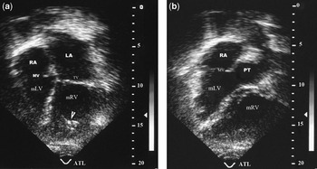
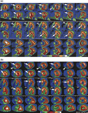
At the age of 22, he joined the Marines without disclosing his cardiac condition, completed basic training, and entered active service. After spending nearly four years in the Marines, he was seen at a Veterans Administration Medical Center for compensation and pension examination. Physical examination detected a heart murmur, but the patient did not reveal his known diagnosis. He was well developed and well nourished, 74 inches tall in height, and weighing 210 lbs. A grade 2 from 6 systolic murmur was noted at the base of the heart. A complete cardiac work-up again confirmed congenitally corrected transposition (Fig. 1). At this time, a dual isotope single photon emission computed tomographic study showed the systemic right ventricle to be moderately enlarged, with increased uptake in the ventricular septum, fixed hypoperfusion in the mid and basal anterior walls, and apical, mid, and basal inferior walls (Fig. 2a). The ejection fraction of the systemic right ventricle was calculated as 52%.
In the 4 chamber view (a), the dilated morphologic right ventricle (mRV) identified by the moderator band (open-arrowhead) placed in the left side of the heart and connected to the left atrium by tricuspid valve (TV); the smaller and (hypoplastic) morphologic left ventricle (mLV) is located in the right side of the heart and connects to the right atrium (RA) by the mitral valve (MV). The parasternal short-axis view (b) shows blood flow entering the right atrium (RA) through the mitral valve (MV) to mLV and pumped to pulmonary trunk (PA) through the pulmonary valve.

In the first study (a) the upper 2 rows are short axis slices: top row, stress; bottom row, rest. Middle 2 rows are horizontal long axis slices: top row, stress; bottom row, rest. Lower 2 rows are vertical long axis slices: top row, stress; bottom row, rest. The morphologically right ventricle is moderately enlarged with increased septal uptake (arrows), fixed hypoperfusion in the mid and basal anterior walls, in the inferior wall, and fixed defect in inferoseptal wall. Part of the morphologically left ventricle as indicated by open-arrowhead is barely visualized. In the second study (b) the upper 2 rows are short axis slices: top row, stress; bottom row, rest. Middle 2 rows are horizontal long axis slices: top row, stress; bottom row, rest. Lower 2 rows are vertical long axis slices: top row, stress; bottom row, rest. The morphologically left ventricle is now moderately enlarged with slightly increased size. As shown on the previous study, there is increased septal uptake; fixed hypoperfusion in the apical and mid anterior walls, and fixed hypoperfusion in the apical and mid anterior walls and defect in the basal anterior wall (with interval expand in size shown on short axis slices), and fixed hypoperfusion in the apical, mid, and basal inferior walls.

Two years later, the patient underwent a second study because of chest pain. He walked for 10 minutes and 4 seconds on a full Bruce protocol, stopping secondary to fatigue, but with no chest pain. The study (Fig. 2b) now showed the systemic right ventricle to be moderately enlarged compared to the prior study. There was still increased septal uptake, reversible hypoperfusion in the apical wall, expanded hypoperfusion in the mid and basal anterior walls, and fixed hypoperfusion in the apical, mid, and basal inferior walls. The ejection fraction was now reduced to 44%. Coronary angiography demonstrated that the morphologically right coronary artery perfused the systemic ventricle, being a large vessel which gave off a high obtuse marginal branch, a large second obtuse marginal branch, and terminated as the inferior interventricular artery. The left coronary artery, a medium sized artery, gave off a diagonal branch and several marginal branches, but did not supply the inferior ventricular walls. There was luminal irregularity in this artery just distal to its orifice, but no changes in the morphologically right coronary artery. The studies revealed duodenogastric reflux as a likely cause of his chest pain. Further examination of the upper gastrointestinal tract showed a hiatal hernia and duodenogastric reflux. Two months after his last visit, the patient collapsed while running after some troublesome adolescents, and died at the scene of his fall. Unfortunately, post mortem examination was not carried out.
Discussion
Congenitally corrected transposition is a rare congenital cardiac malformation. Although patients have been reported to survive well into late adulthood in the absence of associated lesions, several long-term studies of such patients reveal increasing systemic ventricular dysfunction, resulting in clinical congestive heart failure with increasing age.Reference Rutledge, Nihill, Fraser, Smith, Mcmathon and Bezod1, Reference Wu and Child2 In the study of Graham et al.,Reference Graham, Bernard and Mellen3 over three-tenths of patients with no associated significant lesions had developed congestive heart failure by their fifth decade. Insufficiency of the systemic atrioventricular valve is believed to play a major role both in the systemic ventricular dysfunction and the subsequent congestive cardiac failure.Reference Nebyh and Crubb4 In our patient, we speculate that heavy physical activity while in the Marines could have significantly accelerated the hypertrophy of the systemic right ventricle, placing him at risk for myocardial ischaemia. The cause of his sudden death, while not documented, was likely due to a fatal arrhythmia. He had no evidence of congestive heart failure when seen only a few months before this death, and the left-sided atrioventricular valvar insufficiency was no more than mild to moderate.
There are few reports in the literature describing myocardial perfusion by single photon emission computed tomographic imaging in the setting of congenitally corrected transposition,Reference Duarte, Matusmoto, Moises, Tebexreni and Mastrocolla5–Reference Hornung, Bernard, Jaeggi, Howman-Giles, Celermajer and Hawker7 these giving findings similar to those in our patient. Our patient is the first reported to have serial studies, which demonstrated progressive enlargement of the morphologically right ventricle. The findings suggest that when the systemic ventricle is perfused by a morphologically right coronary artery, as is the case in congenitally corrected transposition, there may be inadequate perfusion to supply the progressive and extensive hypertrophy. Further perfusion studies in other patients with this rare condition would be helpful in determining the role, if any, of such hypoperfusion in the development of the known late cardiac dysfunction in patients with a morphologically right ventricle functioning as the systemic ventricle.
Acknowledgements
We express our appreciation to Pranab Das, Helga Miles, Vickie Kiefer, Becky Wierzbinske, and Kelly Gross, and to Estil Robinson, for his help during the preparation of the digital images. We also thank Mark Ingram, our reference librarian, for his excellent and diligent search of the literature during the preparation of this paper.