Introduction
Sialadenitis is an acute or chronic inflammatory and infectious condition of the salivary glands causing swelling and pain of the affected gland. Sialadenitis constitutes one of the most frequent clinical conditions of the salivary glands.Reference Bradley1 Classification of sialadenitis is based on aetiology and pathophysiology, and includes bacterial, viral, immune or granulomatous causes.Reference Seifert2
Obstructive salivary gland diseases such as sialolithiasis are a common course of chronic sialadenitis of the submandibular gland.Reference Seifert2, Reference Bradley3 Obstruction of the duct reduces the flow of saliva. For chronic obstructive sialadenitis, two factors seem to be important: reduced salivary flow, and retrograde contamination of the salivary gland and duct with bacteria from the oral cavity.Reference Bradley3
Bacteria either occur as free-swimming ‘planktonic’ or aggregated ‘sessile’; the latter is also known as biofilm.Reference Marcus, Post, Stoodley, Hall-Stoodley, McGill and Sureshkumar4 Biofilms are increasingly important in chronic infections,Reference Parsek and Singh5 including those of the ENT region.Reference Homoe, Bjarnsholt, Wessman, Sorensen and Johansen6 Micro-organisms are also proposed to contribute to the formation of sialoliths.Reference Teymoortash, Wollstein, Lippert, Peldszus and Werner7 In addition, bacterial biofilms have recently been suggested as a pathogenic factor in sialolithiasis.Reference Fusconi, Petrozza, Schippa, de Vincentiis, Familiari and Pantanella8 However, it is not known whether biofilms are involved or present in chronic obstructive sialadenitis of the submandibular gland.
This descriptive case–control study aimed to test the hypothesis that biofilm is morphologically present in histological submandibular gland sections from patients with chronic obstructive sialadenitis.
Materials and methods
Study population
The study comprised patients who, between 2012 and 2013, suffered from submandibular chronic sialadenitis, had an additional diagnosis of sialolithiasis, and who underwent submandibular excision at one of two hospitals. Patients were selected longitudinally.
The inclusion criteria were: submandibular chronic sialadenitis diagnosis for at least one year, with an additional diagnosis of sialolithiasis showing typical symptoms such as pain and recurrent swelling. Furthermore, endoscopic treatment and/or sialdochotomy in the included patients had failed or had been considered not feasible. The exclusion criteria were: acute infection, age of less than 18 years and antibiotic treatment during the previous 3 months.
The surgical procedure for the gland excision was a classic submandibular adenectomy. The gland was fixated in formalin immediately after the excision. The location of the ex vivo histological section of the gland was randomly chosen, and was taken either from the gland poles or the central parts of the gland.
Within the same study period, from the same two hospitals, ex vivo sections of submandibular glands (adenectomised because of pleomorphic adenoma) were included as a control group, from patients matched by gender and age. These sections were taken solely from the healthy part of the glands (sections did not include the pleomorphic adenoma).
The local ethical committee of the Capital Region of Denmark approved the study (H-4-2011-022).
Biofilm analysis
Biofilm was visualised by conventional light microscopy, as well as fluorescent bacterial probes (peptide nucleic acid fluorescence in situ hybridisation ‘PNA-FISH’) and subsequent confocal laser scanning microscopy. Samples were stained with a peptide nucleic acid fluorescence in situ hybridisation probe UniBac targeting bacteria in general (red) (AdvanDx, Woburn, Massachusetts, USA). To detect DNA, DAPI (4', 6-diamidino-2-phenylindol) was used as a counter stain. The manufacturer's instructions were followed for the staining procedure. Bacterial biofilm was defined as aggregated bacteria, with each bacteria being 1–2 μm in size, morphologically either cocci- or rod-shaped, and fluorescent only in the spectrum of the fluorophore used.
All samples were examined blinded and independently by a physician (SAS) and a specialist (SED) in the peptide nucleic acid fluorescence in situ hybridisation method. Comprehensive microscopic analyses of the sections were made to ensure that the bacteria identified during confocal laser scanning microscopy investigation were within the tissue and not due to contamination from outside the tissue.
Statistics
Fisher's exact test was used to compare the groups, and the level of significance was set to p < 0.05.
Results
Population
The sialadenitis group comprised 10 patients suffering from chronic obstructive sialadenitis of the submandibular gland; 5 were male and 5 were female. The patients’ median age was 41 years (range, 21–65 years). The control group comprised 10 patients with pleomorphic adenoma; 5 were male and 5 were female. Median age was 40 years (range, 22–65 years). The patients’ demographics and treatment are summarised in Table I.
Overview of patient demographics, symptom duration and treatment

Biofilm
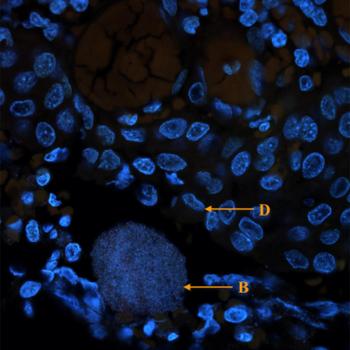
Five of the 10 submandibular glands (50 per cent; 95 per cent confidence interval = 24–76 per cent) from patients with chronic obstructive sialadenitis showed morphological evidence of bacterial biofilm, both in terms of biofilm formation with aggregation of bacteria, and immune response (Figures 1 and 2). There was no discrepancy of the results between the two examiners.
Images show a section of the submandibular gland (sialadenitis group) stained with peptide nucleic acid fluorescence in situ hybridisation (universal bacterial probe (Texas red) and DAPI as a counter stain (blue); magnification ×63). ‘D’ = DAPI-stained nucleus of immunological cells; ‘A’ = glandular acini (golden yellow area); ‘B’ = fluorescent bacterial biofilms (violet and more intense red colour); ‘E’ = erythrocytes

Magnification of a section of the submandibular gland from a different patient (sialadenitis group) showing bacterial biofilm (‘B’) surrounded by DAPI-stained immunological cells (‘D’). The section is stained with peptide nucleic acid fluorescence in situ hybridisation (universal bacterial probe (Texas red) and DAPI as a counter stain (blue); magnification ×63).

Based on size and shape, the bacteria in the biofilms morphologically resembled cocci. In all five glands, the biofilms were found within the tissue of the gland and surrounded by inflammatory cells, primarily polymorphonuclear leukocytes (Figures 1 and 2).
All 10 histological sections from the control group (comprising non-sialadenitis submandibular glands) were without morphological signs of bacterial biofilm. In the sialadenitis group, the number of biofilm positive sections was significantly higher than in the control group (p = 0.03). In four sections (three from sialadenitis patients and one from a control patient), bacteria were found lining the tissue; this was interpreted as contamination because of the location and morphological appearance.
No differences were found between the cases in which biofilm was confirmed and not confirmed in the sialadenitis group with respect to age (p = 0.1), gender (p = 0.2), duration of symptoms (p = 0.4) and endoscopic intervention (p = 1) (Table I).
Discussion
Bacterial biofilm is reported as a factor in many chronic infections.Reference Bjarnsholt, Moser, Jensen and Højby9 In this descriptive study with controls, we demonstrated morphological evidence of bacterial biofilm in half of the histological submandibular sections investigated from the sialadenitis group, but in none from the non-sialadenitis group (Table I).
Morphologically, the bacteria in the biofilms resembled cocci, which is in accordance with non-haemolytic streptococcus being one of the most abundant bacteria of the oral cavityReference Aas, Paster, Stokes, Olsen and Dewhirst10 and Staphylococcus aureus being frequently linked to acute bacterial sialadenitis.Reference Bradley3
Sialadenitis is a common clinical condition of the salivary glands.Reference Bradley1 Bacterial sialadenitis is typically considered a consequence of ascending bacterial infection and reduced salivary flow, and is known to be linked to the obstructive disease sialolithiasis.Reference Bradley3 Thus, sialolithiasis may be considered a consequence of sialadenitis or vice versa.Reference Fusconi, Petrozza, Schippa, de Vincentiis, Familiari and Pantanella8, Reference Marchal, Kurt, Dulguerov and Lehmann11 Biofilm has recently been identified inside a sialolith,Reference Fusconi, Petrozza, Schippa, de Vincentiis, Familiari and Pantanella8 supporting the notion that bacteria in the gland duct are causal to the sialolith rather than secondary to duct obstruction.
Treatment of chronic obstructive sialadenitis is often conservative, with massage, saliva stimulation and antibiotics given for acute exacerbation. However, if a sialolith is present, treatment may include stone removal, either endoscopically or by open approach surgery. Chronic, intractable sialadenitis is managed by surgical excision of the entire gland.Reference Bradley3, Reference Koch, Zenk and Iro12 Recurrent infection is not a rare event, and biofilm in the glands may contribute to this. In the present study, 5 of the 10 sialadenitis patients had previously undergone sialendoscopy and/or sialodochotomy (Table I), and it could be speculated that previous surgical or endoscopic intervention introduced bacteria in the duct system and promoted biofilm formation. However, there was no difference in the number of biofilm cases between patients who had previously undergone endoscopic or surgical treatment and those who had not. Prior antibiotic treatment also did not seem to influence the presence of biofilm (Table I).
The strength of the study is the direct morphological evidence of bacterial biofilm formation in the submandibular gland excised because of chronic sialadenitis with sialolithiasis. The biofilms identified are unlikely to be a result of contamination as they were present within the tissue. It should be stressed, however, that the presence of bacterial biofilm does not constitute proof of causality for chronic sialadenitis.
Regarding the study limitations, the sample size was somewhat small, only one sample was available from each patient and the negative findings in the control group may be a consequence of the limited material available. Furthermore, only a eubacterial probe was used, and no culturing or other molecular methods were utilised to confirm bacteria in the samples.
The observation of biofilm in the submandibular gland suggests that early and aggressive antibiotic treatment, together with stone removal, may prevent and/or postpone recurrent chronic obstructive sialadenitis. Inasmuch as the biofilm per se is challenging to treat with the normal regimen of antibiotics, the presence of biofilm in sialolithiasis may have prognostic implications.
In conclusion, we present morphological evidence of bacterial biofilm formation in histological sections of submandibular glands from patients suffering from chronic obstructive sialadenitis. The results indicate that biofilm may play a role in the pathogenesis, recurrence and/or chronicity of chronic obstructive sialadenitis of the submandibular gland.
• Bacteria and reduced salivary flow are associated with acute sialadenitis
• Bacterial biofilm was found in sections of the submandibular gland with chronic obstructive sialadenitis
• It is speculated that biofilm may play a role in the chronicity of sialadenitis
Acknowledgements
We thank AdvanDx, for generously supplying the peptide nucleic acid fluorescence in situ hybridisation probes, and Sharon Taylor Schrøder, for language revision. This work was supported by: the Research Foundation at Nordsjaellands University Hospital (number E2012); the A P Møller Foundation for the Advancement of Medical Science (number 15–355); and Inge & Per Refshall's Research Grant (number 2016-1840/68KBN).
Competing interests
None declared