




No CrossRef data available.
Article contents
Immunolocalization of Leishmania (Viannia) braziliensis membrane antigens recognized by mAbs SST-2, SST-3, and SST-4
Published online by Cambridge University Press: 17 October 2003
Abstract
The immunolocalization of Leishmania (Viannia) braziliensis stage-specific antigens recognized by mAbs was analysed by transmission electron microscopy. The antigen recognized by mAb SST-2 was present at the surface of promastigotes, including the flagellum and flagellar pocket. The reactivity of SST-2 with isolates of different serodemes showed a pronounced microheterogeneity in terms of the number of reactive bands within the low molecular weight range from 24 to 33 kDa. The 180 kDa glycoprotein recognized by mAb SST-3 was present only in the flagellar membrane. SST-3 also recognized multiple discrete bands from 160 to 200 kDa, as observed in several serodemes. In contrast, mAb SST-4, which recognizes a 98 kDa antigen, showed weak labelling on the promastigote surface by transmission electron microscopy and indirect immunofluorescence. Based on Western blotting, indirect immunofluorescence, and solid-phase radio-immunoassay, the antigens recognized by mAbs SST-2, SST-3 and SST-4 were present in all L. (V.) braziliensis analysed, from 7 different serodemes.
Information
- Type
- Research Article
- Information
- Copyright
- 2003 Cambridge University Press
INTRODUCTION
During their developmental cycle, parasites of the genus Leishmania occur as flagellated promastigotes within the alimentary tract of their insect vector, the phlebotomine sandfly, and as amastigotes in the mononuclear phagocytes of mammalian hosts. Various species and subspecies of Leishmania are responsible for a broad spectrum of cutaneous, mucocutaneous, and visceral leishmaniasis, which occur throughout the world and constitute a major public health problem in tropical areas (Modabber, 1993). Members of the Leishmania mexicana complex and Viannia subgenus are associated with human cutaneous leishmaniasis, and mucocutaneous leishmaniasis, respectively (Lainson & Shaw, 1987; Samady, Janniger & Schwartz, 1996; Herwaldt, 1999). In the New World, mucocutaneous leishmaniasis is caused mainly by Leishmania (Viannia) braziliensis, although some cases have been attributed to other Leishmania species (Barral et al. 1991; Convit et al. 1993; Herwaldt, 1999).
Monoclonal antibodies (mAbs) have been used as tools for immunocharacterization of surface antigens, allowing the identification and classification of New World Leishmania species (McMahon-Pratt & David, 1981; McMahon-Pratt, Bennett & David, 1982; Grimaldi, David & McMahon-Pratt, 1987; Shaw, Ishikawa & Lainson, 1989; Grimaldi et al. 1991; Hanham, Shaw & Lainson, 1991 a; Hanham et al. 1991 b; Grimaldi & McManhon-Pratt, 1996). A panel of specific mAbs were used by Grimaldi et al. (1987) and Shaw et al. (1989) to identify different species of Leishmania. Shaw et al. (1986) defined 7 different L. (V.) braziliensis serodemes based on mAb reactivities to promastigotes.
We recently produced mAbs against membranes of L. (V.) braziliensis, and characterized the antigens recognized by Western blotting, immunoprecipitation of labelled parasites, and immunofluorescence (Silveira et al. 2001). In the present study, we analysed the presence of antigens recognized by mAbs SST-2, SST-3, and SST-4 in several L. (V.) braziliensis serodemes, and their cellular localization by transmission electron microscopy (EM).
MATERIALS AND METHODS
Parasites and cells
Leishmania (Viannia) braziliensis promastigotes were cultured at 26 °C by several passages of log-phase parasites in Medium 199 supplemented with 10% (v/v) heat-inactivated foetal calf serum (Cultilab, Campinas, SP, Brasil). Table 1 shows International codes of the L. (V.) braziliensis used. Hybridoma cell lines SST-2 (IgG1), SST-3 (IgG3), and SST-4 (IgG1) were grown in RPMI-1640 medium supplemented with 10% foetal calf serum at 37 °C.

Cellular radio-immunoassay (RIA)
Parasites were washed with PBS and resuspended in PBS (4×10⁷ parasites/ml). Parasites were adsorbed on 96-well plates pre-coated with 0·1% poly-L-lysine (molecular weight 500000) for 30 min as described by McMahon-Pratt, Jaffe & Grimaldi (1984 b). Promastigotes (2×10⁶/well) were added, plates were centrifuged for 10 min at 850 g, and parasites were fixed for 15 min with 0·5% glutaraldehyde in cold PBS. Plates were washed with PBS, unbound aldehyde groups were blocked by adding 0·1 M glycine, pH 8·0, for 30 min, and plates were washed again with PBS and used for solid-phase RIA as follows. The plates were blocked with 1% bovine serum albumin (BSA) in PBS (200 μl) for 2 h, incubated with mAb culture supernatant for 2 h. The amount of mAb bound was determined by reaction with 50 μl of rabbit anti-mouse IgG (Cappel, ICN Pharmaceuticals, Aurora, OH) diluted 1[ratio ]500 in 1% BSA/PBS. Plates were washed 3 times with PBS, 50 μl 125I-labelled protein A (Pierce, Rockford, IL) in 1% BSA (105 cpm/well) were added, incubated for 1 h, washed 5 times with PBS, and the radioactivity in each well was measured with a gamma-counter (Straus et al. 1993). Negative reactivity was defined as well with counts representing less than 0·1% of added 125I-labelled protein A.
Indirect immunofluorescence
Promastigotes (1×10⁸) were fixed with 1% formaldehyde in PBS for 10 min. Cells were washed, resuspended in 1 ml of PBS, and 20 μl of the solution were added to each cover-slip. Air-dried preparations were flooded for 1 h with PBS containing 5% BSA, and incubated sequentially with mAb culture supernatant (1 h), and fluorescein isothiocyanate (FITC)-conjugated goat anti-mouse IgG (DAKO, Carpinteria, CA) diluted 1[ratio ]200 in 1% BSA/PBS (1 h) (Suzuki et al. 1997). The cover-slips were washed 5 times with PBS after each incubation, then examined with an epifluorescence microscope. In control experiments in the presence of an irrelevant IgG3 mAb, no fluorescence was observed. The criterion used to define negative reactivity was complete lack of fluorescence on the parasites.
Western blotting
From each serodeme 3×10⁸ parasites were solubilized in 1 ml of sample buffer (Tris–HCl 0·123 M, pH 6·8; SDS 0·139 M; glycerol 2·17 M). Samples of 15 μl were applied in 1×5 mm lane, and separated on sodium dodecyl sulfate–polyacrylamide gel electrophoresis (SDS–PAGE) carried out in 8·0% polyacrylamide slab gels (10×8 cm) (Laemmli, 1970) under non-reducing conditions, and transferred onto nitrocellulose (Towbin, Staehelin & Gordon, 1979). The nitrocellulose was blocked with 5% BSA in PBS for 2 h, incubated with mAbs (culture supernatant) for 2 h, rinsed in PBS, then incubated sequentially with goat anti-mouse IgG conjugated to biotin (DAKO, Carpinteria, CA) diluted 1[ratio ]2000 in 1% BSA/PBS (2 h), avidin conjugated to peroxidase (DAKO, Carpinteria, CA) diluted 1[ratio ]1000 in 1% BSA/PBS (1 h), and 4-chloro-1-naphthol (Straus et al. 1996). The reaction was terminated after 5 min by washing with distilled water. Antibody-reactive bands were visualized on nitrocellulose as blue spots.
Transmission EM
Pre-embedding assay.
Promastigotes (3×10⁸) were fixed with 4% paraformaldehyde and 0·05% glutaraldehyde in 0·1 M Na-cacodylate buffer, pH 7·6 (CB), for 20 min at 4 °C. Cells were washed (3400 g for 3 min) in 0·1 M pH 7·4 phosphate buffer (PB) and incubated with 0·05% sodium borohydride and 0·1% glycine in PB for 10 min. Parasites were washed with 0·5% BSA and 0·1% gelatin in PB (AGPB) and blocked with 5% goat serum in AGPB for 1 h. Approximately 1×10⁷ parasites were incubated with 0·5 ml of mAb (culture supernatant for 1·5 h at 25 °C), washed with AGPB, and incubated with gold (10 nm)-conjugated goat anti-mouse IgG (Electron Microscopy Sciences, Fort Washington, PA) diluted 1[ratio ]20 in 1% BSA/PBS (1·5 h at 25 °C). Parasites were washed with AGPB and PB, fixed with 2·5% glutaraldehyde in PB (10 min at 25 °C), washed in PB, and set in 4% agarose. The agarose was cut and the material was contrasted with 2% osmium tetroxide in CB (1 h at 25 °C), washed with water, and incubated with 0·5% uranyl in 0·3 M sucrose (30 min at 25 °C). The material was then washed with water, gradually dehydrated in ethanol (70%, 90%, and absolute), and infiltrated sequentially with propylene oxide (2 times for 15 min), Araldite in propylene oxide (1[ratio ]2; v/v; 1 h); Araldite in propylene oxide (1[ratio ]1; v/v; overnight; agitation); and Araldite (5 h; desiccator). After polymerization, the material was cut (70–90 nm thickness). The grids were contrasted with uranyl acetate (8 min) and with lead citrate (4 min) (Straus et al. 1996), and then examined by JEOL1200 EX-II transmission EM under 80 kV.
Post-embedding assay in cryosections.
In some experiments 1×10⁸ promastigotes were washed with PBS, fixed with 4% paraformaldehyde and 0·1% glutaraldehyde at 4 °C for 30 min, washed with PBS, and incubated with 50 mM ammonium chloride for 30 min in order to block free aldehyde groups. The parasites were washed with PBS and set into 10% gelatin to provide a supporting matrix. Fragments (2–3 mm) were incubated in 2·3 M sucrose, overnight at 4 °C. The specimens were placed onto a metal carrier and frozen in liquid nitrogen. Cryosections (40–50 nm) were mounted on nickel grids, washed with PBS, blocked with PBS containing 1% BSA and 5% goat serum for 30 min, incubated with hybridoma culture supernatant for 90 min, and with colloidal gold (10 nm) conjugated to goat anti-mouse IgG diluted 1[ratio ]20 in 1% BSA/PBS for 90 min. After each incubation the grids were washed 3 times with 1% BSA in PBS. After the last wash with 1% BSA, the sections were washed with distilled water, dried, and stained with a mixture of 3% uranyl acetate and 1·5% methyl-cellulose (1[ratio ]9; v/v) in 2 cycles of 10 min. Excess solution was removed with filter paper, and the sections were examined by JEOL 1200 EX-II transmission EM.
RESULTS
Immunolocalization of antigens recognized by mAbs SST-2, SST-3, and SST-4 was carried out by transmission EM. Antigens recognized by SST-2 were visualized only on the surface of L. (V.) braziliensis promastigotes, including the flagella (Fig. 1A). Post-embedding labelling of cryosections (Fig. 1B) showed stronger labelling on all parasite surfaces and the flagellar pocket, in comparison with pre-embedding labelling. By pre-embedding assay mAb SST-3 labelled only the flagellar membrane (Fig. 2A, B), the recognized antigens were also probed by post-embedding labelling on cryosections, but only very weak labelling was observed, suggesting that these flagellar antigens are better preserved by the pre-embedding method. mAb SST-4 did not show any labelling by the pre-embedding method, and a minimal labelling on the surface of promastigotes was detected by post-embedding labelling on cryosections by transmission EM (Fig. 3).

Fig. 1. Transmission EM of Leishmania (V.) braziliensis promastigotes using mAb SST-2. (A) Promastigotes were fixed, incubated sequentially with SST-2 and gold-conjugated anti-mouse IgG, and set in Araldite. (B) Promastigotes processed for cryosection. Grids were incubated sequentially with SST-2 and with gold-conjugated anti-mouse IgG. FP, flagellar pocket; F, flagellum; M, mitochondria; N, nucleus. Arrows indicate gold particle labelling.

Fig. 2. Transmission EM of Leishmania (V.) braziliensis promastigotes using mAb SST-3. Promastigotes were fixed, incubated sequentially with SST-3 and gold-conjugated anti-mouse IgG, and set in Araldite. F, flagellum; M, mitochondria; L, lipidic vesicles. Arrows indicate gold particle labelling.

Fig. 3. Transmission EM of Leishmania (V.) braziliensis promastigote using mAb SST-4. Grids were incubated sequentially with SST-4 and gold-conjugated anti-mouse IgG. M, mitochondria; N, nucleus. Arrows indicate gold particle labelling.
In order to confirm that mAbs SST-2, SST-3, and SST-4 were reactive with all L. (V.) braziliensis serodemes, we performed an indirect immunofluorescence assay. Strong fluorescence was observed for SST-2 and SST-3 with all L. (V.) braziliensis stocks tested. SST-2 labelled the entire surface of the promastigote, including the flagellum, whereas SST-3 showed strong reactivity only with the flagellum. mAbs SST-2 and SST-3 were also reactive with all serodemes tested by solid-phase RIA. SST-4 showed positive reactivity only by solid-phase RIA; no labelling was detected by indirect immunofluorescence assay. Table 2 summarizes reactivity of the mAbs with different serodemes by indirect immunofluorescence and solid-phase RIA.

The antigens recognized by mAbs SST-2, SST-3, and SST-4 in L. (V.) braziliensis were analysed by Western blotting. mAb SST-2 strongly recognized a 28 kDa component in all serodemes (Fig. 4A). Multiple low molecular weight components (24–32 kDa) were observed, and some serodemes shared common bands. SST-2 also showed weak reactivity with a 72 kDa component of some stocks. SST-3 recognized several bands of ~180 kDa, and showed distinct reactivity patterns for the different serodemes tested, varying from 160 to 200 kDa (Fig. 4B). mAb SST-4 recognized a ~98 kDa antigen present in all serodemes (Fig. 4C).

Fig. 4. Western blot of Leishmania(V.) braziliensis promastigotes with mAbs SST-2 (A), SST-3 (B), and SST-4 (C) after separation of whole cell lysates by SDS–PAGE in the absence of β-mercaptoethanol. Samples equivalent to 4·5×106 promastigotes were loaded in each lane. Lanes 1 through 7 correspond to serodemes 1 through 7, respectively.
DISCUSSION
Few specific L. (V.) braziliensis antigens are described in the literature: a 72 kDa protease in the promastigote surface (Legrand et al. 1987; Kutner et al. 1988; Kutner, Pellerin & Brénière,1990); a 47–54 kDa glycoprotein (Rodríguez, Hernández & Merino, 1983; Misle, Márquez & Hernández, 1985; Nagakura et al. 1986); a 25 kDa antigen by (Misle et al. 1985) and 17 and 24 kDa promastigote stage-specific antigens (McMahon-Pratt, Bennett & Jaffe, 1984 a). We recently described 3 mAbs, SST-2, SST-3, and SST-4, reactive with promastigotes of this species (Silveira et al. 2001). mAb SST-2 is specific for L. (V.) braziliensis, and recognizes a conformational epitope in a doublet peptide of low molecular weight (24 and 28 kDa) on the parasite surface. mAb SST-3 is specific for the L. (V.) braziliensis flagellum and recognizes a carbohydrate epitope with ~180 kDa surface glycoprotein. mAb SST-4 recognizes a conformational epitope in a 98 kDa protein of L (V.) braziliensis, and cross-reacts weakly with a protein of the same molecular weight in L. (V.) panamensis. In the present study we analysed immunolocalization of the antigens recognized by mAbs SST-2, SST-3, and SST-4 in L. (V.) braziliensis promastigotes, and their presence in several L. (V.) braziliensis serodemes.
Transmission EM showed strong SST-2 labelling at the promastigote surface and the flagellar pocket. Labelling was best observed in cryosections. The presence of these antigens in all serodemes was demonstrated by indirect immunofluorescence and solid-phase RIA, and confirmed by Western blotting, whereby the 28 kDa antigen recognized by SST-2 was present in all L. (V.) braziliensis tested. There was microheterogeneity in terms of small molecular weight components recognized by this mAb (1–5 bands from 18 to 32 kDa).
Indirect immunofluorescence showed that mAb SST-3 labelled exclusively the flagellum, in all serodemes analysed. Transmission EM demonstrated that the recognized antigen is present at the flagellar surface. Under Western blotting, SST-3 recognized a family of high molecular weight isoforms (160–200 kDa), varying according to the Leishmania stock used. In most cases the stocks shared 2 or 3 common bands; strongest reactivity was generally observed for the 160 and 180 kDa components.
Inhibition of mAb SST-2 and SST-3 binding by periodate oxidation, as demonstrated by Silveira et al. (2001), suggests that the recognized antigens are glycosylated, and that this glycosylation may be responsible for the microheterogeneity of the L. (V.) braziliensis glycoproteins.
Microheterogeneity in parasite antigens recognized by mAbs was also described by Jaffe, Rachamin & Sarfstein (1990) in various isolates of L. (L.) donovani.
The 98 kDa protein recognized by SST-4 is present in all serodemes of L. (V.) braziliensis tested, as shown by Western blotting and solid-phase RIA. By transmission EM, SST-4 showed little labelling in promastigotes, in agreement with the lack of reactivity observed by indirect immunofluorescence. However, the small amount of 98 kDa protein present on the parasite surface may play an important role in parasite–macrophage interaction, as suggested by our previous study (Silveira et al. 2001).
In conclusion it has been demonstrated that the ~18–32 kDa antigens, 160–180 kDa glycoproteins, and 98 kDa protein, recognized respectively by mAbs SST-2, SST-3, and SST-4, are specific antigens present in all serodemes of L. (V.) braziliensis tested. These mAbs provide useful tools for identification of this parasite species, and for elucidating roles of the respective antigens in promastigote interaction with mammalian host cells or insect vector intestinal epithelium.
The authors thank Maria Vieira Seles for technical assistance, Drs Jeffrey Shaw and Edna Ishikawa from Instituto Evandro Chagas, Belém, Pará, Brazil, for the identification of Leishmania and Dr Stephen Anderson for editing of the manuscript. This work was supported by FAPESP, CNPq, PRONEX and CAPES.
References
REFERENCES
BARRAL, A.,
PEDRAL-SAMPAIO, D.,
GRIMALDI, G. Jr., MOMEN, H.,
McMAHON-PRATT, D.,
JESUS, A. R.,
ALMEIDA, R.,
BADARÓ, R.,
BARRAL-NETTO, M.,
CARVALHO, E. M. &
JOHNSON, W. D. Jr.
(1991).
Leishmaniasis in Bahia, Brazil: Evidence that Leishmania amazonensis produces a wide spectrum of clinical disease.
American Journal of Tropical Medicine and Hygiene 44, 536–546.CrossRefGoogle Scholar
CONVIT, J.,
ULRICH, M.,
FERNÁNDEZ, C. T.,
TAPIA, F. J.,
CÁCERES-DITTMAR, G.,
CASTÉS, M. &
RONDÓN, A. J.
(1993).
The clinical and immunological spectrum of American cutaneous leishmaniasis.
Transactions of the Royal Society of Tropical Medicine and Hygiene 87, 444–448.CrossRefGoogle Scholar
GRIMALDI, G. Jr., DAVID, J. R. &
McMAHON-PRATT, D.
(1987).
Identification and distribution of New World Leishmania species characterized by serodeme analysis using monoclonal antibodies.
American Journal of Tropical Medicine and Hygiene 36, 270–287.CrossRefGoogle Scholar
GRIMALDI, G. Jr. &
McMAHON-PRATT, D.
(1996).
Monoclonal antibodies for the identification of New World Leishmania species.
Memorias do Instituto Oswaldo Cruz 91, 37–42.CrossRefGoogle Scholar
GRIMALDI, G. Jr.,
MOMEN, H.,
NAIFF, R. D.,
McMAHON-PRATT, D. &
BARRET, T. V.
(1991).
Characterization and classification of leishmanial parasites from humans, wild mammals, and sand flies in the Amazon region of Brasil.
American Journal of Tropical Medicine and Hygiene 44, 645–661.CrossRefGoogle Scholar
HANHAM, C. A.,
SHAW, J. J. &
LAINSON, R.
(1991 a).
Monoclonal antibodies that react with Leishmania (Viannia) naiffi.
Journal of Parasitology 77, 27–38.Google Scholar
HANHAM, C. A.,
ZHAO, F.,
SHAW, J. J. &
LAINSON, R.
(1991 b).
Monoclonal antibodies for the identification of New World Leishmania.
Transactions of the Royal Society of Tropical Medicine and Hygiene 85, 220.Google Scholar
JAFFE, C. L.,
RACHAMIN, N. &
SARFSTEIN, R.
(1990).
Characterization of two proteins from Leishmania donovani and their use for vaccination against visceral leishmaniasis.
Journal of Immunology 144, 699–702.Google Scholar
KUTNER, S.,
PELLERIN, P. &
BRÉNIÈRE, S.
(1990).
Identification of cytoplasmic antigens related to the major surface antigens of Leishmania braziliensis braziliensis and L. donovani chagasi.
Parasitology Research 76, 185–191.CrossRefGoogle Scholar
KUTNER, S.,
PELLERIN, P.,
BRÉNIÈRE, S. F.,
DESJEUX, P. &
DEDET, J. P.
(1988).
Identification and purification of a 72 kDa antigen of Leishmania braziliensis braziliensis present on the surface and in the cytoplasm of the promastigotes and its specific recognition by sera from mucocutaneous leishmaniasis patients.
Memorias do Instituto Oswaldo Cruz 83, 431–433.CrossRefGoogle Scholar
LAEMMLI, U. K.
(1970).
Cleavage of structural proteins during the assembly of the head of bacteriophage T4.
Nature, London 227, 680–685.CrossRefGoogle Scholar
LAINSON, R. &
SHAW, J. J.
(1987).
Evolution, classification and geographical distribution. In
The Leishmaniasis in Biology and Medicine
(ed. Peters, W. E. & Killick-Kendrick, R.
), pp. 1–120. Academic Press, London.
LEGRAND, D.,
DESJEUX, P.,
PONT, F.,
BRÉNIÈRE, S. F.,
LEMESRE, J. P.,
SANTORO, F. &
CAPRON, A.
(1987).
Identification of a major 72 kilodalton surface antigen in twelve isolates of Leishmania braziliensis braziliensis.
Molecular and Biochemical Parasitology 24, 117–124.CrossRefGoogle Scholar
McMAHON-PRATT, D.,
BENNETT, E. &
DAVID, J. R.
(1982).
Monoclonal antibodies that distinguish subspecies of Leishmania braziliensis.
Journal of Immunology 129, 926–927.Google Scholar
McMAHON-PRATT, D.,
BENNETT, E. &
JAFFE, C. L.
(1984 a).
Stage specific antigens of Leishmania.
Federation Proceedings 43, 1628.Google Scholar
McMAHON-PRATT, D. &
DAVID, J. R.
(1981).
Monoclonal antibodies that distinguish between New World species of Leishmania.
Nature, London 291, 581–583.CrossRefGoogle Scholar
McMAHON-PRATT, D.,
JAFFE, C. L. &
GRIMALDI, G.
(1984 b).
Application of monoclonal antibodies to the classification of Leishmania species. In
Genes and Antigens of Parasites. A Laboratory Manual (ed. Morel, C. M.), pp. 133–135. Fundação Oswaldo Cruz, Rio de Janeiro.
MISLE, J. A.,
MÁRQUEZ, M. E. &
HERNÁNDEZ, A. G.
(1985).
Solubilization and partial purification of a cell surface component of Leishmania braziliensis. Zeitschrift für Parasitenkunde 71, 419–428.CrossRefGoogle Scholar
MODABBER, F.
(1993).
Leishmaniasis. In Tropical Disease Research: Progress 1991–92: Eleventh Programme Report of the UNDP/WHO Special Programme for Research and Training in Tropical diseases (TDR), pp. 77–91. WHO, Geneva.
NAGAKURA, K.,
TACHIBANA, H.,
KANEDA, Y. &
NAKAE, T.
(1986).
Leishmania braziliensis; localization of glycoproteins in promastigotes. Experimental Parasitology 61, 335–342.CrossRefGoogle Scholar
RODRÍGUEZ, N.,
HERNÁNDEZ, A. G. &
MERINO, F.
(1983).
Effect of a purified excreted factor from Leishmania braziliensis on macrophage activity.
International Archives on Allergy Application to Immunology 72, 206–210.CrossRefGoogle Scholar
SAMADY, J. A.,
JANNIGER, C. K. &
SCHWARTZ, R. A.
(1996).
Cutaneous and mucocutaneous leishmaniasis.
Pediatrics, Dermatology 57, 13–20.Google Scholar
SHAW, J. J.,
ISHIKAWA, E. A. Y. &
LAINSON, R.
(1989).
A rapid and sensitive method for the identification of Leishmania with monoclonal antibodies using fluorescein-labelled avidin.
Transactions of the Royal Society of Tropical Medicine and Hygiene 83, 783–784.CrossRefGoogle Scholar
SHAW, J. J.,
LAINSON, R.,
McMAHON-PRATT, D. &
DAVID, J. R.
(1986).
Serodemes of the Leishmania braziliensis complex. In
Leishmania Taxonomie et Phylogenèse. Applications Éco-épidémiologiques (ed. Rioux, J.), pp. 179–183. Colloque International, 2–6 July, 1984, Montpellier,
IMEE.
SILVEIRA, T. G. V.,
SUZUKI, E.,
TAKAHASHI, H. K. &
STRAUS, A. H.
(2001).
Inhibition of macrophage invasion by monoclonal antibodies specific to Leishmania (Viannia) braziliensis promastigotes and characterisation of their antigens.
International Journal for Parasitology 31, 1451–1458.CrossRefGoogle Scholar
STRAUS, A. H.,
LEVERY, S. B.,
JASIULIONIS, M. G.,
SALYAN, M. E. K.,
STEELE, S. J.,
TRAVASSOS, L. R.,
HAKOMORI, S. &
TAKAHASHI, H. K.
(1993).
Stage-specific glycosphingolipids from amastigote forms of Leishmania (L.) amazonensis.
Journal of Biological Chemistry 268, 13723–13730.Google Scholar
STRAUS, A. H.,
FREYMULLER, E.,
TRAVASSOS, L. R. &
TAKAHASHI, H. K.
(1996).
Immunochemical and subcellular localization of the 43 kDa glycoprotein antigen of Paracoccididioicles brasiliensis with monoclonal antibodies.
Journal of Medical and Veterinary Mycology 34, 181–186.CrossRefGoogle Scholar
SUZUKI, E.,
TOLEDO, M. S.,
TAKAHASHI, H. K. &
STRAUS, A. H.
(1997).
A monoclonal antibody directed to terminal residue of β-galactofuranose of a glycolipid antigen isolated from Paracoccidioides brasiliensis: cross-reactivity with Leishmania major and Trypanosoma cruzi.
Glycobiology 7, 463–468.CrossRefGoogle Scholar
TOWBIN, H.,
STAEHELIN, T. &
GORDON, J.
(1979).
Electrophoretic transfer of proteins from polyacrylamide gels to nitrocellulose sheets: procedure and some applications.
Proceedings of the National Academy of Sciences, USA 76, 4350–4354.CrossRefGoogle Scholar

Table 1. Leishmania (Viannia) braziliensis International codes

Fig. 1. Transmission EM of Leishmania (V.) braziliensis promastigotes using mAb SST-2. (A) Promastigotes were fixed, incubated sequentially with SST-2 and gold-conjugated anti-mouse IgG, and set in Araldite. (B) Promastigotes processed for cryosection. Grids were incubated sequentially with SST-2 and with gold-conjugated anti-mouse IgG. FP, flagellar pocket; F, flagellum; M, mitochondria; N, nucleus. Arrows indicate gold particle labelling.
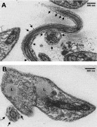
Fig. 2. Transmission EM of Leishmania (V.) braziliensis promastigotes using mAb SST-3. Promastigotes were fixed, incubated sequentially with SST-3 and gold-conjugated anti-mouse IgG, and set in Araldite. F, flagellum; M, mitochondria; L, lipidic vesicles. Arrows indicate gold particle labelling.
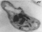
Fig. 3. Transmission EM of Leishmania (V.) braziliensis promastigote using mAb SST-4. Grids were incubated sequentially with SST-4 and gold-conjugated anti-mouse IgG. M, mitochondria; N, nucleus. Arrows indicate gold particle labelling.
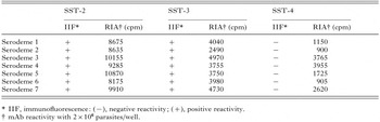
Table 2. Reactivity of mAbs SST-2, SST-3, and SST-4 with various Leishmania braziliensis serodemes, by indirect immunofluorescence (IIF) and solid-phase radioimmunoassay (RIA)
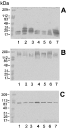
Fig. 4. Western blot of Leishmania(V.) braziliensis promastigotes with mAbs SST-2 (A), SST-3 (B), and SST-4 (C) after separation of whole cell lysates by SDS–PAGE in the absence of β-mercaptoethanol. Samples equivalent to 4·5×106 promastigotes were loaded in each lane. Lanes 1 through 7 correspond to serodemes 1 through 7, respectively.
 You have
Access
- 5
- Cited by