COVID-19 cardiac manifestations are various, including asymptomatic myocardial injury, acute coronary syndrome, mild-to-fulminant myocarditis, stress cardiomyopathy and cardiogenic shock. Despite this variety, the mechanism of cardiac involvement has not yet been clarified. Reference Clerkin, Fried and Raikhelkar1 In this case report, we present a young child with COVID-19 infection who had complete atrioventricular block with severe right ventricular dysfunction.
Case presentation
An 8-year-old boy was admitted to the hospital with abdominal pain and distention that lasted for several days. The patient’s vital signs were within normal limits, except for marked bradycardia. Cardiac murmur, diminished breath sounds and hepatomegaly were also noted. He had neither personal nor a family history of congenital heart disease, immunodeficiency or autoimmune disease. Echocardiography that was performed 2 years ago due to a heart murmur was normal. All the members of the family had COVID-19 infections 4 months ago and received home treatment. Results of COVID-19 immunoglobulin (Ig) M and IgG antibodies were positive. The patient was started on dopamine, milrinone and furosemide infusions. He received intravenous immunoglobin twice at a dose of 1 g/kg within the first 48 hours of hospitalisation as an empirical treatment for potential myocarditis. Immunomodulatory therapy (methylprednisolone, 2 mg/kg once per day), antiviral therapy (favipiravir, 600 mg daily, 5 days in total) and therapeutic anticoagulation with low-molecular weight heparin was initiated. A temporary pacemaker was implanted in another centre on the 7th day of hospitalisation because of a complete heart block and severe bradycardia, and the patient was referred to our centre for further treatment.
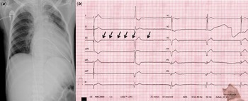
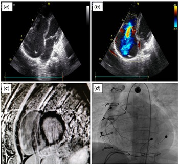
His laboratory findings (complete blood count, inflammatory markers, troponin I, creatine kinase-myocardial band (CK-MB)) were normal. High-sensitivity brain natriuretic peptide (1922 pg/mL; ref.: 0–125 pg/mL) was elevated. The chest radiography was consistent with cardiomegaly (Fig 1a). While briefly changing the temporary battery, the electrocardiogram showed a complete atrioventricular block with a ventricular escape rhythm at a rate of 30 bpm (Fig 1b). The echocardiogram revealed dilatation of the inferior vena cava and hepatic veins, enlarged right atrium and right ventricle (Fig 2a), severe tricuspid regurgitation and severe right ventricular systolic dysfunction with tricuspid annular plane systolic excursion 6 mm and fractional area change 20% (Fig 2b). Near-normal left ventricular systolic function was detected with an ejection fraction of 55%.
( a ) Chest X-ray shows marked cardiomegaly and a temporary pacemaker lead at the right ventricular apex. ( b ) Complete atrioventricular block and ventricular escape rhythm are seen (ventricular rate: 30 bpm) at the 12 lead electrocardiogram (black arrows indicate atrial activity).

( a ) Enlarged right atrium and right ventricle at the echocardiogram. ( b ) Severe tricuspid regurgitation at the echocardiogram. ( c ) Late gadolinium enhacement was detected in the free wall of the right ventricle indicative of diffuse fibrosis in magnetic resonance imaging. ( d ) Hypokinetic right ventricle and narrow right main coronary artery are seen at cardiac catheterisation.

In MRI, the right ventricle was enlarged and globally hypokinetic. Late gadolinium enhancement was detected in the free wall of the right ventricle indicative of diffuse fibrosis (Fig 2c). However, oedema and hyperaemia supporting acute myocarditis were not observed in T1 and T2-weighted images. Evaluation for common infections associated with myocarditis and atrioventricular block identified no other active infectious cause. In addition, there were no signs of rheumatologic disease with cardiac involvement. Cardiac catheterisation revealed a hypokinetic right ventricle and a very narrow right main coronary artery (Fig 2d).
An epicardial, dual-chamber pacemaker was surgically implanted and tricuspid valvuloplasty was performed in our centre because the complete atrioventricular block continued on the 19th day of hospitalisation. An endomyocardial biopsy that was taken during surgery also supported the diagnosis of fibrosis microscopically, which was detected by cardiac magnetic resonance.
Discussion
COVID-19 is an ongoing pandemic caused by SARS-CoV-2, which is thought to infect host cells via angiotensin-converting enzyme 2 receptors and cause COVID-19, but the mechanisms involved in myocardial interaction are not yet clear. Myocardial damage and ventricular dysfunction due to acute myocardial injury are thought to be responsible for the cardiac involvement. Reference Clerkin, Fried and Raikhelkar1 In the latest Chinese report, heart involvement was defined in 12% of adult patients. Reference Huang, Wang and Li2 The most common cardiovascular complications identified in adult patients with COVID-19 are myocardial infarction, rapidly developing fulminant myocarditis with reduced left ventricular function, cardiomyopathies, arrhythmias and venous thromboembolism. Reference Azevedo, Botelho and Hollanda3 However, there are insufficient data about cardiac involvement in children. In this case report, a child who developed right ventricular dysfunction and third-degree atrioventricular block in the chronic phase of COVID-19 infection is presented in order to contribute to the literature.
In the adult literature, atrioventricular block was noticed in the acute phase during hospitalisation. It was emphasised that hypoxia or electrolyte imbalance could trigger cardiac arrhythmias. In most patients, the arrhythmias were temporary and resolved in the following days. Reference Kir, Mohan and Sancassani4,Reference El-Assaad, Hood-Pishchany and Kheir5 However, the mechanism that causes arrhythmia development in children, as in our patient, is unclear, and it is unknown whether the arrhythmia will be temporary. Reference Lakkireddy, Chung and Gopinathannair6 Based on adult data, we waited 2 weeks for the atrioventricular node to resume but complete atrioventricular block continued. We hypothesised that both the hypoperfusion of the AV node and right myocardial cells was due to the very narrow right coronary artery shown in cardiac catheterisation. Therefore, the atrioventricular node damage became permanent.
Acute myocarditis was reported with COVID-19 infection in adults when acute myocardial damage or fulminant myocarditis developed due to COVID-19 infection; troponin-I, CK-MB and high-sensitivity brain natriuretic peptide levels may be elevated. Reference Zeng, Liu and Yuan7 However, troponin results within normal limits cannot exclude myocarditis, especially for atypical forms such as giant cell myocarditis or patients in the chronic phase of COVID-19 infections. Reference Siripanthong, Nazarian and Muser8 Although troponin levels were within normal limits in our patient, the detection of fibrosis in biopsy suggested an inflammatory process that predominantly involved the right ventricle and affected the atrioventricular node. The fact that the patient’s previous echocardiographic examinations were normal also supported this hypothesis.
Cardiac MRI has evolved to become the preferred imaging modality for non-invasive evaluation in acute myocarditis. Reference Hundley and Bluemke9,Reference Friedrich, Larose and Patton10 T2-weighted imaging, early gadolinium enhancement, late gadolinium enhancement, signal intensities and mapping of T1–T2 relaxation times are used to investigate pathophysiologic changes due to oedema, hyperaemia and fibrosis, and identify both acute or chronic changes of myocardial inflammation. In our patient, cardiac MRI failed to show diffuse high signal intensity in T2-weighted images suggesting myocardial wall oedema. In addition to these MRI findings, normal CK-MB and troponin I values helped us to exclude acute myocarditis. Therefore, it was concluded that the patient was in the chronic phase of the disease.
Conclusion
The long-term consequences of cardiovascular effects in children infected with COVID-19 are unknown. This report is the first description of permanent complete heart block and severe right ventricular dysfunction due to COVID-19 in a child. Cardiac MRI, catheterisation and endomyocardial biopsy may guide the differential diagnosis. Additional studies are needed in terms of cardiovascular involvement in the child with COVID-19 infection.
Supplementary material
For supplementary material accompanying this paper visit https://doi.org/10.1017/S1047951121004248
Acknowledgements
We thank our paediatric cardiac surgery team for their valuable support.
Financial support
This research received no specific grant from any funding agency, commercial or not-for-profit sectors.
Conflicts of interest
None.
Ethical standards
The authors assert that this work complies with the ethical standards of the relevant national guidelines on human experimentation and with the Helsinki Declaration of 1975, as revised in 2008. This case was approved by the patient’s family.