INTRODUCTION
The international recommendations of quality factor depend on linear energy transfer (LET) of radiation and increases from 1 to 20 1-Reference Kliauga and Dvorak3. LET, on the other hand increases from 3.5 to 200 keV/µmReference Filyuskkin4. The quality factor assigned for x-rays, gamma rays and electrons as 1, irrespective of the energy. The radiation quality factor represents the degree of radiation injury. In clinical practice, we use energy from 20 kV to 25 MV, which is indeed a wide range of energy. To assume that 25 MV x-rays produce the same biological effect as 20 kV is based on hypothetical assumption and no data is provided as how we have arrived at the quality factor of 1. The accepted quality factor according to FilyushhinReference Filyuskkin4 has no radiobiological basis and certainly need a revision.
In this paper, a new approach is presented to assess the radiation quality factor of x-rays, gamma rays and electrons in mitotic phase of the HeLa cells.
MATERIALS AND METHODS
Experimental data
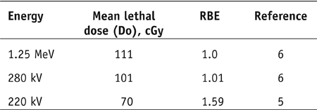
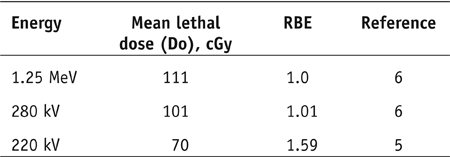
We have abstracted the data on HeLa cells S-3 irradiated in different phases of the cell cycles from the literatureReference Terashima and Tolmach5,Reference Sapozink6. Since the mitotic phase is the most sensitive to x-rays and gamma rays, it is therefore, most appropriate to assess the quality factor of x-rays and gamma rays in the mitotic phase. The mitotic phase of the HeLa cells S-3 at 220 kV, 280 kV and 1.25 MeV are abstracted from the literature along with their mean lethal radiation dose (Do) of 70, 101, and 111 cGy (Table 1), respectively.
RESULTS
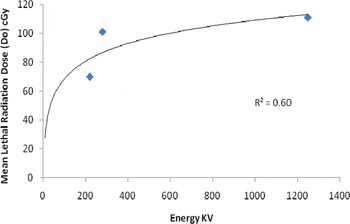
In Figure 1, it is interesting to note that as the energy decreases, the Do value decreases as well. Extrapolating the curve it appears that at lower energy, Do decreases and become more potent. It is interesting to point out that at higher energy such as used in radiation therapy, the induction of secondary cancer is very lowReference Xu, Bednarz and Paganetti7. On the other hand, in mammography (20-35 kVp) the induction of cancer with radiation at low energy is much higher. For example, in fluoroscopy, the women who had multiple fluoroscopic studies for pulmonary tuberculosis ended up with breast cancer and the energy used were 60-120 kVp8-12. On the other hand, during breast treatment in radiation oncology, the contra lateral breast receives 5% of the total radiation dose (6000 cGy) which comes out to be 300 cGy. This does not include the port film dose. However, the induction of cancer in the contra lateral breast is not supported by the published resultsReference Xu, Bednarz and Paganetti7.
Correlation between mean lethal radiation dose (Do) and Energy (kV) in the mitotic phase of the HeLa cells S-3 yielding a correlation of 0.60.

It is well known and generally accepted that 200 kVp x-rays is biologically more effective than 1.25 MeV gamma rays (Co-60)Reference Malone, Porter and Hendry13. Frankenberg et al.Reference Frankenberg, Kelnhofer and Bar14 observed from the experimental study in human hybrid cell line (CGLI) that the RBE of mammography x-rays relative to 200 kVp x-rays was determined to be about four for doses 0.5 Gy. In conclusion they noted, “Both the data available in the literature and the results of present study strongly suggest an increase of RBE (M) for carcinogenesis in animals, neoplastic cells transformation and clastogenic effects with decreasing photon energy or increasing LET to an RBE (M) approximately 8 for mammography x-rays relative to Co-60 gamma rays.”
Supporting the argument of Frankenberg et al.Reference Frankenberg, Kelnhofer and Bar14, the calculated RBE from the present data increases to 1.59 for energy of 220 kV relative to Co-60 gamma rays (Table 1)
DISCUSSION
The question is why the low energy has more potent effect compared to high energy. Chen et al.Reference Chen and Watt15 pointed out that the optimum damage occurs when the mean free path between ionizations is equivalent to the strand separation in the double stranded DNA. This criterion is met by low energy only.
Ionizing radiation in cells is generally attributed to direct impact of high energy quanta and production of OH species. Sanche et al.Reference Sanche16 questioned this age old notion. They therefore, suggested that DNA lesions are indeed induced by the lower energy secondary electrons generated by the primary ionizing radiation. Sanche et al.Reference Sanche16 showed that low energy electrons induce both single and double strand breaks at energy well below the ionization limit of DNA (7.5 eV). Additionally, they revealed that the single or double bond strand breaks are one to two orders of magnitude larger for electrons than for photon of corresponding energy.
It is therefore important that the identification of radiation quality in biological systems within specific energy range should be assigned an appropriate quality factor. This would be most appropriate if a typical cell line (such as HeLa cells S-3) should be chosen and either the RBE or Do values should be assessed in the mitotic phase. Based on these measurements, an appropriate quality factor should be assigned.
Acknowledgements
I would like to thank Ms. Tamara L. Lemon for editing the manuscript.